. 2005 Mar 9;24(5):1033-45.
doi: 10.1038/sj.emboj.7600560. Epub 2005 Feb 10.

The yeast EDC1 mRNA undergoes deadenylation-independent decapping stimulated by Not2p, Not4p, and Not5p

Affiliations

The yeast EDC1 mRNA undergoes deadenylation-independent decapping stimulated by Not2p, Not4p, and Not5p

Denise Muhlrad et al. EMBO J. .

Abstract

A major mechanism of eukaryotic mRNA degradation initiates with deadenylation followed by decapping and 5' to 3' degradation. We demonstrate that the yeast EDC1 mRNA, which encodes a protein that enhances decapping, has unique properties and is both protected from deadenylation and undergoes deadenylation-independent decapping. The 3' UTR of the EDC1 mRNA is sufficient for both protection from deadenylation and deadenylation-independent decapping and an extended poly(U) tract within the 3' UTR is required. These observations highlight the diverse forms of decapping regulation and identify a feedback loop that can compensate for decreases in activity of the decapping enzyme. Surprisingly, the decapping of the EDC1 mRNA is slowed by the loss of Not2p, Not4p, and Not5p, which interact with the Ccr4p/Pop2p deadenylase complex. This indicates that the Not proteins can affect decapping, which suggests a possible link between the mRNA deadenylation and decapping machinery.


Figures

Figure 1
Figure 1
The EDC1 mRNA poly (A) tail has a uniform distribution at steady state. (A) Cartoon of the EDC1 mRNA with pertinent sequences and oligonucleotide locations (probe and o3′) marked. The Northern is probed with oRP1211 (Table III). Distances from the 3′ end of the oligonucleotides used for RNaseH reactions to the 3′ end of the mRNA are bracketed. (B) Polyacrylamide gel of the EDC1 mRNA in wild-type strain yRP683 cut with several combinations of oligonucleotides. Above each lane is the oligonucleotides with which the mRNAs in each lane were cut as shown in part A. Oligo 3′ (o3′)=oRP1209 (Table III). Simultaneous RNaseH cleavage with o3′ and dT and labeled (+) and serial cleavage are designated with an arrow. The markers and their sizes are shown to the left of the left-most lane. Cartooned to each side of the panel are the species of mRNA present in each band. Asterisks denote background bands. Lanes are labeled below for reference. (C) The MFA2pG mRNA from a wild-type strain yRP840 without (−) or with oligo(dT) cleavage. Size markers are shown on the right with their sizes labeled. The bracket designates the distribution of the poly(A) tail.
Figure 2
Figure 2
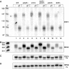
The poly(A) tail of the EDC1 mRNA does not change in strains deficient in mRNA deadenylation. (A) A polyacrylamide Northern gel probed with oRP1211 is shown after cleavage of the EDC1 mRNA with oligo o3′ (oRP1209) without (−) or with (dT) subsequent oligo(dT) cleavage. Deletion strains are marked across the top above each bracket set. WT=yRP840; pop2Δ=yRP1920; ccr4Δ=yRP1616; ccr4Δ pan2Δ=yRP1620; pan2Δ=yRP1619; ski2Δ=yRP1195. M represents the marker lane the band sizes of which are shown on the left. Lanes are numbered across the bottom for reference. (B) The MFA2 mRNA probed with oRP140 in the same deletion strains as panel A. Marker sizes are given on the left. (C, D) The 7s standard probed with oRP100 for Northerns in parts A and B, respectively.
Figure 3
Figure 3
Decay products from the EDC1pG mRNA possess long poly(A) tails. This panel shows a Northern blot of the EDC1pG mRNA (pRP1173) in a wild-type strain yRP683 following different RNaseH- and oligo-directed cleavages specified as follows: no oligo (−), lane 1; oligo(dT) (dT), lane 2; EDC1 oligo (o3′) oPR1209, lane 3; serial EDC1 oRP1209 and oligo(dT) (o3′ → dT), lane 4. M designates the marker lane with the sizes given on the right next to the lane. Simple cartoons to each side show the species of mRNA present in the band designated with an arrow. pG represents the poly(G) structure and pU indicates the 3′ UTR poly(U) sequence.
Figure 4
Figure 4
Analysis of decay and deadenylation of the EDC1pG mRNA (pRP1173) in wild-type strain yRP683 (WT) (left panel) and dcp2Δ strain yRP1358 (right panel). Strains were grown in galactose media and transcription was repressed at zero time by the addition of glucose. Samples were collected at various times after repression marked in minutes above each panel. To allow analysis on a polyacrylamide Northern of the poly(A) tail lengths on both full-length mRNA and the decay fragment, the mRNA was cleaved with an oligo 5′ of the poly(U) sequence, oRP1208, and RNaseH prior to analysis on the Northern gel. The zero minute time point in each panel was also cleaved with oligo(dT) (0dT) and is shown in the left-most lanes. M stands for the markers and their sizes are next to the lanes at the left of each panel. Arrows point to the corresponding mRNA species present in each band, which are cartooned between the Northern panels. pG represents the poly(G) structure and pU indicates the 3′ UTR poly(U) sequence. Below each panel is shown the 7s standard for the respective Northern.
Figure 5
Figure 5
The EDC1 3′ UTR dictates its novel decay properties. (A) The sequence of the region of the 3′ UTR of the EDC1 mRNA. The stop codon is shaded gray. The approximate 3′ end of the mRNA as mapped is shown with an arrow. Areas deleted for analysis are marked within brackets and labeled with the number of bases deleted or ΔU for removal of the poly(U) sequence. (B) Polyacrylamide gel of the parental MFA2pGE transcript (pRP1169) and deletion constructs (pRP1170-Δ50, pRP1171-Δ72, pRP1172-ΔU) with oligo(dT) cleavage (dT) and without (−) in a wild-type strain yRP683. To allow analysis of the poly(A) tail lengths on both full-length mRNA and the decay fragment, the mRNA was cleaved with oligo oRP59 5′ of the poly(U) sequence and RNaseH prior to running the Northern gel. The species present in each band in the two outside lanes are labeled next to these lanes. FL is the full-length message and FRAG is the poly(G) containing decay intermediate. Marker sizes are shown on the left of the marker lane M. Constructs are given above each lane set and correspond to the sequences in part A. Lane numbers are given below the panel for reference. (C) Cleavage of mRNAs MFA2pGE (pRP1168) and EDC1pG (pRP1173) with oligo o3′ (oRP1209), which cleaves 3′ of the poly(U) sequence. Lanes are designated as follows: (−) no oligo cleavage; (o3′) oligo o3′ cleavage; (o3′ → dT) for serial cleavage. Markers (M) are shown on the left with the sizes given and lane numbers are given below the panel for reference.
Figure 6
Figure 6
Mutations in decapping factors affect the EDC1 mRNA. Wild-type (WT) and strains deleted for specific mRNA decapping factors were analyzed on polyacrylamide gels showing the levels and adenylation status of the EDC1 mRNA. Deletion strains are marked above each set of bracketed lanes. Strains are as follows: WT=yRP840; lsm1Δ=yRP1410; pat1Δ=yRP1372; dhh1Δ=yRP1560; xrn1Δ=yRP1359; dcp1Δ=yRP1211; dcp2Δ=yRP1358. All lanes are cleaved with an oligo 5′ of the poly(U) oligo RP1208 (−). Oligo(dT) cleavage in addition to oligo oRP1208 lanes is marked above as (dT). Markers are shown in lane M and the sizes are given on the left. The lower panel is the same Northern probed with oRP100, the 7s loading standard.
Figure 7
Figure 7
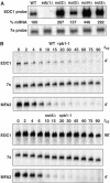
The not2Δ, not4Δ, and not5Δ strains show alterations in the decay of the EDC1 mRNA. (A) Steady-state levels of the EDC1 mRNA in wild-type (WT) (yRP840), not2Δ (yRP1670), not3Δ (yRP1671), not4Δ (yRP1672), and not5Δ (yRP1673) strains. Strains are given across the top of the agarose Northern. The upper panel is probed for the EDC1 mRNA, and the lower panel shows the standardizing 7s probe. Values determined for the %mRNA based on multiple experiments are given between the two panels. (B) Half-life measurements in temperature-sensitive RNA polymerase II (rpb1-1) mutant strains. Wild-type (WT) yRP582 or not4Δ yRP1806 strains containing a temperature-sensitive rpb1-1 mutation were shifted to 37°C and RNA was analyzed at various times in minutes following the shift, which are labeled above each panel set. The top panel shows the half-life of the EDC1 mRNA. The middle panel gives the 7s standardizing mRNA. The bottom panel shows the endogenous MFA2 mRNA to demonstrate the effectiveness of the rpb1-1 shut off. Half-life values for each set of experiments are given on the right of the respective panels.
Figure 8
Figure 8
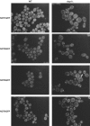
The NOT proteins localize to P-bodies in dcp1Δ strains. The respective NOT1–4-GFP fusion proteins are designated on the left of each row of pictures. Wild-type (WT) strains are shown in the left column for each fusion protein: not1=yRP1817; not2=yRP1818; not3=yRP1819; not4=yRP1820. The dcp1Δ strains are in the right column of pictures: not1=yRp1915; not2=yRP1916; not3=yRP1917; not4=yRP1918. All pictures have identical gain and normalization settings.

Similar articles

Cited by

References

    1. Badis G, Saveanu C, Fromont-Racine M, Jacquier A (2004) Targeted mRNA degradation by deadenylation-independent decapping. Mol Cell 15: 5–15 - PubMed
    1. Beelman CA, Stevens A, Caponigro G, LaGrandeur TE, Hatfield L, Fortner DM, Parker R (1996) An essential component of the decapping enzyme required for normal rates of mRNA turnover. Nature 382: 642–646 - PubMed
    1. Bonnerot C, Boeck R, Lapeyre B (2000) The two proteins Pat1p (Mrt1p) and Spb8p interact in vivo, are required for mRNA decay, and are functionally linked to Pab1p. Mol Cell Biol 20: 5939–5946 - PMC - PubMed
    1. Bouveret E, Rigaut G, Shevchenko A, Wilm M, Seraphin B (2000) A Sm-like protein complex that participates in mRNA degradation. EMBO J 19: 1661–1671 - PMC - PubMed
    1. Brown CE, Sachs AB (1998) Poly (A) tail length control in Saccharomyces cerevisiae occurs by message-specific deadenylation. Mol Cell Biol 18: 6548–6559 - PMC - PubMed

Publication types

MeSH terms