

Cryo-electron tomography of vaccinia virus
- PMID: 15699328
- PMCID: PMC549483
- DOI: 10.1073/pnas.0409825102
Cryo-electron tomography of vaccinia virus
Abstract
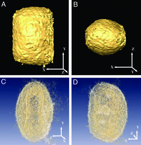
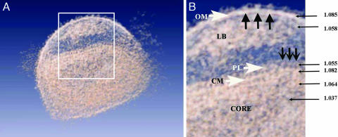
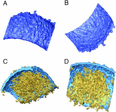
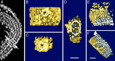
The combination of cryo-microscopy and electron tomographic reconstruction has allowed us to determine the structure of one of the more complex viruses, intracellular mature vaccinia virus, at a resolution of 4-6 nm. The tomographic reconstruction allows us to dissect the different structural components of the viral particle, avoiding projection artifacts derived from previous microscopic observations. A surface-rendering representation revealed brick-shaped viral particles with slightly rounded edges and dimensions of approximately 360 x 270 x 250 nm. The outer layer was consistent with a lipid membrane (5-6 nm thick), below which usually two lateral bodies were found, built up by a heterogeneous material without apparent ordering or repetitive features. The internal core presented an inner cavity with electron dense coils of presumptive DNA-protein complexes, together with areas of very low density. The core was surrounded by two layers comprising an overall thickness of approximately 18-19 nm; the inner layer was consistent with a lipid membrane. The outer layer was discontinuous, formed by a periodic palisade built by the side interaction of T-shaped protein spikes that were anchored in the lower membrane and were arranged into small hexagonal crystallites. It was possible to detect a few pore-like structures that communicated the inner side of the core with the region outside the layer built by the T-shaped spike palisade.
Figures


Similar articles
-
Palisade structure in intact vaccinia virions.mBio. 2024 Feb 14;15(2):e0313423. doi: 10.1128/mbio.03134-23. Epub 2024 Jan 3. mBio. 2024. PMID: 38171004 Free PMC article.
-
Cryo-X-ray tomography of vaccinia virus membranes and inner compartments.J Struct Biol. 2009 Nov;168(2):234-9. doi: 10.1016/j.jsb.2009.07.009. Epub 2009 Jul 16. J Struct Biol. 2009. PMID: 19616103
-
Structure of intracellular mature vaccinia virus observed by cryoelectron microscopy.J Virol. 1994 Mar;68(3):1935-41. doi: 10.1128/JVI.68.3.1935-1941.1994. J Virol. 1994. PMID: 8107253 Free PMC article.
-
Structure of complex viruses and virus-infected cells by electron cryo tomography.Curr Opin Microbiol. 2006 Aug;9(4):437-42. doi: 10.1016/j.mib.2006.06.016. Epub 2006 Jul 10. Curr Opin Microbiol. 2006. PMID: 16829161 Review.
-
Electron microscopy and atomic force microscopy studies of chromatin and metaphase chromosome structure.Micron. 2011 Dec;42(8):733-50. doi: 10.1016/j.micron.2011.05.002. Epub 2011 May 12. Micron. 2011. PMID: 21703860 Review.
Cited by
-
Multiscale perspectives of virus entry via endocytosis.Virol J. 2013 Jun 5;10:177. doi: 10.1186/1743-422X-10-177. Virol J. 2013. PMID: 23734580 Free PMC article. Review.
-
Palisade structure in intact vaccinia virions.mBio. 2024 Feb 14;15(2):e0313423. doi: 10.1128/mbio.03134-23. Epub 2024 Jan 3. mBio. 2024. PMID: 38171004 Free PMC article.
-
The Vaccinia virion: Filling the gap between atomic and ultrastructure.PLoS Pathog. 2019 Jan 7;15(1):e1007508. doi: 10.1371/journal.ppat.1007508. eCollection 2019 Jan. PLoS Pathog. 2019. PMID: 30615658 Free PMC article.
-
Neutralization Determinants on Poxviruses.Viruses. 2023 Dec 8;15(12):2396. doi: 10.3390/v15122396. Viruses. 2023. PMID: 38140637 Free PMC article. Review.
-
The structure of a putative scaffolding protein of immature poxvirus particles as determined by electron microscopy suggests similarity with capsid proteins of large icosahedral DNA viruses.J Virol. 2007 Oct;81(20):11075-83. doi: 10.1128/JVI.00594-07. Epub 2007 Aug 1. J Virol. 2007. PMID: 17670837 Free PMC article.
References
Publication types
MeSH terms
LinkOut - more resources
Full Text Sources
Other Literature Sources
