. 2005 Feb 14;168(4):575-85.
doi: 10.1083/jcb.200407124. Epub 2005 Feb 7.

DRhoGEF2 regulates actin organization and contractility in the Drosophila blastoderm embryo

Affiliations

DRhoGEF2 regulates actin organization and contractility in the Drosophila blastoderm embryo

Mojgan Padash Barmchi et al. J Cell Biol. .

Abstract

Morphogenesis of the Drosophila melanogaster embryo is associated with a dynamic reorganization of the actin cytoskeleton that is mediated by small GTPases of the Rho family. Often, Rho1 controls different aspects of cytoskeletal function in parallel, requiring a complex level of regulation. We show that the guanine triphosphate (GTP) exchange factor DRhoGEF2 is apically localized in epithelial cells throughout embryogenesis. We demonstrate that DRhoGEF2, which has previously been shown to regulate cell shape changes during gastrulation, recruits Rho1 to actin rings and regulates actin distribution and actomyosin contractility during nuclear divisions, pole cell formation, and cellularization of syncytial blastoderm embryos. We propose that DRhoGEF2 activity coordinates contractile actomyosin forces throughout morphogenesis in Drosophila by regulating the association of myosin with actin to form contractile cables. Our results support the hypothesis that specific aspects of Rho1 function are regulated by specific GTP exchange factors.


Figures
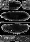
Figure 1.
Figure 1.
Expression of DRhoGEF2 during embryonic development. (A–C) DRhoGEF2 protein accumulates apically in cells of the invaginating ventral furrow (A and C, arrows), in cells of the posterior midgut primordium (B, arrow), and in cells in the cephalic fold (C, arrowhead). (D) At late stage 11, DRhoGEF2 is apically localized in all epidermal cells. (E) At late stage 14, DRhoGEF2 is enriched in a periodic pattern in the epidermis (arrows) and in commissural axon tracts (arrowhead). (F) DRhoGEF2 is enriched in longitudinal and commissural axon tracts in the central nervous system. (G) During dorsal closure of the epidermis, DRhoGEF2 is highly enriched at the apical cortex of leading edge cells. Anterior is to the left. B–E show lateral views; A and F show ventral views; and G shows dorsal view. Bars: (A–C) 10 μm; (D–G) 50 μm.
Figure 2.
Figure 2.
Distribution of DRhoGEF2 during blastoderm cellularization. DRhoGEF2 is shown in green; actin is shown in red, and DNA is shown in blue. (A) At the interphase of syncytial divisions DRhoGEF2 is concentrated in apical actin caps. (B) During syncytial nuclear cycles DRhoGEF2 redistributes to the metaphase furrows. The DRhoGEF2 domain is narrower than the actin domain (arrow). DRhoGEF2 is also observed in large cytoplasmic vesicles. (C) At the onset of cellularization, DRhoGEF2 redistributes to the furrow canal (arrow). Apical DRhoGEF2 levels are low. (D) DRhoGEF2 remains concentrated at the base of the furrow canal throughout membrane invagination. Apical DRhoGEF2 levels increase during the slow phase. (E) During basal closure, DRhoGEF2 is enriched at the basal-most edge of blastoderm cells in the region where actin rings are located (arrowheads). Left three panels show sagittal views; right panels show grazing view. Bars, 10 μm.
Figure 3.
Figure 3.
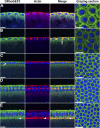
Phenotype of DRhoGEF2 mutants during blastoderm cellularization. Actin is shown in red; and DNA is shown in blue. (A and A′) Metaphase furrows at cycle 13 in the wild type are shown. (B and B′) Same stage in DRhoGEF2 l(2)04291 is shown. Metaphase furrows invaginate to variable depths (B, arrow) and actin is irregularly distributed. Sometimes furrow formation fails (B′, arrow). Abnormal nuclei are eliminated from the cell surface (B′, arrowhead; and Video 1). (C and C′) Slow phase in the wild type is shown. (D and D′) Same stage in DRhoGEF2 l(2)04291 is shown. Significant amounts of actin fail to redistribute to the furrow canal, but remain apical (D, F, and H, arrows). Nuclei are not squeezed by the furrow and are wider than in the wild type. Actin rings have a rounded rather than a hexagonal shape, are irregular in size, and are frequently multinucleated (D′). (E and E′) Onset of the fast phase in the wild type is shown. (F and F′) Same stage in DRhoGEF2 l(2)04291 is shown. The furrow canal does not expand and no constriction of actin rings is observed. (G and G′) Shown is the basal closure in the wild type. (H and H′) Same stage in DRhoGEF2 l(2)04291. Actin rings are irregular in size and some remain very large. A–H show sagittal views at the same magnification; A′–H′ show grazing views at the same magnification. Bars, 10 μm.
Figure 4.
Figure 4.
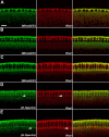
Localization of DRhoGEF2 with respect to Rho1 and β-Heavy spectrin. (A–C) DRhoGEF2 (green) and Rho1 (red) colocalize precisely at the furrow canal during membrane invagination. (D and E) β-Heavy spectrin (green) and Rho1 (red) are localized in adjacent nonoverlapping membrane subdomains (compare arrowheads). (right) Merge of the left two panels. All panels have the same magnification. Bar, 10 μm.
Figure 5.
Figure 5.
Localization of DRhoGEF2 in Rho1L3, dia5 and, bnk mutants. (A) Rho1 L3 embryo stained for actin (red) and DRhoGEF2 (green). Example of severe phenotype. Although actin structures are severely disrupted, DRhoGEF2 is concentrated at actin-rich structures (compare arrows). (B) Rho1 L3 embryo stained for Rho1 (red) and DRhoGEF2 (green). Example of less severe phenotype (possibly caused by paternal rescue; Fig. S3). DRhoGEF2 localization is unaffected although Rho1 is reduced to background levels (compare arrows). (C) DRhoGEF2 l(2)04291 embryo stained for Rho1 (red) and Dia (green). Concentration of Rho1 at the furrow canal is abrogated (compare arrows). Localization of Dia at the furrow canal is not affected. (D) dia 5 embryo stained for actin (red) and DRhoGEF2 (green). DRhoGEF2 concentration at the furrow canal (arrow) is unaffected. (E) Df(3R)tll-e embryo (bnk mutant) stained for actin (red) and DRhoGEF2 (green). Actin rings contract prematurely pinching the nuclei. DRhoGEF2 distribution is not affected by the absence of bnk function. DNA is stained in blue. (right) Merge of left two panels. Bars, 10 μm.
Figure 6.
Figure 6.
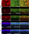
Localization of Bnk in DRhoGEF2 mutants and phenotype of DRhoGEF2-bnk double mutants. (A–D) Bnk. Insets depict the same image with Bnk in green and actin in red. (E–H) Actin is shown in red; and DNA is shown in blue. (A′–H′) Corresponding grazing views of A–H. (A) Wild type at the slow phase is shown. (B) Same stage as A in DRhoGEF2 l(2)04291 is shown. Bnk concentration at the furrow canal is reduced (compare arrows in A and B). (C) Wild type at the end of the slow phase is shown. (D) Same stage as C in DRhoGEF2 l(2)04291 is shown. Bnk is not detectable at the furrow canal. (E–H) DRhoGEF2-bnk double mutants. (E) Metaphase furrows invaginate nonuniformly and occasionally fail to form (E′, arrow). Nuclei drop out of the cortical layer (E, arrowhead) and leave behind empty pseudocells (E′, arrowhead). (F) Early slow phase. Between some nuclei the furrow canal fails to invaginate (F, arrow) leading to multinucleated actin rings (F′). (G) Late slow phase. The actin network progressively disintegrates. Remaining actin rings do not constrict as in bnk mutants (arrow). (H) At later stages (as judged by the increased depth at which actin is found), actin forms randomly distributed aggregates. A–H show sagittal views at same magnification; A′–H′ show grazing views at same magnification. Bars, 10 μm.
Figure 7.
Figure 7.
Rescue of DRhoGEF2 mutants and ectopic DRhoGEF2 expression. (A) DRhoGEF2 l(2)04291 cuticle showing ventral hole. (B) Cuticle of DRhoGEF2 l(2)04291 larva ubiquitously expressing UAS-DRhoGEF2-RE. The ventral open phenotype is rescued. (C) prd-Gal4, UAS-DRhoGEF2-RE embryo at stage 10 is shown. Bnk (red) accumulates in prd-domains in the epidermis. (C′) Same as in C showing DRhoGEF2 in green and Bnk in red. Bars, 50 μm. Anterior is to the left. Lateral views are shown, except for A, which is a ventro-lateral view.
Figure 8.
Figure 8.
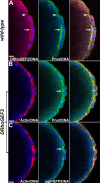
Localization of DRhoGEF2 during pole cell formation. (A) Wild type. DRhoGEF2 (red) colocalizes with Pnut (green) at the base of polar buds (arrowhead) and at contractile rings at the base of pole cells (arrow). (B) DRhoGEF2 l(2)04291. Localization of Pnut (green) at the contractile ring is unaffected (arrow). (C) DRhoGEF2 l(2)04291. Localization of myosin II (sqh-GFP, green) at the contractile ring is unaffected (arrow). (B and C) Actin is shown in red. (right) Merge of left two panels. Bars, 10 μm.
Figure 9.
Figure 9.
Role of DRhoGEF2 during pole cell formation. Actin is shown in red; and DNA is shown in blue. (A) Actin is irregularly distributed in polar buds. (B) Cortical actin of pole cells fails to separate from the syncytium during interphase. (C) At metaphase, actin is disorganized and furrow formation fails in the pole cell region. (D) At the onset of cellularization, actin surrounding the pole cells is disorganized and nuclei accumulate in the yolk at the posterior pole (arrow). (E) During the slow phase, pole cells remain embedded in the somatic nuclear layer. (F) The invaginating cellularization front obliterates the pole cells. (G) At the fast phase, most pole cells have been lost. All panels same magnification. Bar, 10 μm.

Similar articles

Cited by

References

    1. Afshar, K., B. Stuart, and S.A. Wasserman. 2000. Functional analysis of the Drosophila diaphanous FH protein in early embryonic development. Development. 127:1887–1897. - PubMed
    1. Banerjee, J., and P.B. Wedegaertner. 2004. Identification of a novel sequence in PDZ-RhoGEF that mediates interaction with the actin cytoskeleton. Mol. Biol. Cell. 15:1760–1775. - PMC - PubMed
    1. Barrett, K., M. Leptin, and J. Settleman. 1997. The Rho GTPase and a putative RhoGEF mediate a signaling pathway for the cell shape changes in Drosophila gastrulation. Cell. 91:905–915. - PubMed
    1. Bate, M., and A. Martinez Arias. 1993. The Development of Drosophila melanogaster. Cold Spring Harbor Laboratory Press, Cold Spring Harbor, NY. 1558.
    1. Brand, A.H., and N. Perrimon. 1993. Targeted gene expression as a means of altering cell fates and generating dominant phenotypes. Development. 118:401–415. - PubMed

Publication types

MeSH terms