. 2005 Feb 7;33(3):807-15.
doi: 10.1093/nar/gki197. Print 2005.

Catalytic mechanism of Escherichia coli ribonuclease III: kinetic and inhibitor evidence for the involvement of two magnesium ions in RNA phosphodiester hydrolysis

Affiliations

Catalytic mechanism of Escherichia coli ribonuclease III: kinetic and inhibitor evidence for the involvement of two magnesium ions in RNA phosphodiester hydrolysis

Weimei Sun et al. Nucleic Acids Res. .

Abstract

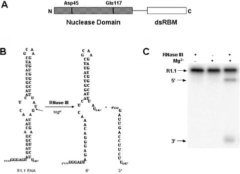
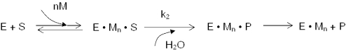
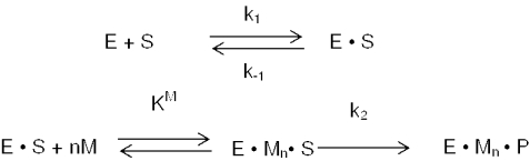
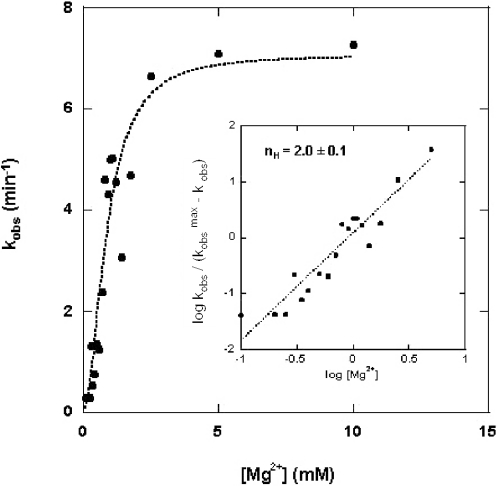
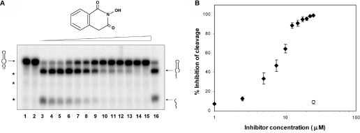
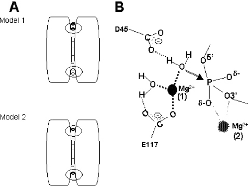
Escherichia coli ribonuclease III (RNase III; EC 3.1.24) is a double-stranded(ds)-RNA-specific endonuclease with key roles in diverse RNA maturation and decay pathways. E.coli RNase III is a member of a structurally distinct superfamily that includes Dicer, a central enzyme in the mechanism of RNA interference. E.coli RNase III requires a divalent metal ion for activity, with Mg2+ as the preferred species. However, neither the function(s) nor the number of metal ions involved in catalysis is known. To gain information on metal ion involvement in catalysis, the rate of cleavage of the model substrate R1.1 RNA was determined as a function of Mg2+ concentration. Single-turnover conditions were applied, wherein phosphodiester cleavage was the rate-limiting event. The measured Hill coefficient (n (H)) is 2.0 +/- 0.1, indicative of the involvement of two Mg2+ ions in phosphodiester hydrolysis. It is also shown that 2-hydroxy-4H-isoquinoline-1,3-dione--an inhibitor of ribonucleases that employ two divalent metal ions in their catalytic sites--inhibits E.coli RNase III cleavage of R1.1 RNA. The IC50 for the compound is 14 microM for the Mg2+-supported reaction, and 8 microM for the Mn2+-supported reaction. The compound exhibits noncompetitive inhibitory kinetics, indicating that it does not perturb substrate binding. Neither the O-methylated version of the compound nor the unsubstituted imide inhibit substrate cleavage, which is consistent with a specific interaction of the N-hydroxyimide with two closely positioned divalent metal ions. A preliminary model is presented for functional roles of two divalent metal ions in the RNase III catalytic mechanism.


Figures

Figure 1
Figure 1
E.coli ribonuclease III and the phage T7 substrate, R1.1 RNA. (A) Domain structure of the E.coli RNase III polypeptide. The positions of several conserved residues in the nuclease domain are indicated. (B) Sequence and structure of R1.1 RNA. The secondary structure is that originally proposed by Dunn and Studier (31). The cleavage site is indicated by the arrow, with the two products of cleavage 47 and 13 nt in size. (C) Gel electrophoretic pattern of R1.1 RNA cleavage, showing the requirement for Mg2+. Experimental conditions involved incubation of internally 32P-labeled R1.1 RNA (synthesized using [α-32P]UTP) with RNase III for 5 min at 37°C (see Materials and Methods). The reaction products were separated by electrophoresis in a 15% polyacrylamide gel and then visualized by phosphorimaging. The partial reaction displayed is similar to the gel electrophoretic patterns analyzed in the kinetic analyses (see Results).
Scheme 1
Scheme 1
Scheme 2
Scheme 2
Figure 2
Figure 2
Mg2+ concentration dependence of the rate of substrate cleavage. Cleavage reactions involved internally 32P-labeled R1.1 RNA (1.1 μM) and RNase III (790 nM) (see Materials and Methods). Cleavage rates were based on the production of the 47 nt product (see Figure 1B and C). The kobs values (min−1) were determined and plotted as a function of the Mg2+ concentration according to Equation 1 (see Results). The best-fit curve used an n value of 2. The inset displays the Hill analysis of the kinetic data. Here, the kobs values were plotted as a function of the log of the Mg2+ concentration according to Equation 2 (see Results). The slope of the best-fit line is 2.0, with a standard error of ±0.1.
Figure 3
Figure 3
N-hydroxyimide inhibition of R1.1 RNA cleavage by RNase III in the presence of Mg2+ ion. (A) Cleavage assay carried out in the presence of increasing concentrations of 2-hydroxy-4H-isoquinoline-1,3-dione (structure shown above gel image). The amount of internally 32P-labeled R1.1 RNA (synthesized using [α-32P]CTP) was 10–40 nmol, and the amount of RNase III was ∼100 fmol. MgCl2 (10 mM final concentration) was added to initiate the reaction, with a reaction time of 2 min at 37°C. Reactions were electrophoresed in a 15% polyacrylamide gel and visualized by phosphorimaging (see Materials and Methods for additional information). Lane 1 displays a reaction where substrate was incubated with RNase III in the absence of Mg2+. Lane 2 displays a reaction where substrate was incubated with Mg2+ in the absence of RNase III. Lane 3 is the complete reaction, but without added inhibitor. Lanes 4–13 display reactions carried out in the presence of 2.5, 5, 7.5, 10, 12.5, 15, 17.5, 20, 22.5 and 25 μM inhibitor, respectively. Lane 14 is a control reaction lacking inhibitor, but containing the same amount of ethanol as in the reaction in lane 13. The position of R1.1 RNA is shown on the left and the positions of the two product fragments are on the right. The asterisks indicate positions of small amounts of nonenzymatic breakdown products. (B) Cleavage assay carried out in the presence of increasing concentrations of 2-methoxy-1,3(2H,4H)-isoquinolinedione (structure shown above the gel image). The reactions in lanes 1–3 correspond to those of lanes 1–3 in the experiment shown in (A) (see above), and the reactions in lanes 4–8 contain 5, 10, 25, 100 and 250 μM of the compound, respectively. Lane 9 is a control reaction lacking the compound, but containing the same amount of ethanol as in the reaction in lane 8. (C) Graphic representation of the inhibitory action of the N-hydroxyimide. The solid triangles represent the percentage inhibition of cleavage by the N-hydroxyimide. The solid squares indicate inhibition by the O-methylated N-hydroxyimide. The open circle represents the effect of ethanol alone on the cleavage reaction.
Figure 4
Figure 4
Noncompetitive inhibitory behavior of 2-hydroxy-4H-isoquinoline-1,3-dione. The initial rate of cleavage of 5′-32P-labeled R1.1 RNA was measured in the presence of several concentrations of the N-hydroxyimide. The RNase III concentration was 10 nM and the buffer consisted of 160 mM NaCl, 10 mM MgCl2, 30 mM Tris–HCl (pH 7.9), 0.1 mM EDTA, 0.1 mM DTT, 5% glycerol and 5 μg/ml tRNA. The reaction was initiated by adding MgCl2 and quenched by adding EDTA (20 mM) final concentration. Reaction times were 0.5 and 1 min (37°C), and the N-hydroxyimide concentrations were 0, 10 and 20 μM (designated by the filled circles, open circles and filled triangles, respectively). The best-fit lines in the double-reciprocal plot exhibit different y-intercepts but share the same x-intercept, consistent with a noncompetitive (mixed) mode of inhibition (36,37) (see also Discussion). The Km and kcat values for the reaction in the absence of inhibitor are 14 nM and 2.5 min−1, respectively. These can be compared with values of 42 nM and 1.16 min−1, as determined in a separate study of R1.1 RNA cleavage kinetics (24).
Figure 5
Figure 5
2-hydroxy-4H-isoquinoline-1,3-dione inhibition of R1.1 RNA cleavage by RNase III in the presence of Mn2+ ion. (A) Cleavage assay carried out in the presence of increasing concentrations of 2-hydroxy-4H-isoquinoline-1,3-dione (structure shown above gel image). The amount of internally 32P-labeled R1.1 RNA (synthesized using [α-32P]UTP) was 10–40 nmol and the amount of RNase III was 100 fmol. MnCl2 (2 mM final concentration) was added to initiate the reaction, with a reaction time of 2 min. Reactions were electrophoresed in a 15% polyacrylamide gel and visualized by phosphorimaging (see Materials and Methods for additional information). Lane 1 displays a reaction where substrate was incubated with RNase III in the absence of Mn2+. Lane 2 displays a reaction where substrate was incubated with Mn2+ in the absence of RNase III. Lane 3 is the complete reaction, lacking inhibitor. Lanes 4–15 display reactions carried out in the presence of 0.5, 1, 2.5, 5, 7.5, 10, 12.5, 15, 17.5, 20, 22.5 and 25 μM inhibitor, respectively. Lane 16 displays a control reaction lacking inhibitor, but containing the same amount of ethanol as in the reaction in lane 15. The position of R1.1 RNA is shown on the left and the positions of the two product fragments are indicated on the right. (B) Graphic representation of the inhibitory action of the N-hydroxyimide on the Mn2+-supported cleavage reaction. The solid rhombuses (average of three experiments) represent inhibition by the N-hydroxyimide. The open circle represents the effect of ethanol alone on cleavage of substrate (reaction in lane 16).
Figure 6
Figure 6
Model for a two-metal-ion-dependent catalytic mechanism for RNase III. (A) Alternative binding modes for two Mg2+ ions to the nuclease domain. The diagram depicts the homodimeric structure of the nuclease domain and the location of the binding sites for Mg2+ (or Mn2+) at each end of the subunit interface (20,26). The dsRBDs of each subunit are not shown. Metal ions are indicated by the filled circles. A rectangle indicates a functional active site; and a circle indicates a nonfunctional active site, with site functionality defined here by metal ion occupancy. The assumption in the two models is that, in the absence of substrate, RNase III already carries a single Mg2+ ion at each site, as observed in the crystal structures (20,26). A catalytic requirement for two Mg2+ ions either would indicate a requirement for double occupancy of at least one of the two sites (Model 1, Mg2+ ions denoted by filled circles), or single occupancy of both sites (Model 2). Note that Model 2 would not require the binding of additional metal ions. The experimental data support Model 1 (see Results and Discussion). A modified Model 1 would include the binding of a second Mg2+ ion to the second active site and would be invoked if a substrate is destined to be cleaved in a concomitant manner at two target sites. (B) Proposed involvement of two Mg2+ ions in the catalytic mechanism. The diagram derives from Model 1 (A) and is based on structural (20) and enzymological (24,34,56) data. The Mg2+ ion in binding site 1 (observed in the crystal structure) would activate the water nucleophile (see Discussion). For simplicity, the figure shows only two (D45 and E117) of the four (E41, D45, D114, E117) carboxyl side chains that have been shown in A.aeolicus RNase III to coordinate the Mg2+ ion in binding site 1 (20,26). Moreover, a third metal-bound water molecule is not shown. The Mg2+ ion in binding site 2 (proposed placement) could (i) enhance the electrophilicity of the phosphorus and neutralize the negative charge of the pentacoordinate intermediate, and (ii) protonate or otherwise increase the acidity of the 3′-oxygen leaving group. Note that binding of substrate would be involved in the creation of binding site 2. The identity(ies) of the protein moieties that coordinate the second Mg2+ ion are not known.

Similar articles

Cited by

References

    1. Court D.L. RNA processing and degradation by RNase III. In: Belasco J.G., Brawerman G., editors. Control of Messenger RNA Stability. New York: Academic Press; 1993. pp. 71–116.
    1. LaMontagne B., Larose S., Boulanger J., AbouElela S. The RNase III family: a conserved structure and expanding functions in eukaryotic dsRNA metabolism. Curr. Issues Mol. Biol. 2001;3:71–78. - PubMed
    1. Nicholson A.W. The ribonuclease superfamily: forms and functions in RNA maturation, decay, and gene silencing. In: Hannon G.J., editor. RNAi: A Guide to Gene Silencing. Cold Spring Harbor, New York: Cold Spring Harbor Laboratory Press; 2003. pp. 149–174.
    1. Bernstein E., Caudy A.A., Hammond S.M., Hannon G.J. Role for a bidentate nuclease in the initiation step of RNA interference. Nature. 2001;409:363–366. - PubMed
    1. Provost P., Dishart D., Doucet J., Frendewey D., Samuelsson B., Radmark O. Ribonuclease activity and RNA binding of recombinant human dicer. EMBO J. 2002;21:5864–5874. - PMC - PubMed

Publication types