. 2005 Feb 23;24(4):813-23.
doi: 10.1038/sj.emboj.7600527. Epub 2005 Feb 3.

Perinuclear Mlp proteins downregulate gene expression in response to a defect in mRNA export

Affiliations

Perinuclear Mlp proteins downregulate gene expression in response to a defect in mRNA export

Patrizia Vinciguerra et al. EMBO J. .

Abstract

The mRNA export adaptor Yra1p/REF contributes to nascent mRNP assembly and recruitment of the export receptor Mex67p. yra1 mutants exhibit mRNA export defects and a decrease in LacZ reporter and certain endogenous transcripts. The loss of Mlp1p/Mlp2p, two TPR-like proteins attached to nuclear pores, rescues LacZ mRNA levels and increases their appearance in the cytoplasm, without restoring bulk poly(A)+ RNA export. Chromatin immunoprecipitation, FISH and pulse-chase experiments indicate that Mlps downregulate LacZ mRNA synthesis in a yra1 mutant strain. Microarray analyses reveal that Mlp2p also reduces a subset of cellular transcripts in the yra1 mutant. Finally, we show that Yra1p genetically interacts with the shuttling mRNA-binding protein Nab2p and that loss of Mlps rescues the growth defect of yra1 and nab2 but not other mRNA export mutants. We propose that Nab2p and Yra1p are required for proper mRNP docking to the Mlp platform. Defects in Yra1p prevent mRNPs from crossing the Mlp gate and this block negatively feeds back on the transcription of a subset of genes, suggesting that Mlps link mRNA transcription and export.


Figures
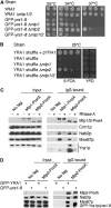
Figure 1
Figure 1
(A) Deletion of MLP2 or MLP1 rescues the ts phenotype of GFP-yra1-8. In all, 10-fold dilutions of wt YRA1 or GFP-yra1-8 strains as such, or in combination with Δmlp1, Δmlp2 or Δmlp1Δmlp2, were spotted on YEPD plates and incubated at 25 (two last dilutions only), 34 or 37°C for 3 days. (B) Deletion of MLP2 bypasses the requirement for Yra1p. The YRA1 shuffle strain (yra1::HIS3, pURA3-YRA1) as such, or combined with Δmlp1, Δmlp2 or Δmlp1/2, was spotted on a control plate (YEPD; two last dilutions only) or on medium containing 5-FOA to select against pURA3-YRA1. The YRA1 shuffle strain transformed with a wt YRA1 plasmid (pFS1877) served as positive control. (C) Mlp proteins physically interact with Yra1p and Mex67p in an RNA-dependent manner. Extracts prepared from a nontagged strain (FSY1026) or from strains expressing Mlp1-ProtA (FSY1567) or Mlp2-ProtA (FSY1351) fusions were treated (+) or not treated (−) with RNase A and purified on Pan Mouse IgG Dynabeads. Total extracts (input, left) or affinity-purified (IgG, right) extracts were analysed by Western blotting with antibodies against ProtA or the indicated proteins. Two different exposures are shown for the immunoprecipitation of Yra1p, expressed from the YRA1 cDNA. (D) The GFP-yra1-8 mutation affects mRNP composition and interaction with Mlp2p. Extracts prepared from nontagged or Mlp2-ProtA-tagged strains expressing wt GFP-Yra1p or GFP-yra1-8 and shifted to 37°C for 2.5 h were purified on Pan Mouse IgG Dynabeads. Total or affinity-purified extracts were analysed by Western blotting as above. GFP-yra1-8 migrates slightly more slowly than GFP-Yra1p.
Figure 2
Figure 2
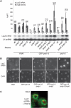
(A) Loss of Mlp1p and Mlp2p restores LacZ mRNA levels in GFP-yra1-8. Strains expressing wt Yra1p (FSY1485) or mutant GFP-yra1-8 (FSY1486) or yra1-8 (FSY1786) proteins as such, or in combination with the indicated Δmlp1, Δmlp2 or Δrrp6 simple, double or triple disruptions, were transformed with the reporter plasmid pLGSD5 (Legrain and Rosbash, 1989) encoding LacZ from a galactose-inducible promoter. Transformants were pregrown in selective medium containing 2% glycerol/2% lactate/0.05% glucose, and LacZ expression was induced by the addition of 3% galactose/1% raffinose for 2.5 h at 37°C. LacZ mRNA levels (light grey bars), defined by primer extension analysis of total RNA, were quantified and normalized to the U1 snRNA internal control. The same strains were analysed for β-galactosidase (β-gal) activity (dark grey bars). LacZ mRNA levels and β-galactosidase enzymatic activities were expressed as a percentage of wt and compared on the same histogram. Error bars have been derived from three independent experiments. (B) In situ localization of LacZ transcripts. The indicated strains transformed with pLGSD5 were induced with galactose as above and processed for in situ hybridization with Cy3-conjugated oligonucleotide probes specific for LacZ mRNA (panels a–e). DAPI staining indicates the position of the nucleus (panels f–j). RNA distribution was examined in fixed cells using a Zeiss axioplan fluorescent microscope with × 100 objective. Exposure time was of 4 s in panels a–d and only 1 s in panel e. (C) LacZ transcripts are located at the nuclear periphery. wt cells transformed with pLGSD5 were induced and prepared as above. Fixed cells were hybridized with Cy3-conjugated LacZ probes and subsequently immunostained with anti-Nsp1p monoclonal and FITC-labelled secondary antibodies. Samples were analysed and images taken with a Zeiss LSM510 confocal microscope.
Figure 3
Figure 3
LacZ mRNA decay rates differ in GFP-yra1-8 and GFP-yra1-8Δmlp2. Exponential cultures of wt YRA1, YRA1Δmlp2, GFP-yra1-8 and GFP-yra1-8Δmlp2 strains transformed with the pGAL1-LacZ reporter construct were induced in medium containing 3% galactose/1% raffinose for 2.5 h at 37°C, transferred to preheated medium containing 2% glucose and collected after various times in glucose at 37°C. LacZ transcripts were quantified by primer extension and their levels expressed as a percentage of wt at time 0. LacZ mRNA half-lives were calculated as described (Materials and methods).
Figure 4
Figure 4
ChIP analysis of TBP binding to pGAL1-LacZ promoter. The indicated strains transformed with the pGAL1-LacZ reporter were induced with galactose for 2.5 h at 37°C prior to crosslinking with formaldehyde. Crosslinked and sonicated extracts were immunoprecipitated with antibodies against TBP, and copurifying DNA was amplified by real-time PCR with primers specific for the GAL1 promoter. The relative abundance of the GAL1 promoter segment in each immunoprecipitate was expressed as a fold increase with respect to a nontranscribed intergenic region value set to 1. TBP binding in the different strains was then expressed as a percentage of binding in wt YRA1. In wt YRA1, a 20-fold (±5) increase of TBP was measured at the GAL1 promoter in the presence of galactose. Values were derived from four independent experiments.
Figure 5
Figure 5
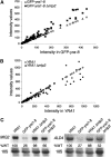
Mlp2p downregulates a subset of cellular transcripts in GFP-yra1-8 but not in wt YRA1. (A) The DNA microarray intensity values of the 194 transcripts decreased in GFP-yra1-8 (open triangles), and their values in GFP-yra1-8Δmlp2 (closed squares) were plotted as a function of the intensity of these 194 transcripts in GFP-yra1-8. (B) The intensity values of the same 194 selected transcripts in wt YRA1 (open diamonds) and YRA1Δmlp2 (closed circles) were plotted as a function of transcript intensity in wt YRA1. (C) Mlp2p downregulates MIG2 and ALD4 transcripts in GFP-yra1-8 but not in wt YRA1. Exponentially growing cultures of the indicated strains were shifted for 2.5 h to 37°C. Total RNA was extracted and examined by Northern blot analysis with MIG2- and ALD4-specific probes. The same blots were rehybridized with a probe specific for 18S RNA to correct for unequal loading.
Figure 6
Figure 6
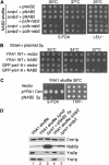
Nab2p genetically interacts with Mlp proteins and Yra1p. (A) Deletion of MLP1 or MLP2 rescues the ts phenotype of ΔN-nab2. The NAB2 shuffle strain (nab2::HIS3, pURA3-NAB2) as such, or deleted for MLP1 or MLP2, was transformed with wt NAB2 (pAC717) or ΔN-nab2 (pAC1152) plasmids. Transformants were spotted as 10-fold dilutions on 5-FOA to select against the wt pURA3-NAB2 plasmid or on a LEU− selective plate to control for cell number, and incubated for 5 days at 25, 30 or 37°C as indicated. (B) Overexpression of Nab2p partially rescues the GFP-yra1-8 ts phenotype. Wt YRA1 and GFP-yra1-8 mutant strains were transformed with empty vector or a high-copy plasmid expressing wt NAB2 (pFS2764). Transformants were spotted on selective plates and incubated for 3 days at 25, 34 or 37°C. (C) Overexpression of Nab2p bypasses the requirement for Yra1p. The YRA1 shuffle strain (yra1::HIS3, pURA3-YRA1) was transformed with an empty vector, a wt YRA1 plasmid (pFS1878) or a high-copy plasmid expressing NAB2 (pFS2764). Transformants were spotted as 10-fold serial dilutions on 5-FOA or on a selective plate as above. Plates were incubated for 5 days at 30°C. (D) The loss of Mlp2p or overexpression of Nab2p does not bypass the requirement for Yra1p by inducing Yra2p overexpression. The indicated strains were grown at 25°C in YEPD and proteins from equal amounts of cells were extracted and analysed by Western blotting with antibodies against the indicated proteins.
Figure 7
Figure 7
Model. Mlp proteins establish a link between mRNA synthesis and export. In wt cells, the mRNA-binding proteins Yra1p and Nab2p are essential for mRNP docking to the Mlp export gate at the nuclear periphery. mRNP complexes produced in the GFP-yra1-8 mutant strain are retained by the Mlp selective filter and mRNP stalling negatively feeds back on mRNA synthesis. Loss of Mlp1p or Mlp2p alleviates the negative effect on mRNA synthesis and allows a fraction of transcripts to reach the cytoplasm. Whether Mlp proteins physically interact with nascent transcripts has to be determined.

Similar articles

Cited by

References

    1. Bangs P, Burke B, Powers C, Craig R, Purohit A, Doxsey S (1998) Functional analysis of Tpr: identification of nuclear pore complex association and nuclear localization domains and a role in mRNA export. J Cell Biol 143: 1801–1812 - PMC - PubMed
    1. Bousquet-Antonelli C, Presutti C, Tollervey D (2000) Identification of a regulated pathway for nuclear pre-mRNA turnover. Cell 102: 765–775 - PubMed
    1. Burkard KT, Butler JS (2000) A nuclear 3′–5′ exonuclease involved in mRNA degradation interacts with Poly(A) polymerase and the hnRNA protein Npl3p. Mol Cell Biol 20: 604–616 - PMC - PubMed
    1. Casolari JM, Brown CR, Komili S, West J, Hieronymus H, Silver PA (2004) Genome-wide localization of the nuclear transport machinery couples transcriptional status and nuclear organization. Cell 117: 427–439 - PubMed
    1. Chavez S, Beilharz T, Rondon AG, Erdjument-Bromage H, Tempst P, Svejstrup JQ, Lithgow T, Aguilera A (2000) A protein complex containing Tho2, Hpr1, Mft1 and a novel protein, Thp2, connects transcription elongation with mitotic recombination in Saccharomyces cerevisiae. EMBO J 19: 5824–5834 - PMC - PubMed

Publication types

MeSH terms