. 2005 Feb 23;24(4):717-29.
doi: 10.1038/sj.emboj.7600559. Epub 2005 Feb 3.

PACS-2 controls endoplasmic reticulum-mitochondria communication and Bid-mediated apoptosis

Affiliations

PACS-2 controls endoplasmic reticulum-mitochondria communication and Bid-mediated apoptosis

Thomas Simmen et al. EMBO J. .

Erratum in

  • EMBO J. 2005 Mar 23;24(6):1301

Abstract

The endoplasmic reticulum (ER) and mitochondria form contacts that support communication between these two organelles, including synthesis and transfer of lipids, and the exchange of calcium, which regulates ER chaperones, mitochondrial ATP production, and apoptosis. Despite the fundamental roles for ER-mitochondria contacts, little is known about the molecules that regulate them. Here we report the identification of a multifunctional sorting protein, PACS-2, that integrates ER-mitochondria communication, ER homeostasis, and apoptosis. PACS-2 controls the apposition of mitochondria with the ER, as depletion of PACS-2 causes BAP31-dependent mitochondria fragmentation and uncoupling from the ER. PACS-2 also controls formation of ER lipid-synthesizing centers found on mitochondria-associated membranes and ER homeostasis. However, in response to apoptotic inducers, PACS-2 translocates Bid to mitochondria, which initiates a sequence of events including the formation of mitochondrial truncated Bid, the release of cytochrome c, and the activation of caspase-3, thereby causing cell death. Together, our results identify PACS-2 as a novel sorting protein that links the ER-mitochondria axis to ER homeostasis and the control of cell fate, and provide new insights into Bid action.


Figures
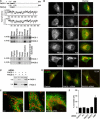
Figure 1
Figure 1
Identification and characterization of PACS-2, a sorting protein found on the ER and mitochondria. (A) Top: Schematic and Kyte–Doolittle hydrophobicity plot of the human PACS-1 and PACS-2 proteins. FBR, cargo/adaptor-binding region; ARR, atrophin-1-related region. Radiation hybrid and genome database analyses mapped the PACS-1 gene to chromosome 11q13.1 (Genbank AY320283) and the PACS-2 gene to chromosome 14q32.33 (Genbank AY320284). Bottom: Northern blot analysis of the tissue distribution for PACS-1 and PACS-2 transcripts. (B) Confocal immunofluorescence of endogenous PACS-1 and PACS-2 in A7 cells. PACS-1/PACS-2 were visualized with Alexa488 (green) and markers were visualized with Alexa546 or mitotracker (red). Scale bar, 10 μm. (C) A7 cells transfected with PACS-1 or PACS-2 siRNAs were analyzed by Western blot 48 h post-transfection. (D) A7 cells were transfected or not with PACS-1 or PACS-2 siRNAs. After 48 h, cells were processed for immunofluorescence microscopy using anti-CI-MPR (green) and anti-TGN46 (red). (E) A7 cells were transfected with control (scrambled), PACS-1, or PACS-2 siRNAs for 48 h and processed for confocal immunofluorescence localization of mitochondria (mitotracker, red) and ER (PDI, green). (F) A7 cells were transfected with the corresponding siRNAs and assayed for cell death by Annexin V/propidium iodide staining and FACS analysis. Treatment of cells with the proapoptotic CtBP siRNA served as a positive control (Zhang et al, 2003). Error for all graphs=s.d.
Figure 2
Figure 2
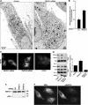
PACS-2 depletion disrupts mitochondrial structure. (A) A7 cells were transfected or not with the PACS-2 siRNA and processed for electron microscopy. Magnification, × 4500. (B) The extent of uncoupling of mitochondria from the ER of either control cells or PACS-2-depleted cells was quantified using morphometric analysis (see Materials and methods). (C) Cells were incubated with 100 nM TMRM for 30 min before microscopic analysis. Control cells were treated with 10 μM FCCP for 30 min, which uncouples the ΨM and blocks TMRM loading. (D) MAMs were isolated from crude homogenates (HMG) of control cells (trans) and PACS-2- and PACS-2Admut-expressing cells by Percoll gradient fractionation, and identified by Western blot using an anti-PSS-1 Ab, which is specific for the MAM fraction (Stone and Vance, 2000). The effect of PACS-2 or PACS-2Admut on the localization of MAM-associated FACL4 was determined by Western blot. Right: Quantitation of MAM-associated FACL4 (n=3). (E) Lysates from control and siRNA-transfected A7 cells were analyzed by Western blot using anti-BAP31. (F) A7 cells were transfected with crBAP31-flag and subsequently transfected with the PACS-2 siRNA for 48 h. Cells were then processed for immunofluorescence with anti-Flag mAb to detect crBAP31-expressing cells (right panel) and mitotracker (left panel). Arrows, rod-like mitochondria. Arrowheads, fragmented mitochondria.
Figure 3
Figure 3
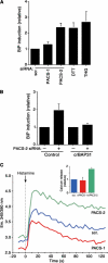
PACS-2 depletion disrupts ER homeostasis. (A) siRNA-treated A7 cells were lysed 48 h post-transfection and the amount of BiP was determined by Western blot. BiP amounts in cells treated with 1 mM DTT or 5 mM thapsigargin (THG) for 16 h served as positive controls. All values are normalized to control cells transfected with scrambled siRNA. (B) Control and HeLa(KB)-crBAP31 cells were transfected or not with PACS-2 siRNA and analyzed for BiP expression by Western blot. (C) A7 cells depleted of PACS-1 or PACS-2 and control cells (transfected with scrambled siRNA) were loaded with Fura-2 and treated with histamine to stimulate calcium release from the ER through IP3R. Inset: Relative amounts of histamine-releasable ER calcium (n=3).
Figure 4
Figure 4
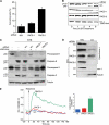
STS-induced cell death depends on PACS-2. (A) A7 cells transfected with PACS-1, PACS-2 or scrambled siRNAs for 48 h were incubated with 1.2 μM STS for an additional 24 h, and the percentage of viable cells was quantified by FACS analysis using anti-annexinV/propidium iodide staining. (B) A7 cells were transfected with PACS-specific or scrambled siRNAs, or treated with oligofectamine alone (control). Apoptosis was induced with 1.2 μM STS for 0, 1, 2, 4, and 6 h in the absence or presence of 10 μM zVAD-fmk and lysates were analyzed by Western blot to detect PARP cleavage to the p85 product. (C) A7 cells were transfected with siRNAs as indicated, treated with 1.2 μM STS for 4 h in the absence or presence of 10 μM zVAD-fmk, harvested, and pro- and activated caspase-8 and -3 or α-tubulin (loading control) were analyzed by Western blot. (D) Post-nuclear supernatants from A7 cells treated with 1.2 μM STS for 4 h were resolved by sedimentation into the cytosol and mitochondria-containing heavy membrane fractions, followed by Western blot with anti-cytochrome c, anti-cytochrome c oxidase I (mitochondria marker) or anti-α-tubulin (cytosol marker). (E) A7 cells depleted of PACS-1 or PACS-2 and control cells (transfected with scrambled siRNA) were incubated with 1.2 μM STS for 4 h and then loaded with Fura-2. Following histamine stimulation, the calcium release was monitored as in Figure 3C.
Figure 5
Figure 5
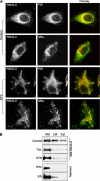
Apoptosis induction redirects PACS-2 onto mitochondria. (A) A7 cells were either untreated (control) or treated with 1.2 μM STS for 1 h and incubated with primary antibodies and species-specific secondary antibodies to detect PACS-2 (green) and PDI (red) or stained with mitotracker (red). (B) Control and tunicamycin (Tun) or STS-treated cells were homogenized and post-nuclear supernatants were resolved by sedimentation into the cytosol, light and heavy (mitochondria fraction) membranes, followed by Western blot with anti-PACS-2, anticytochrome c oxidase I (mitochondria) or anti-BiP (ER).
Figure 6
Figure 6
Death signals stimulate PACS-2-mediated trafficking of Bid to mitochondria. (A) Top: GST-PACS-2FBR was incubated with His6-Bid and tBid and bound Bid molecules were detected by Western blot using anti-Bid antibodies. Bottom: GST-PACS-2FBR was incubated with either His6-Bid or CK2-phosphorylated His6-Bid and bound His6-Bid was detected by Western blot using anti-Bid antibodies. (B) Control HeLa cells or cells expressing PACS-2-ha were treated or not for 2 h with 1.2 μM STS or with 0.1 μg/ml Fas Ab and 5 μg/ml cycloheximide. PACS-2-ha was immunoprecipitated from cell lysates and co-immunoprecipitating Bid was detected by Western blot using an anti-Bid Ab. (C) MCF-7:Bid-GFP cells were either transfected with the PACS-2 siRNA for 48 h or infected with Ad:trans (trans, expressing the tet transactivator) alone or together with Ad:PACS-2 for 24 h and then treated with Fas Ab (1 μg/ml) and cycloheximide (20 μg/ml) for 2 h, followed by a 30-min loading with mitotracker. Fixed cells were scored for Fas Ab-mediated recruitment of Bid-GFP to the mitochondria. Top: bar graph normalized to the control (Oligofectamine). Middle: fluorescence microscopy showing Bid-GFP and mitotracker (red) staining under the various conditions. Large fields of the coverslips containing 150–450 cells were scored for translocation of GFP-Bid to the mitochondria. Bottom: Fas Ab stimulates PACS-2 and Bid to translocate to the mitochondria. MCF-7:Bid-GFP treated with Fas Ab for 2 h were processed for immunofluorescence microscopy. (D) Top: HeLa cells transfected with control (scr.) or PACS-2 siRNAs were treated or not with Fas Ab (1 μg/ml) and cycloheximide (20 μg/ml) for 2 h. Bid and tBid in the mitochondria and cytosolic fractions (top panels) or from the total lysates (bottom panels) were detected by Western blot.

Similar articles

Cited by

References

    1. Arvidson B, Seeds J, Webb M, Finlay L, Barklis E (2003) Analysis of the retrovirus capsid interdomain linker region. Virology 308: 166–177 - PubMed
    1. Belden WJ, Barlowe C (2001) Deletion of yeast p24 genes activates the unfolded protein response. Mol Biol Cell 12: 957–969 - PMC - PubMed
    1. Berridge MJ (2002) The endoplasmic reticulum: a multifunctional signaling organelle. Cell Calcium 32: 235–249 - PubMed
    1. Blagoveshchenskaya AD, Thomas L, Feliciangeli SF, Hung CH, Thomas G (2002) HIV-1 Nef downregulates MHC-I by a PACS-1- and PI3K-regulated ARF6 endocytic pathway. Cell 111: 853–866 - PubMed
    1. Boatright KM, Salvesen GS (2003) Mechanisms of caspase activation. Curr Opin Cell Biol 15: 725–731 - PubMed

Publication types

MeSH terms

Substances