. 2005 Feb;166(2):409-19.
doi: 10.1016/s0002-9440(10)62264-x.

Interferon-gamma and tumor necrosis factor-alpha synergize to induce intestinal epithelial barrier dysfunction by up-regulating myosin light chain kinase expression

Affiliations

Interferon-gamma and tumor necrosis factor-alpha synergize to induce intestinal epithelial barrier dysfunction by up-regulating myosin light chain kinase expression

Fengjun Wang et al. Am J Pathol. 2005 Feb.

Abstract

Numerous intestinal diseases are characterized by immune cell activation and compromised epithelial barrier function. We have shown that cytokine treatment of epithelial monolayers increases myosin II regulatory light chain (MLC) phosphorylation and decreases barrier function and that these are both reversed by MLC kinase (MLCK) inhibition. The aim of this study was to determine the mechanisms by which interferon (IFN)-gamma and tumor necrosis factor (TNF)-alpha regulate MLC phosphorylation and disrupt epithelial barrier function. We developed a model in which both cytokines were required for barrier dysfunction. Barrier dysfunction was also induced by TNF-alpha addition to IFN-gamma-primed, but not control, Caco-2 monolayers. TNF-alpha treatment of IFN-gamma-primed monolayers caused increases in both MLCK expression and MLC phosphorylation, suggesting that MLCK is a TNF-alpha-inducible protein. These effects of TNF-alpha were not mediated by nuclear factor-kappaB. However, at doses below those needed for nuclear factor-kappaB inhibition, sulfasalazine was able to prevent TNF-alpha-induced barrier dysfunction, MLCK up-regulation, and MLC phosphorylation. Low-dose sulfasalazine also prevented morphologically evident tight junction disruption induced by TNF-alpha. These data show that IFN-gamma can prime intestinal epithelial monolayers to respond to TNF-alpha by disrupting tight junction morphology and barrier function via MLCK up-regulation and MLC phosphorylation. These TNF-alpha-induced events can be prevented by the clinically relevant drug sulfasalazine.


Figures
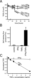
Figure 1
Figure 1
IFN-γ and TNF-α synergize to reduce barrier function in Caco-2 cell monolayers. A: Caco-2 monolayers were incubated with the indicated cytokine(s) (IFN-γ = 10 ng/ml, TNF-α = 2.5 ng/ml) added to media in the basal chamber. Conditions were no cytokines (control), continuous IFN-γ (IFN-γ), continuous TNF-α (TNF-α), continuous IFN-γ and TNF-α (IFN-γ + TNF-α), TNF-α for 24 hours followed by IFN-γ (TNF-α → IFN-γ), or IFN-γ for 24 hours followed by TNF-α (IFN-γ → TNF-α). TER fell only in cells exposed to IFN-γ and TNF-α continuously or IFN-γ followed by TNF-α (n = 3 in this representative experiment). B: Caco-2 monolayers were incubated with the indicated cytokine(s) added to the basal chamber, as above. Conditions were no cytokines (control), continuous IFN-γ for 32 hours (IFN-γ), no cytokines for 24 hours followed by TNF-α for 8 hours (TNF-α), or IFN-γ for 24 hours followed by TNF-α for 8 hours (IFN-γ/TNF-α). At that time monolayers were assayed for permeability to 3kD fluorescein isothiocyanate-dextran. Paracellular permeability was increased only in cells exposed to IFN-γ followed by TNF-α (P < 0.001) (n = 6 in this representative experiment). C: Caco-2 monolayers were incubated with (filled circles) or without (open circles) IFN-γ (10 ng/ml) for 24 hours, washed, and transferred to media with TNF-α at indicated concentrations. TER shown is 48 hours after TNF-α addition. TNF-α caused dose-dependent TER decreases in IFN-γ-primed monolayers, but not in monolayers without IFN-γ pretreatment (n = 3 in this representative experiment).
Figure 2
Figure 2
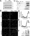
IFN-γ- and TNF-α-induced barrier dysfunction is associated with apoptosis-independent tight junction disruption. A: Caco-2 monolayers were treated with IFN-γ (10 ng/ml) for 24 hours followed by TNF-α (2.5 ng/ml) for times indicated. Cell lysates were analyzed for cleavage of caspase-3, caspase-8, and PARP as markers of apoptosis. No increases in cleaved caspase-3, caspase-8, or PARP were detected in Caco-2 cells treated with the indicated cytokine(s). The positions of intact (I) and cleaved (C) proteins are shown. Data are representative of four similar experiments, each performed in triplicate. B: Caco-2 monolayers were treated with IFN-γ (10 ng/ml) for 24 hours followed by TNF-α (2.5 ng/ml) for 8 hours. Tight junction proteins (ZO-1, occludin, and claudin-1) were detected by immunofluorescence microscopy. Treatment of IFN-γ-primed monolayers with TNF-α caused obvious disruptions of the distribution of tight junction proteins. The tight junction staining was decreased in intensity and became irregular. Increased intracellular pools of occludin and claudin-1 were also apparent. Data are representative of six similar experiments, each performed in duplicate. C: Caco-2 monolayers were treated with IFN-γ (10 ng/ml) for 24 hours followed by TNF-α (2.5 ng/ml) for 8 hours, and harvested at 4°C in TBS with 1% Triton X-100. Cell lysates (adjusted to 40% sucrose) were loaded under 0 to 40% continuous sucrose gradients and centrifuged at 180,000 × g for 18 hours. SDS-PAGE immunoblots for occludin and claudin-1 were performed. Fractions from 1.06 to 1.16 g/ml are shown, with lipid raft and load regions designated. Treatment with IFN-γ and TNF-α caused both occludin and claudin-1 to be removed from the raft fraction. Data are representative of three similar experiments, each performed in duplicate. D: Immunoblots detecting occludin (Figure 2C) were analyzed quantitatively. The amount of occludin present in each fraction is shown as a percentage of the sum of occludin detected in all fractions. The peak occludin content within raft fractions was detected at a density of 1.082 g/ml and was 9.9% of total in control monolayers (open symbols) but only 2.9% of total in monolayers treated with IFN-γ and TNF-α (filled symbols). Data are representative of three similar experiments, each performed in duplicate. E: Immunoblots detecting claudin-1 (Figure 2C) were analyzed quantitatively. The amount of claudin-1 present in each fraction is shown as a percentage of the sum of claudin-1 detected in all fractions. The peak claudin-1 content within raft fractions was detected at a density of 1.082 g/ml and was 3.6% of total in control monolayers (open symbols) but only 1.6% of total in monolayers treated with IFN-γ and TNF-α (filled symbols). Data are representative of three similar experiments, each performed in duplicate. F: Caco-2 monolayers were treated with IFN-γ (10 ng/ml) for 24 hours or IFN-γ for 24 hours followed by TNF-α (2.5 ng/ml) for 18 hours. Cell lysates were analyzed for the expression of tight junction proteins ZO-1, occludin, and claudin-1 by SDS-PAGE immunoblot. Expression of these tight junction proteins was not significantly changed by cytokine treatment. Data are representative of four similar experiments, each performed in triplicate.
Figure 3
Figure 3
IFN-γ- and TNF-α-dependent TER decreases are accompanied by increases in MLC phosphorylation and MLCK expression. A: Caco-2 monolayers were incubated in media with or without IFN-γ (10 ng/ml) for 24 hours and then transferred to media with or without TNF-α (2.5 ng/ml), as indicated. In the designated conditions, 200 μmol/L PIK, a highly specific membrane-permeant inhibitor of MLC kinase, was added to the apical media 2 hours after TNF-α addition. As we have shown previously, PIK alone induced a small increase in TER. However, in monolayers treated sequentially with IFN-γ and TNF-α, PIK was able to nearly completely prevent TER loss (P < 0.02) (n = 3 in this representative experiment). B: Caco-2 monolayers were incubated in media with or without IFN-γ (10 ng/ml) for 24 hours and then transferred to media with or without TNF-α (2.5 ng/ml), as indicated. Monolayers were harvested 8 hours after TNF-α addition. MLC phosphorylation was assessed by SDS-PAGE immunoblot. TNF-α (2.5 ng/ml) caused a small increase in MLC phosphorylation in monolayers without IFN-γ (10 ng/ml) pretreatment (P > 0.05), but a significant increase in MLC phosphorylation in cells with IFN-γ pretreatment (P < 0.05) (n = 2 in this representative experiment). C: Caco-2 monolayers were incubated in media with or without IFN-γ (10 ng/ml) for 24 hours and then transferred to media with or without TNF-α (2.5 ng/ml), as indicated. Monolayers were harvested 8 hours after TNF-α addition. MLCK protein expression was analyzed by SDS-PAGE immunoblot. TNF-α (2.5 ng/ml) caused a slight increase MLCK expression in monolayers without IFN-γ (10 ng/ml) pretreatment (P > 0.05), but a significant increase in MLCK expression occurred after TNF-α treatment of monolayers primed with IFN-γ (P < 0.05) (n = 2 in this representative experiment).
Figure 4
Figure 4
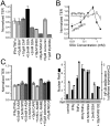
Barrier defects induced by TNF-α and IFN-γ are not abrogated by NF-κB inhibition. A: Caco-2 monolayers were cultured with IFN-γ (10 ng/ml) for 24 hours and transferred to media with 10 μmol/L BAY 11-7085 (BAY), 10 μmol/L capsaicin (CAP), 0.5 mmol/L SSA, 2 mmol/L SSA, 18 μmol/L SN50, 30 μmol/L MG132, 5 μmol/L curcumin, or 1 μmol/L triptolide with or without TNF-α (2.5 ng/ml). None of the NF-κB inhibitors significantly altered TER without TNF-α treatment. TER of monolayers treated with TNF-α is normalized to identical monolayers, treated with the same NF-κB inhibitor, but without TNF-α. SSA (0.5 mmol/L) prevented TER decreases, but 2 mmol/L SSA, 18 μmol/L SN50, 30 μmol/L MG132, and 1 μmol/L triptolide exacerbated TER decreases. BAY 11-7085 (10 μmol/L), 10 μmol/L capsaicin, and 5 μmol/L curcumin had no significant effects (n = 3 in this representative experiment). B: Caco-2 monolayers were cultured with (closed circles) or without (open circles) IFN-γ (10 ng/ml) for 24 hours and transferred to TNF-α (2.5 ng/ml). SSA was also included at indicated doses (0.01 to 2 mmol/L). Maximal barrier protection occurred at 0.5 mmol/L. Higher SSA doses were not protective and, as shown in A, 2 mmol/L SSA exacerbated TER loss (n = 3 in this representative experiment). C: Caco-2 monolayers were cultured with IFN-γ (10 ng/ml) for 24 hours and transferred to TNF-α (2.5 ng/ml) with 0.5 mmol/L SSA, 0.5 mmol/L 5-aminosalicylic acid (5-ASA), 0.5 mmol/L sulfapyridine (SPD), 0.5 mmol/L 4-aminosalicylic acid (4-ASA), 10 μmol/L caffeic acid phenethyl ester, 1 mmol/L and 10 mmol/L NG-nitro-l-arginine-methyl-ester (L-NAME), 10 μmol/L adenosine, and 10 μmol/L 5′-N-ethylcarboxamidoadenosine (NECA). TER of monolayers treated with TNF-α is normalized to identical monolayers, treated with the same drug, but without TNF-α. Only SSA prevented barrier dysfunction induced by IFN-γ and TNF-α (n = 3 in this representative experiment). D: NF-κB RelA p65 (closed bars) was detected by SDS-PAGE immunoblot of nuclear fractions isolated 10 minutes after TNF-α (2.5 ng/ml) and drug addition to monolayers with or without IFN-γ (10 ng/ml) pretreatment for 24 hours, as indicated. A representative immunoblot of nuclear fractions is shown below the graph. NF-κB (RelA p65) nuclear translocation was inhibited by 2 mmol/L SSA and 30 μmol/L MG132, but not by 0.5 mmol/L SSA [n = 2 in this experiment which is representative of nine similar experiments, each with duplicate samples (total n = 18)]. NF-κB-dependent luciferase expression (open bars) was assessed in Caco-2 cells transiently transfected with pNFκB-TA-Luc reporter construct. Monolayers were then treated identically to the translocation assay, but harvested 8 hours after TNF-α addition to allow for luciferase synthesis. NF-κB-dependent luciferase expression was inhibited by 2 mmol/L SSA and 30 μmol/L MG132, but not by 0.5 mmol/L SSA [n = 2 in this experiment which is representative of four similar experiments, each with duplicate samples (total n = 8)].
Figure 5
Figure 5
Low dose SSA prevents morphological tight junction disruption, increased MLC phosphorylation, and increased MLCK expression. A: Caco-2 monolayers were treated with IFN-γ (10 ng/ml) for 24 hours followed by TNF-α (2.5 ng/ml) and 0.5 mmol/L SSA for 8 hours. Tight junction proteins (ZO-1, occludin, and claudin-1) were detected by immunofluorescence microscopy. 0.5 mmol/L SSA completely prevented ZO-1, occludin, and claudin-1 redistribution in IFN-γ-primed TNF-α-treated monolayers. Data are representative of three similar experiments, each performed in triplicate. B: Caco-2 monolayers were incubated with IFN-γ (10 ng/ml) for 24 hours and then transferred to media with TNF-α (2.5 ng/ml) and 0.5 mmol/L SSA, 2 mmol/L SSA, or 30 μmol/L MG132, as indicated. Monolayers were harvested 8 hours after TNF-α addition. Lysates were assayed for phosphorylated MLC by SDS-PAGE immunoblot. SSA (0.5 mmol/L) prevented increases in MLC phosphorylation induced by IFN-γ and TNF-α (P < 0.01). In contrast, 2 mmol/L SSA and 30 μmol/L MG132 failed to prevent increases in MLC phosphorylation (P > 0.05 for 2 mmol/L SSA, P = 0.04 for 30 μmol/L MG132) (n = 2 in this representative experiment). C: Caco-2 monolayers were incubated with IFN-γ (10 ng/ml) for 24 hours and then transferred to media with TNF-α (2.5 ng/ml) and 0.5 mmol/L SSA, 2 mmol/L SSA, or 30 μmol/L MG132, as indicated. Monolayers were harvested 8 hours after TNF-α addition. Lysates were assayed for MLCK by SDS-PAGE immunoblot. SSA (0.5 mmol/L) prevented increases in MLCK expression induced by IFN-γ and TNF-α (P < 0.02). In contrast, 2 mmol/L SSA and 30 μmol/L MG132 failed to prevent increases in MLCK expression (P > 0.05 for 2 mmol/L SSA or 30 μmol/L MG132) (n = 2 in this representative experiment).

Similar articles

Cited by

References

    1. Clayburgh DR, Shen L, Turner JR. A porous defense: the leaky epithelial barrier in intestinal disease. Lab Invest. 2004;84:282–291. - PubMed
    1. Irvine EJ, Marshall JK. Increased intestinal permeability precedes the onset of Crohn’s disease in a subject with familial risk. Gastroenterology. 2000;119:1740–1744. - PubMed
    1. Wyatt J, Vogelsang H, Hubl W, Waldhoer T, Lochs H. Intestinal permeability and the prediction of relapse in Crohn’s disease. Lancet. 1993;341:1437–1439. - PubMed
    1. Yacyshyn BR, Meddings JB. CD45RO expression on circulating CD19+ B cells in Crohn’s disease correlates with intestinal permeability. Gastroenterology. 1995;108:132–137. - PubMed
    1. Madara JL. Loosening tight junctions. J Clin Invest. 1989;83:1089–1094. - PMC - PubMed

Publication types

MeSH terms

LinkOut - more resources