

Ultrastructural changes in dysferlinopathy support defective membrane repair mechanism
- PMID: 15677541
- PMCID: PMC1770568
- DOI: 10.1136/jcp.2004.018978
Ultrastructural changes in dysferlinopathy support defective membrane repair mechanism
Abstract
Background: The dysferlin gene has recently been shown to be involved in limb girdle muscular dystrophy type 2B and its allelic disease, Miyoshi myopathy, both of which are characterised by an active muscle degeneration and regeneration process. Dysferlin is known to play an essential role in skeletal muscle fibre repair, but the process underlying the pathogenetic mechanism of dysferlinopathy is not completely understood.
Aims: To define both specific alterations of muscle fibres and a possible sequential mechanism of myopathy development.
Methods: A histological, immunohistochemical, and ultrastructural analysis of 10 muscle biopsies from patients with molecularly diagnosed dysferlinopathy.
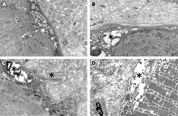
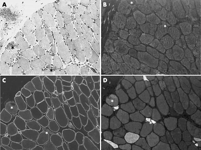
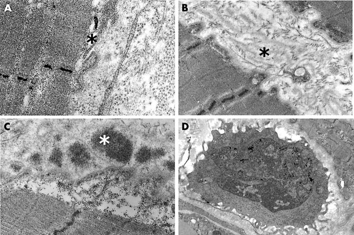
Results: An inflammatory response was seen in most of the muscle biopsies. The immunohistochemical pattern demonstrated active regeneration and inflammation. Non-necrotic fibres showed alterations at different submicroscopic levels, namely: the sarcolemma and basal lamina, subsarcolemmal region, and sarcoplasmic compartment. In the subsarcolemmal region there were prominent aggregations of small vesicles, probably derived from the Golgi apparatus, which consisted of empty, swollen cisternae. In the sarcolemma there were many gaps and microvilli-like projections, whereas the basal lamina was multilayered.
Conclusions: The histopathological, immunohistochemical, and ultrastructural data show that dysferlinopathy is characterised by a very active inflammatory/degenerative process, possibly associated with an inefficient repair and regenerative system. The presence of many crowded vesicles just beneath the sarcolemma provides submicroscopical proof of a defective resealing mechanism, which fails to repair the sarcolemma.
Figures
Similar articles
-
Defective membrane repair in dysferlin-deficient muscular dystrophy.Nature. 2003 May 8;423(6936):168-72. doi: 10.1038/nature01573. Nature. 2003. PMID: 12736685
-
The earliest pathologic alterations in dysferlinopathy.Neurology. 2001 Jun 12;56(11):1472-81. doi: 10.1212/wnl.56.11.1472. Neurology. 2001. PMID: 11402103
-
Attenuated muscle regeneration is a key factor in dysferlin-deficient muscular dystrophy.Hum Mol Genet. 2009 Jun 1;18(11):1976-89. doi: 10.1093/hmg/ddp121. Epub 2009 Mar 13. Hum Mol Genet. 2009. PMID: 19286669 Free PMC article.
-
Dysferlin and the plasma membrane repair in muscular dystrophy.Trends Cell Biol. 2004 Apr;14(4):206-13. doi: 10.1016/j.tcb.2004.03.001. Trends Cell Biol. 2004. PMID: 15066638 Review.
-
Sarcolemmal proteins and the spectrum of limb-girdle muscular dystrophies.Semin Pediatr Neurol. 2002 Jun;9(2):81-99. doi: 10.1053/spen.2002.33795. Semin Pediatr Neurol. 2002. PMID: 12139001 Review.
Cited by
-
Recessive mutations in the putative calcium-activated chloride channel Anoctamin 5 cause proximal LGMD2L and distal MMD3 muscular dystrophies.Am J Hum Genet. 2010 Feb 12;86(2):213-21. doi: 10.1016/j.ajhg.2009.12.013. Epub 2010 Jan 21. Am J Hum Genet. 2010. PMID: 20096397 Free PMC article.
-
Tetraspanin CD82 Associates with Trafficking Vesicle in Muscle Cells and Binds to Dysferlin and Myoferlin.Adv Biol (Weinh). 2023 Dec;7(12):e2300157. doi: 10.1002/adbi.202300157. Epub 2023 Jul 12. Adv Biol (Weinh). 2023. PMID: 37434585 Free PMC article.
-
The inflammatory pathology of dysferlinopathy is distinct from calpainopathy, Becker muscular dystrophy, and inflammatory myopathies.Acta Neuropathol Commun. 2022 Feb 8;10(1):17. doi: 10.1186/s40478-022-01320-z. Acta Neuropathol Commun. 2022. PMID: 35135626 Free PMC article.
-
Dysferlin stabilizes stress-induced Ca2+ signaling in the transverse tubule membrane.Proc Natl Acad Sci U S A. 2013 Dec 17;110(51):20831-6. doi: 10.1073/pnas.1307960110. Epub 2013 Dec 3. Proc Natl Acad Sci U S A. 2013. PMID: 24302765 Free PMC article.
-
Annexin A2 Mediates Dysferlin Accumulation and Muscle Cell Membrane Repair.Cells. 2020 Aug 19;9(9):1919. doi: 10.3390/cells9091919. Cells. 2020. PMID: 32824910 Free PMC article.
References
-
- Zatz M, de Paula F, Starling A, et al. The 10 autosomal recessive limb-girdle muscular dystrophies. Neuromuscul Disord 2003;13:532–44. - PubMed
-
- Laval SH, Bushby KM. Limb-girdle muscular dystrophies—from genetics to molecular pathology. Neuropathol Appl Neurobiol 2004;30:91–105. - PubMed
-
- Campanaro S, Romualdi C, Fanin M, et al. Gene expression profiling in dysferlinopathies using a dedicated muscle microarray. Hum Mol Genet 2002;11:3283–98. - PubMed
-
- Fanin M, Angelini C. Muscle pathology in dysferlin deficiency. Neuropathol Appl Neurobiol 2002;28:461–70. - PubMed
-
- Bansal D, Miyake K, Vogel SS, et al. Defective membrane repair in dysferlin-deficient muscular dystrophy. Nature 2003;423:168–72. - PubMed
Publication types
MeSH terms
Substances
Grants and funding
LinkOut - more resources
Full Text Sources