. 2005 Feb;25(3):1100-12.
doi: 10.1128/MCB.25.3.1100-1112.2005.

Eukaryotic translation initiation factor 4E activity is modulated by HOXA9 at multiple levels

Affiliations

Eukaryotic translation initiation factor 4E activity is modulated by HOXA9 at multiple levels

Ivan Topisirovic et al. Mol Cell Biol. 2005 Feb.

Abstract

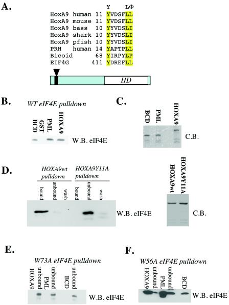
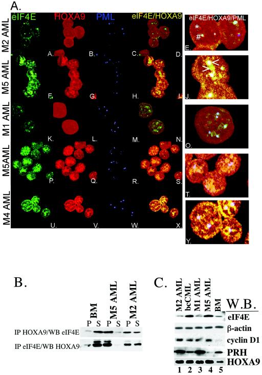
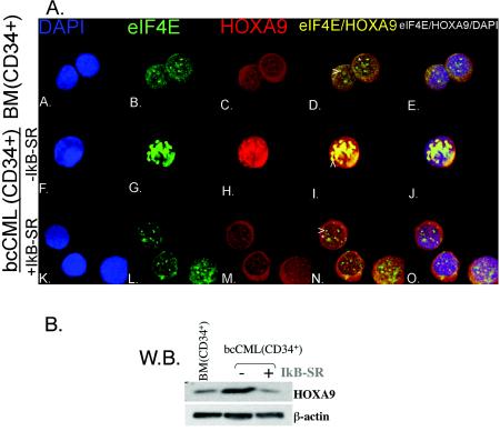
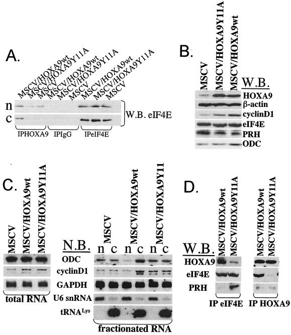
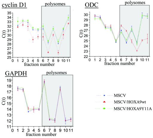
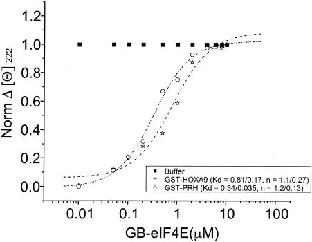
The eukaryotic translation initiation factor 4E (eIF4E) alters gene expression on multiple levels. In the cytoplasm, eIF4E acts in the rate-limiting step of translation initiation. In the nucleus, eIF4E facilitates nuclear export of a subset of mRNAs. Both of these functions contribute to eIF4E's ability to oncogenically transform cells. We report here that the homeodomain protein, HOXA9, is a positive regulator of eIF4E. HOXA9 stimulates eIF4E-dependent export of cyclin D1 and ornithine decarboxylase (ODC) mRNAs in the nucleus, as well as increases the translation efficiency of ODC mRNA in the cytoplasm. These activities depend on direct interactions of HOXA9 with eIF4E and are independent of the role of HOXA9 in transcription. At the biochemical level, HOXA9 mediates these effects by competing with factors that repress eIF4E function, in particular the proline-rich homeodomain PRH/Hex. This competitive mechanism of eIF4E regulation is disrupted in a subset of leukemias, where HOXA9 displaces PRH from eIF4E, thereby contributing to eIF4E's dysregulation. In regard to these results and our previous finding that approximately 200 homeodomain proteins contain eIF4E binding sites, we propose that homeodomain modulation of eIF4E activity is a novel means through which this family of proteins implements their effects on growth and development.


Figures

FIG. 1.
FIG. 1.
HOXA9 directly binds eIF4E with a conserved eIF4E binding site. (A) Sequence alignment of HOXA9 from a variety of species. Residues that are part of the conserved eIF4E binding site are highlighted in yellow; the “Φ” symbol indicates residues that are hydrophobic. Numbers indicate the position in the amino acid sequence. eIF4G, PRH, and Bicoid, two homeodomain-containing proteins that interact and alter eIF4E activity, are included for comparison. The schematic below indicates the relative positions of the eIF4E binding site (indicated by the arrowhead) and the homeodomain (HD) in HOXA9. Accession numbers sequences are as follows: p31269 (human), P09631 (mouse), q6pwd5 (striped bass), q9IA26 (horn shark), 042506 (puffer fish), NP002720 (human PRH), p09081 (Bicoid), and NP937884 (eIF4G). (B to F) GST pull-down analysis of HOXA9. HOXA9 or the Y11A HOXA9 mutant was immobilized on glutathione-agarose, and the ability to bind wild-type or mutant forms of eIF4E, monitored by Western blotting (W.B.) and Coomassie blue staining (C.B.), indicates equal loading. In panel B, only the bound fractions are shown. In panels D to F, the bound (i.e., the fraction associating with the beads) and unbound fractions are shown. In addition, in panel D, the supernatant from the sixth wash (wash) of the beads is shown.
FIG. 2.
FIG. 2.
HOXA9 associates with eIF4E and colocalizes with eIF4E nuclear bodies in the subset of myeloid leukemia specimens that show disruption of eIF4E-PRH interaction and facilitated eIF4E-dependent mRNA transport. (A) Confocal micrographs of cells obtained from the bone marrow of patients with the indicated FAB subtypes of AML. Cells were stained with anti-eIF4E-FITC conjugated antibody (green), anti-HOXA9 antibody (red), and anti-PML monoclonal antibody 5E10 (blue). The HOXA9-eIF4E overlay is shown in yellow (indicated on subpanel J by the “>” symbol), the eIF4E-PML overlay is shown in light blue (indicated on subpanel E by the “#” symbol), and the triple HOXA9-eIF4E-PML overlay is shown in white (indicated on subpanel J by the “<” symbol). The objective lens magnification was ×100, and the images were further magnified twofold except for subpanels K to O, which were magnified threefold. An example of HOXA9 aggregate that is distinct from eIF4E nuclear body is shown in subpanel E (in red, indicated by an asterisk). (B) Whole-cell extracts, from the indicated specimens, were immunoprecipitated with anti-eIF4E antibody and analyzed by Western blotting with anti-HOXA9 antibody (IP eIF4E/WB HOXA9). Conversely, extracts obtained from the same set of cells were immunoprecipitated with anti-HOXA9 antibody and probed with anti-eIF4E antibody (IP HOXA9/WB eIF4E). A total of 80% of the immunoprecipitated fraction (P) and 20% of the unbound fraction (S) were subjected to SDS-PAGE on 12 or 15% polyacrylamide gels. (C) Western blot analysis of whole-cell extracts obtained from the indicated specimens. Western blots were probed with anti-eIF4E, anti-cyclinD1, anti-PRH, or anti-HOXA9 antibody. In addition, β-actin is shown as a control for protein loading.
FIG. 3.
FIG. 3.
IκB-SR induced loss of NF-κB activity in CD34+ cells derived from bcCML patients leads to the normalization of HOXA9 protein levels and correlates with the restoration of nuclear architecture. (A) Confocal micrographs of the CD34+ cells derived from the bone marrow of apparently healthy individuals (BM) and from bcCML patients. bcCML CD34+ cells were either transduced with empty vector (−IκB-SR), or with IκB-SR (+IκB-SR). Cells were stained with DAPI (blue), anti-eIF4E antibody (green), and anti-HOXA9 antibody (red). The HOXA9-eIF4E overlay is shown in yellow. The objective lens magnification was ×100, and the images were further magnified by twofold. Note that there is substantial colocalization of the HOXA9 protein with the eIF4E bodies in the CD34+ cells derived from the bcCML patients (subpanel I, shown in yellow, indicated by a “>” symbol). Expression of IκB-SR leads to the loss of colocalization of HOXA9 protein with eIF4E nuclear bodies in the aforementioned cells, with results resembling the staining pattern observed in CD34+ cells derived from the healthy individual (subpanels N and D; the eIF4E nuclear body, shown in green, and HOXA9, shown in red, are labeled with a “>” symbol and an asterisk, respectively). (B) HOXA9 Western blot analysis of whole-cell extracts isolated from the same specimens. β-Actin is shown as a control for protein loading.
FIG. 4.
FIG. 4.
HOXA9 requires integrity of its eIF4E-binding to associate with eIF4E and to facilitate nuclear export of ODC and cyclin D1 transcripts. (A) The same cells were fractionated into cytoplasmic (c) and nuclear (n) fractions. The protein extracts from each fraction were immunoprecipitated with anti-eIF4E (IPeIF4E), IgG (IPIgG), or anti-HOXA9 (IPHOXA9) antibody. A total of 50% of the immunoprecipitated fraction was subjected to SDS-15% PAGE, and the consequent Western blot was probed with anti-eIF4E antibody (W.B. eIF4E). (B) Western blot analysis of whole-cell protein extracts of U937 cells overexpressing the indicated constructs. Western blots were probed with anti-eIF4E, anti-cyclin D1, anti-PRH, anti-ODC, or anti-HOXA9 antibody. In addition, β-actin is shown as a control for protein loading. The normalized relative intensities of the bands were as follows: 1.00 ± 0.14 (vector), 2.22 ± 0.22 (HOXA9Y11A mutant), and 4.80 ± 0.10 (HOXA9 wild type) for cyclin D1 and 1.00 ± 0.13 (vector), 1.00 ± 0.14 (HOXA9Y11A mutant), and 3.21 ± 0.16 (HOXA9 wild type) for ODC. The results are from average values ± the standard deviations from three independent experiments. The quantification was performed with ImageQuant Software (Molecular Dynamics). Area quantitation report values obtained for cyclin D1 or ODC were normalized against the corresponding area quantitation report values obtained for β-actin, and the value obtained for control, empty-vector-transduced cells was set at 1. (C) Total cellular RNA (left panel) or RNA obtained from nuclear (n) or cytoplasmic (c) fractions (right panel) of these cells analyzed by Northern analysis (N.B.). tRNALys and U6snRNA were used as markers for the cytoplasmic and nuclear fractions, respectively. GAPDH is shown as a control for RNA loading. (D) HOXA9 competes with PRH for eIF4E binding. Whole-cell protein extracts were immunoprecipitated with anti-eIF4E (IPeIF4E) or anti-HOXA9 (IPHOXA9) antibody. A total of 50% of the immunoprecipitated fraction was analyzed by SDS-PAGE on 12 or 15% gels, and the resulting Western blot was probed with anti-HOXA9, anti-eIF4E, or anti-PRH antibody.
FIG. 5.
FIG. 5.
HOXA9 facilitates translational initiation of ODC, but not of cyclinD1 and GAPDH mRNA. Polysomes from U937 cells transduced with empty vector (MSCV), U937 cells overexpressing wild-type HOXA9 (MSCV/HOXA9wt) and U937 cells overexpressing HOXA9 mutant that does not bind eIF4E (MSCV/HOXA9Y11A) were prepared and resolved by sedimentation on continuous 10 to 40% Sepharose gradients. Ribosomal profiles of GAPDH, cyclin D1 and ODC transcripts were determined by quantitative real-time PCR analysis. To graphically present the ribosomal profiles of these transcripts, average CT values obtained from two independent experiments both carried out in triplicate were plotted against the number of the fraction. CT values for the control U937 cells (MSCV) are shown in blue, CT values for cells overexpressing wild-type HOXA9 (HOXA9) are shown in red, and CT values for the Y11A mutant are shown in green. Bars represent the standard deviation.
FIG. 6.
FIG. 6.
Affinity of HOXA9 and PRH for eIF4E are calculated by monitoring conformational changes upon binding as observed by using CD spectroscopy. Far UV CD spectra were collected, and normalized molar ellipticity ([θ]) at 222 nM was monitored as a function of eIF4E-GB concentration with 15 nM GST-HOXA9 or 15 nM GST-PRH. Buffer alone shows no effects, as expected. PRH and HOXA9 bound eIF4E with submicromolar affinities. HOXA9 (dashed line)- and PRH (dash-dotted line)-binding isotherms represent fits to a heuristic single-site binding expression.

Similar articles

Cited by

References

    1. Borden, K. L. 2002. Pondering the promyelocytic leukemia protein (PML) puzzle: possible functions for PML nuclear bodies. Mol. Cell. Biol. 22:5259-5269. - PMC - PubMed
    1. Calvo, K. R., D. B. Sykes, M. Pasillas, and M. P. Kamps. 2000. Hoxa9 immortalizes a granulocyte-macrophage colony-stimulating factor-dependent promyelocyte capable of biphenotypic differentiation to neutrophils or macrophages, independent of enforced Meis expression. Mol. Cell. Biol. 20:3274-3285. - PMC - PubMed
    1. Carlile, G. W., W. G. Tatton, and K. L. Borden. 1998. Demonstration of a RNA-dependent nuclear interaction between the promyelocytic leukaemia protein and glyceraldehyde-3-phosphate dehydrogenase. Biochem. J. 335:691-696. - PMC - PubMed
    1. Clemens, M. J., and U. A. Bommer. 1999. Translational control: the cancer connection. Int. J. Biochem. Cell Biol. 31:1-23. - PubMed
    1. Cohen, N., M. Sharma, A. Kentsis, J. M. Perez, S. Strudwick, and K. L. Borden. 2001. PML RING suppresses oncogenic transformation by reducing the affinity of eIF4E for mRNA. EMBO J. 20:4547-4559. - PMC - PubMed

Publication types

MeSH terms