. 2005 Feb;25(3):865-78.
doi: 10.1128/MCB.25.3.865-878.2005.

Direct and indirect interactions between calcineurin-NFAT and MEK1-extracellular signal-regulated kinase 1/2 signaling pathways regulate cardiac gene expression and cellular growth

Affiliations

Direct and indirect interactions between calcineurin-NFAT and MEK1-extracellular signal-regulated kinase 1/2 signaling pathways regulate cardiac gene expression and cellular growth

Bastiano Sanna et al. Mol Cell Biol. 2005 Feb.

Abstract

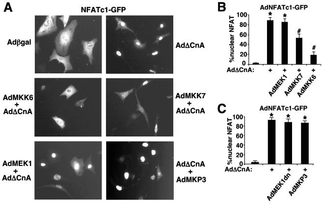
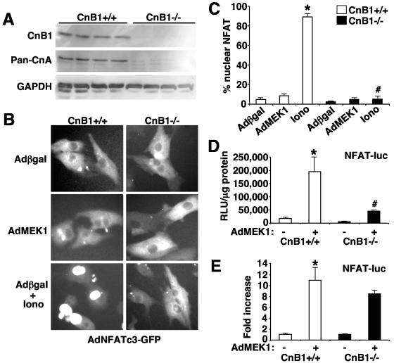
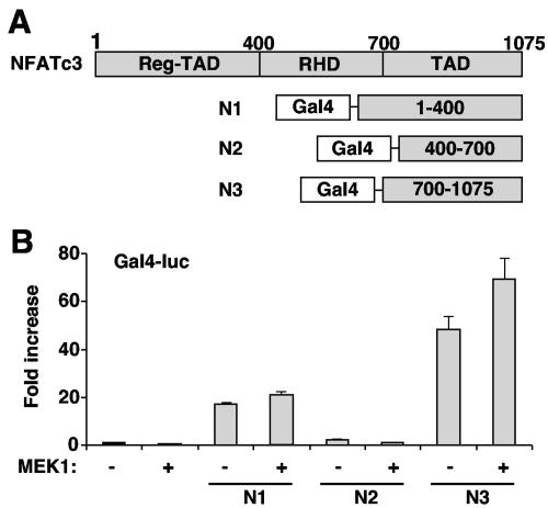
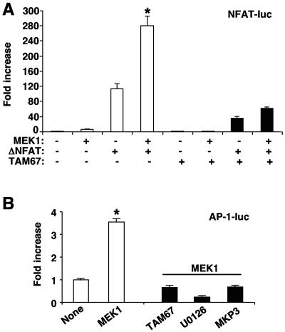
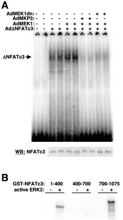
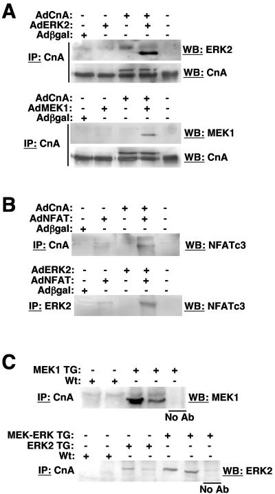
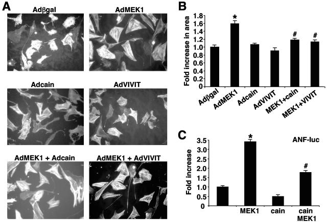
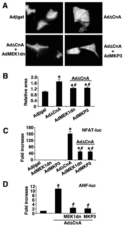
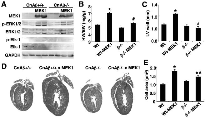
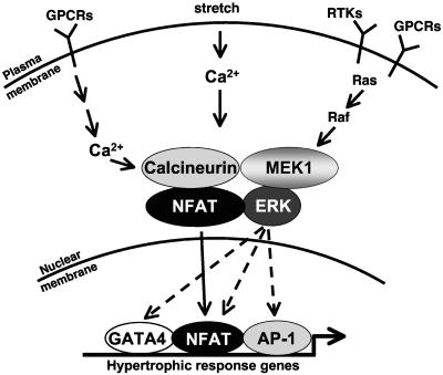
MEK1, a member of the mitogen-activated protein kinase (MAPK) cascade that directly activates extracellular signal-regulated kinase (ERK), induces cardiac hypertrophy in transgenic mice. Calcineurin is a calcium-regulated protein phosphatase that also functions as a positive regulator of cardiac hypertrophic growth through a direct mechanism involving activation of nuclear factor of activated T-cell (NFAT) transcription factors. Here we determined that calcineurin-NFAT and MEK1-ERK1/2 signaling pathways are interdependent in cardiomyocytes, where they directly coregulate the hypertrophic growth response. For example, genetic deletion of the calcineurin Abeta gene reduced the hypertrophic response elicited by an activated MEK1 transgene in the heart, while inhibition of calcineurin or NFAT in cultured neonatal cardiomyocytes also blunted the hypertrophic response driven by activated MEK1. Conversely, targeted inhibition of MEK1-ERK1/2 signaling in cultured cardiomyocytes attenuated the hypertrophic growth response directed by activated calcineurin. However, targeted inhibition of MEK1-ERK1/2 signaling did not directly affect calcineurin-NFAT activation, nor was MEK1-ERK1/2 activation altered by targeted inhibition of calcineurin-NFAT. Mechanistically, we show that MEK1-ERK1/2 signaling augments NFAT transcriptional activity independent of calcineurin, independent of changes in NFAT nuclear localization, and independent of alterations in NFAT transactivation potential. In contrast, MEK1-ERK1/2 signaling enhances NFAT-dependent gene expression through an indirect mechanism involving induction of cardiac AP-1 activity, which functions as a necessary NFAT-interacting partner. As a second mechanism, MEK1-ERK1/2 and calcineurin-NFAT proteins form a complex in cardiac myocytes, resulting in direct phosphorylation of NFATc3 within its C terminus. MEK1-ERK1/2-mediated phosphorylation of NFATc3 directly augmented its DNA binding activity, while inhibition of MEK1-ERK1/2 signaling reduced NFATc3 DNA binding activity. Collectively, these results indicate that calcineurin-NFAT and MEK1-ERK1/2 pathways constitute a codependent signaling module in cardiomyocytes that coordinately regulates the growth response through two distinct mechanisms.


Figures

FIG. 1.
FIG. 1.
MEK1 induces NFAT transcriptional activity. (A) Rat neonatal ventricular cardiomyocytes were plated in serum-free medium and were infected 48 h after plating with AdNFAT-luc together with AdNFATc3, AdMEK1, or Adβgal adenovirus. Cell extracts were collected after 48 h, and luciferase assays were performed (*, P < 0.05 versus AdMEK1). (B) Neonatal cardiomyocytes were infected with AdMEK1, and after 2 h they were transfected with either a plasmid containing NFAT-luc or an identical construct lacking the NFAT binding sites (TATA-luc). (C) NFAT-luc transgenic (n = 3) and NFAT-luc-MEK1 double transgenic (n = 3) mice were sacrificed at 6 weeks of age. Wild-type mice were used as background (Bkgnd) controls (n = 3) (*, P < 0.05 versus NFAT-luc transgenic). Each point was performed in triplicate, and the graph is representative of more than three experiments (A and C).
FIG. 2.
FIG. 2.
MEK1 does not affect NFAT nuclear localization. (A) Neonatal cardiomyocytes were infected with AdNFATc1-GFP and the indicated recombinant adenoviruses. After 48 h, cells were fixed and photographed. (B and C) Nuclear localization of NFATc1 was quantified by counting four randomly selected fields containing ∼100 cells (400 cells for each experimental point). Cells were considered positive when they essentially showed exclusive nuclear localization. The pictures and graphs are representative of three independent experiments (*, P < 0.05 versus AdNFATc1-GFP alone; #, P < 0.05 versus AdNFATc1-GFP plus AdΔCnA).
FIG. 3.
FIG. 3.
MEK1-induced NFAT transcriptional activity is independent of calcineurin. (A) CnB1-deleted mouse embryonic fibroblasts were obtained as described in Materials and Methods. Cell extracts were analyzed by Western blotting with specific antibodies for CnB1. The same membrane was stripped and reprobed with antibodies for pan-CnA and GAPDH. (B and C) CnB1+/+ and CnB1/ MEFs were infected with AdNFATc3-GFP, with or without AdMEK1, and treated with either ionomycin (iono) (1 μM) or solvent (dimethyl sulfoxide) for 30 min. MEFs were fixed and analyzed for NFAT nuclear localization (*, P < 0.05 versus Adβgal in CnB+/+ MEFs; #, P < 0.05 versus ionomycin in CnB1+/+ MEFs). (D) CnB1+/+ and CnB1/ MEFs were infected with AdNFAT-luc in the presence or absence of AdMEK1. Luciferase data are expressed as total relative light units (RLU)/microgram of protein normalized internally to a β-galactosidase expression vector (E) or as fold increase compared to their controls. Each point was performed in triplicate, and the graph is representative of at least three experiments (*, P < 0.05 for CnB1+/+ AdMEK1 versus CnB1+/+ Adβgal; #, P < 0.05 for CnB1/ AdMEK1 versus CnB1+/+ AdMEK1).
FIG. 4.
FIG. 4.
MEK1 does not increase NFAT transactivation potential. (A) Schematic representation of NFATc3 and the different Gal4 fusion proteins that were generated. (B) A Gal4-dependent luciferase reporter construct (1.0 μg) was cotransfected into neonatal cardiomyocytes with expression vectors (0.25 μg) encoding Gal4DBD-NFATc3 N1-N3 fusion mutants with or without a plasmid encoding constitutively active MEK1 (0.25 μg). After 48 h, luciferase assays were performed. Each point was performed in triplicate, and the graph is representative of at least three experiments.
FIG. 5.
FIG. 5.
MEK1 activates NFAT through AP-1 signaling. (A) Neonatal cardiomyocytes were infected with AdNFAT-luc to assay for NFAT transcriptional activity, together with an adenovirus encoding constitutively active NFATc3 (ΔNFAT) in the presence or absence of AdMEK1. The same experimental design was repeated in the presence of an AP-1 dominant-negative TAM67-expressing adenovirus (*, P < 0.05 for MEK1/caNFAT versus caNFAT). (B) Cardiomyocytes were infected with the indicated viruses, and after 2 h they were transfected with an AP-1-luciferase reporter vector. After 48 h, luciferase assays were performed. Each point was performed in triplicate, and the graph is representative of at least three experiments. U0126 (20 μM) was added 6 h before harvesting the cells.
FIG. 6.
FIG. 6.
NFATc3 DNA binding activity is regulated by ERK1/2. (A) Gel mobility shift assay with an NFAT-specific DNA binding site from the interleukin-4 promoter, incubated with cardiomyocyte protein extracts generated from cells infected 24 h prior with the indicated adenoviruses. The lower panel displays a control Western blot (WB) for the activated NFATc3 mutant protein that was overexpressed in each of the indicated reactions. (B) SDS-PAGE of an in vitro kinase reaction between bacterially purified and activated ERK2 and the indicated GST-NFAT fusion protein fragments, also generated in bacteria. Equivalent amounts of protein were loaded in all reactions as assessed by Coomassie brilliant blue staining (data not shown). Identical results were obtained in three independent experiments.
FIG. 7.
FIG. 7.
Calcineurin-NFATc3-MEK1-ERK2 form a complex in vivo. (A) Western blots (WB) for ERK2, CnA, or MEK1 following immunoprecipitation (IP) of CnA from the indicated cardiomyocyte cell extracts, generated from cell cultures previously subjected to adenoviral mediated gene transfer of the indicated constructs. The last lane contains a nonspecific immunoglobulin G (IgG) antibody (Ab). (B) Western blots for NFATc3, following immunoprecipitation of CnA or ERK2 from the indicated cardiomyocyte cell extracts, generated from cell cultures previously subjected to adenovirus-mediated gene transfer of the indicated constructs. The last lane contains a nonspecific IgG antibody. (C) Western blot for activated MEK1 or ERK2 following immunoprecipitation for endogenous CnA from hearts of mice that were either wild-type (Wt), ERK2 transgenic (TG), or ERK2-MEK1 double transgenic. The position of a no-antibody control is also shown. Similar results were observed in three independent experiments.
FIG. 8.
FIG. 8.
Calcineurin-NFAT signaling is necessary for MEK1-induced hypertrophy in vitro. (A) Representative immunocytochemical analysis of actin-stained (phalloidin) neonatal cardiomyocytes treated as indicated. (B) Measurement of cell surface area of neonatal cardiomyocytes infected with the indicated adenoviruses in serum-free media for 48 h (n = 3 independent experiments, at least 400 cells measured in each experiment). (C) ANF promoter activity from cultured cardiomyocytes infected with the indicated adenovirus for 48 h. (*, P < 0.05 versus Adβgal; #, P < 0.05 versus AdMEK1).
FIG. 9.
FIG. 9.
MEK1-ERK1/2 signaling is necessary for a full calcineurin-induced hypertrophic response. (A) Representative immunocytochemical analyses of actin-stained (phalloidin) neonatal cardiomyocytes treated as indicated. (B) Measurement of cell surface area of neonatal cardiomyocytes infected with the indicated adenoviruses in serum-free media for 48 h (n = 3 independent experiments, at least 400 cells measured in each experiment). (C) Neonatal cardiomyocytes were infected with AdNFAT-luc and the indicated adenoviruses, and luciferase assays were performed 48 h afterwards. (D) ANF promoter activity was measured after transfection with an ANF-luciferase promoter vector (0.25 μg), followed by infection with the indicated adenoviruses. (*, P < 0.05 versus Adβgal; #, P < 0.05 versus AdΔCnA alone).
FIG. 10.
FIG. 10.
CnAβ gene targeting reduces MEK1-induced cardiac growth. (A) Western blot analysis of MEK1-ERK1/2 pathway phosphorylation in the hearts of 2-month-old MEK1 transgenic mice (FVB strain) crossed into the CnAβ wild-type (Wt) or null backgrounds. (B) Measurement of heart-to-body weight ratios and (C) echocardiography-measured left ventricular posterior wall thickness (LV wall) show a reduction of cardiac hypertrophy by CnAβ gene targeting. Four or five mice were analyzed in each group. (D) Gross histological analysis and (E) myocyte cross-sectional areas measurements (n = 400 cells per section) also revealed a reduction in hypertrophic growth associated with expression of activated MEK1 in the heart by CnAβ gene disruption. (*, P < 0.05 versus wild type; #, P < 0.05 versus Wt-MEK1).
FIG. 11.
FIG. 11.
Model of the calcineurin-NFAT and MEK1-ERK1/2 signaling module. While calcineurin-NFAT and MEK1-ERK1/2 receive input from stimuli that either mobilize intracellular calcium or result in Ras activation, the full effectiveness of either pathway likely requires simultaneous input from G-protein-coupled receptors (GPCRs), receptor tyrosine kinases (RTKs), and other ill-defined stimuli. All four proteins can form a complex in cardiomyocytes, likely serving as a means of coordinating regulation through at least one direct mechanism involving ERK-mediated phosphorylation of NFAT, which does not alter nuclear translocation but instead influences DNA binding activity of NFAT. Once in the nucleus through the sole action of calcineurin, NFAT can regulate gene expression in coordination with ERK signaling through direct effects on AP-1, GATA4, and other cofactors. It is uncertain if the entire four-protein complex can translocate to the nucleus with NFAT or if only ERK becomes associated with NFAT localized at specific DNA binding sites, permitting additional ERK-regulated events on transcription.

Similar articles

Cited by

References

    1. Alani, R., P. Brown, B. Binetruy, H. Dosaka, R. K. Rosenberg, P. Angel, M. Karin, and M. J. Birrer. 1991. The transactivating domain of the c-Jun proto-oncoprotein is required for cotransformation of rat embryo cells. Mol. Cell. Biol. 11:6286-6295. - PMC - PubMed
    1. Antos, C. L., T. A. McKinsey, N. Frey, W. Kutschke, J. McAnally, J. M. Shelton, J. A. Richardson, J. A. Hill, and E. N. Olson. 2002. Activated glycogen synthase-3 beta suppresses cardiac hypertrophy in vivo. Proc. Natl. Acad. Sci. USA 99:907-912. - PMC - PubMed
    1. Aramburu, J., M. B. Yaffe, C. Lopez-Rodriguez, L. C. Cantley, P. G. Hogan, and A. Rao. 1999. Affinity-driven peptide selection of an NFAT inhibitor more selective than cyclosporin A. Science 285:2129-2133. - PubMed
    1. Beals, C. R., C. M. Sheridan, C. W. Turck, P. Gardner, and G. R. Crabtree. 1997. Nuclear export of NF-ATc enhanced by glycogen synthase kinase-3. Science 275:1930-1934. - PubMed
    1. Braz, J. C., O. F. Bueno, Q. Liang, B. J. Wilkins, Y. S. Dai, S. Parsons, J. Braunwart, B. J. Glascock, R. Klevitsky, T. F. Kimball, T. E. Hewett, and J. D. Molkentin. 2003. Targeted inhibition of p38 MAPK promotes hypertrophic cardiomyopathy through upregulation of calcineurin-NFAT signaling. J. Clin. Investig. 111:1475-1486. - PMC - PubMed

Publication types

MeSH terms