. 2005 Jan 17;168(2):281-90.
doi: 10.1083/jcb.200404008.

ARFGAP1 plays a central role in coupling COPI cargo sorting with vesicle formation

Affiliations

ARFGAP1 plays a central role in coupling COPI cargo sorting with vesicle formation

Stella Y Lee et al. J Cell Biol. .

Abstract

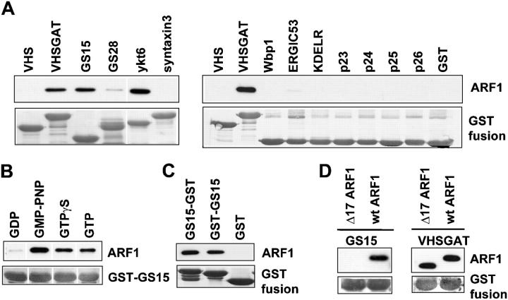
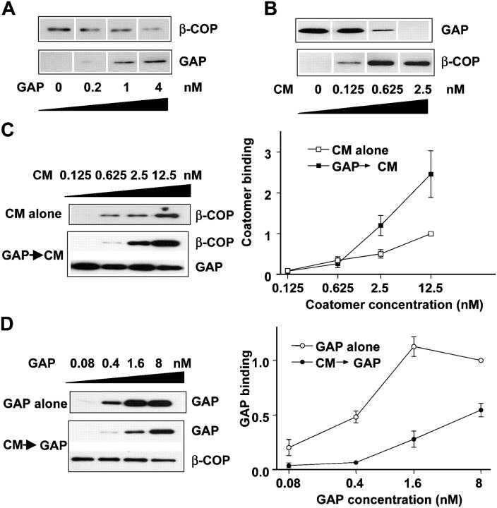
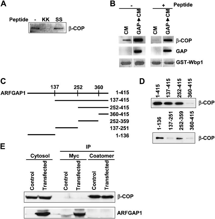
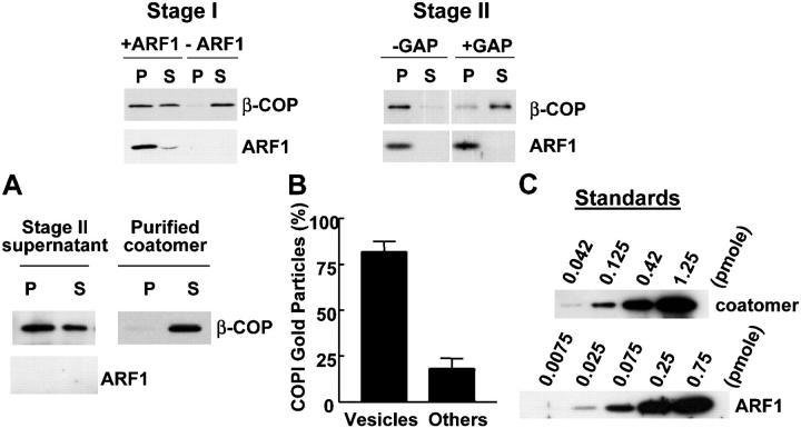
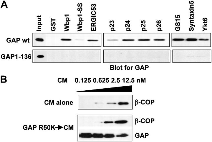
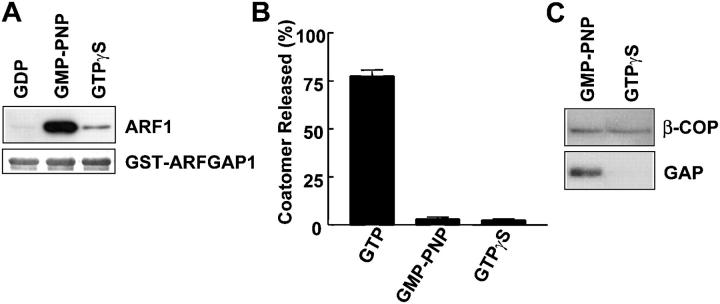
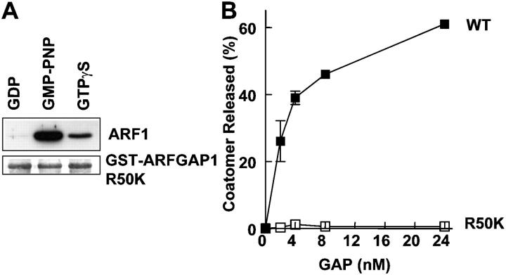
Examining how key components of coat protein I (COPI) transport participate in cargo sorting, we find that, instead of ADP ribosylation factor 1 (ARF1), its GTPase-activating protein (GAP) plays a direct role in promoting the binding of cargo proteins by coatomer (the core COPI complex). Activated ARF1 binds selectively to SNARE cargo proteins, with this binding likely to represent at least a mechanism by which activated ARF1 is stabilized on Golgi membrane to propagate its effector functions. We also find that the GAP catalytic activity plays a critical role in the formation of COPI vesicles from Golgi membrane, in contrast to the prevailing view that this activity antagonizes vesicle formation. Together, these findings indicate that GAP plays a central role in coupling cargo sorting and vesicle formation, with implications for simplifying models to describe how these two processes are coupled during COPI transport.


Figures

Figure 1.
Figure 1.
Activated ARF1 binds selectively to SNARE cargo proteins. (A) Activated ARF1 binds selectively to SNARE cargo proteins. ARF1 loaded with GMP-PNP was incubated with the cytoplasmic domain of different cargo proteins expressed as GST fusion proteins on beads for pull-down assays. Beads were then assessed for bound proteins as indicated. GST fusion protein that contained VHSGAT served as positive control, whereas one that contained VHS served as negative control. (B) Only activated ARF1 binds to the cytoplasmic domain of GS15. ARF1 loaded with different nucleotides as indicated was incubated with GST-GS15 on beads for pull-down assays. Beads were then assessed for bound proteins as indicated. (C) Activated ARF1 binds to the cytoplasmic domain of GS-15 regardless of its orientation of fusion to GST. ARF1 loaded with GMP-PNP was incubated with different fusion proteins (with GS15 appended to GST either at the carboxy terminus [GST-GS15] or amino terminus [GS15-GST]) on beads for pull-down assays. Beads were then assessed for bound proteins as indicated. (D) An activated form of ARF1 that lacks the first 17 amino-terminal residues no longer binds efficiently to the cytoplasmic domain of GS15. Different forms of ARF1 loaded with GMP-PNP were incubated with GST fusion proteins as indicated for pull-down assays. Beads were then assessed for bound proteins as indicated. White lines indicate that intervening lanes have been spliced out.
Figure 2.
Figure 2.
Binding of coatomer to cargo proteins is directly enhanced by GAP. (A) Simultaneous incubation of GAP and coatomer inhibits the binding of coatomer to cargo tail. GST-Wbp1 on beads was incubated with a fixed amount of coatomer (0.625 nM) and simultaneously with an increasing amount of GAP as indicated for pull-down assays. Beads were then assessed for bound proteins as indicated. (B) Simultaneous incubation of GAP and coatomer inhibits the binding of GAP to cargo tail. GST-Wbp1 on beads was incubated with a fixed amount of GAP (1 nM) and simultaneously with an increasing amount of coatomer as indicated for pull-down assays. Beads were then assessed for bound proteins as indicated. (C) Sequential incubation of GAP and then coatomer leads to optimal binding of coatomer to cargo tails. GST-Wbp1 on beads was incubated with either increasing levels of coatomer alone or sequentially with GAP followed by increasing levels of coatomer for pull-down assays. Beads were then assessed for bound proteins as indicated in gel images. For the graph, the level of coatomer on beads was then quantified, normalized to the level of GST fusion proteins, and then expressed as a fraction of the binding seen for the highest concentration of coatomer alone. The mean and standard error were derived from three independent experiments. (D) Sequential incubation of coatomer and then GAP inhibits the binding of GAP to cargo tails. GST-Wbp1 on beads was incubated with either increasing levels of GAP alone or sequentially with coatomer followed by increasing levels of GAP for pull-down assays. Beads were then assessed for bound proteins as indicated in gel images. For the graph, the level of GAP on beads was assessed similarly as done for coatomer above (C).
Figure 3.
Figure 3.
Elucidating how the sequential binding of GAP followed by coatomer results in their cooperative binding to cargo proteins. (A) Coatomer prebound with the Wbp1-free peptide does not bind to GST-Wbp1 on beads. Coatomer was incubated with a peptide that consists of either the cytoplasmic domain of Wbp1 (indicated as KK) or one with the di-lysines substituted with di-serines (indicated as SS) at 4°C for 1 h. GST-Wbp1 on beads was then added for a further incubation at 4°C for 1 h for pull-down assays, with the excess unbound free peptide still present to ensure that coatomer was fully saturated with the Wbp1-free peptide. (B) GAP supports the binding of saturated coatomer (with the Wbp1-free peptide) to GST-Wbp1 on beads. Coatomer, either unsaturated (indicated by no peptide added) or saturated (indicated by peptide added), were incubated with GST-Wbp1 on beads for pull-down assays, which involved either coatomer alone or sequentially with GAP followed by coatomer. In both cases, excess unbound free peptide was present to ensure that coatomer was fully saturated with the Wbp1-free peptide. Beads were then assessed for bound proteins as indicated. (C) Schematic of the different truncation mutants of GAP generated. (D) Two distinct domains in GAP mediating its direct binding to coatomer. Coatomer was incubated with different truncation mutants of GAP expressed as GST fusion proteins on beads for pull-down assays. Beads were then assessed for bound proteins as indicated. (E) GAP and coatomer do not interact with each other in cytosol. Co-precipitation experiments (lanes indicated by IP) were performed on cytosol that expressed myc-tagged ARFGAP1. The anti-ARFGAP1 antibody was not sensitive enough to detect endogenous ARFGAP1, as indicated by direct immunoblotting of cytosol (first two lanes).
Figure 4.
Figure 4.
Effect of ARF1 in complexes that contain cargo proteins, GAP, and coatomer. (A) Prior incubation with ARF1 does not further promote the GAP-enhanced binding of coatomer to GS15. One set of experiments (indicated by GST-GS15) involved GST-GS15 on beads incubated with coatomer alone, GAP alone, both GAP and coatomer in simultaneous incubation, or GAP followed by coatomer in sequential incubation. Another set of experiments (indicated by GST-GS15 + ARF1) involved activated ARF1 bound to GST-GS15 on beads being incubated with GAP alone, coatomer alone, both GAP and coatomer in simultaneous incubation, or GAP followed by coatomer in sequential incubation (B) Addition of GAP releases activated ARF1 bound to GS15. ARF1 preloaded with either GTP or GMP-PNP was bound to GST-GS15 on beads for 1 h, and then incubated with or without GAP (200 nM) for another hour. Beads were then assessed for bound proteins as indicated. (C) GAP does not affect the binding of activated ARF1 to cargo proteins. The different cargo proteins as GST fusions on beads were incubated either with or without GAP and then incubated with activated ARF1 that had been previously loaded with GMP-PNP. Beads were then assessed for bound proteins as indicated. (D) GAP cannot induce truncated ARF1 (Δ17) to bind GS15. GST-GS15 on beads was incubated either with or without GAP followed by incubation with the truncated ARF1 that had been loaded with GMP-PNP. Beads were then assessed for bound proteins as indicated. Incubation with GST-VHSGAT confirms that this truncated ARF1 represents an activated form. Incubation of activated wild-type ARF1 with GST-GS15 served as another positive control.
Figure 5.
Figure 5.
The level of ARF1 released from Golgi membrane is substoichiometric to the level of coatomer released as COPI vesicles. (A) A rapid and quantitative method of assessing the level of coatomer on reconstituted coated vesicles. The two-stage incubation system (indicated by Stage I and Stage II) was performed. After the second-stage incubation that contained GAP, the supernatant fraction was subjected to ultracentrifugation (200,000 g for 1 h) followed by immunoblotting of pellet (P) and supernatant (S) fractions for proteins as indicated. Ultracentrifugation of purified coatomer served as a control. (B) The pellet fraction after the ultracentrifugation contains membranes that are mostly COPI vesicles. The pellet derived from the ultracentrifugation of the supernatant fraction after the second-stage incubation (as described above in A) was analyzed by immunogold EM using anti-COPI antibodies. Quantitation was performed by selecting random fields at 60,000×, counting the gold particles on vesicles versus those on other membranes, and then calculating the fraction on these two respective membranes. The mean and standard error were derived from three independent experiments. (C) Quantitation of purified proteins. Increasing levels of purified proteins as indicated were immunoblotted.
Figure 6.
Figure 6.
The noncatalytic domain of GAP mediates its participation in COPI priming complexes. (A) A truncated GAP, consisting of residues 1–136, does not interact with COPI cargo proteins. 6x-his tagged GAP, either wild-type or truncated, was incubated with different GST fusion proteins on beads for pull-down assays. Beads were then assessed for bound proteins using an anti-6x-his antibody. (B) A catalytic dead GAP still mediates enhanced binding of coatomer to cargo proteins. GST-Wbp1 on beads was incubated with either coatomer alone or sequentially with mutant GAP followed by coatomer for pull-down assays. Beads were then assessed for bound proteins as indicated.
Figure 7.
Figure 7.
Differential effects of GTP analogs on interaction between GAP and ARF1 and on COPI vesicle formation. (A) Effect of different nucleotides on the interaction between GAP and ARF1. ARFGAP1 as a GST fusion protein on beads was incubated with ARF1 loaded with different nucleotides as indicated for pull-down assays. Beads were then assessed for bound proteins as indicated. (B) ARF1 loaded with GMP-PNP inhibits the release of coatomer from Golgi membrane in the vesicle reconstitution assay. The first-stage incubation was performed in the presence of different guanine nucleotides as indicated followed by the second-stage incubation using GAP. The level of coatomer released into the supernatant was quantified and then expressed as a fraction of total. The mean and standard error were derived from three independent experiments. (C) Reconstituted vesicles in the presence of GTPγS have decreased level of GAP as compared with reconstituted vesicles using GMP-PNP. The two-stage incubation system was performed in the presence of either GMP-PNP or GTPγS followed by pipette shearing to release vesicles from Golgi membrane, and then ultracentrifugation to collect coated vesicles in the pellet fraction.
Figure 8.
Figure 8.
A point mutation in GAP (R50K) that selectively abrogates its catalytic activity blocks vesicle formation. (A) Interaction between the point mutant GAP (R50K) and ARF1 bound to different nucleotides. The point mutant GAP (R50K) as GST fusion protein on beads was incubated with different forms of ARF1 previously loaded with nucleotides as indicated for pull-down assays. Beads were then assessed for bound proteins as indicated. (B) The R50K point mutant GAP blocks the release of coatomer from Golgi membrane in the vesicle reconstitution assay. The two-stage incubation was performed using different GAPs at varying concentrations as indicated. The level of coatomer released into the supernatant was quantified and then expressed as a fraction of total. The mean and standard error were derived from three independent experiments.

Similar articles

Cited by

References

    1. Allan, V.J., and T.E. Kreis. 1986. A microtubule-binding protein associated with membranes of the Golgi apparatus. J. Cell Biol. 103:2229–2239. - PMC - PubMed
    1. Bigay, J., P. Gounon, S. Robineau, and B. Antonny. 2003. Lipid packing sensed by ArfGAP1 couples COPI coat disassembly to membrane bilayer curvature. Nature. 426:563–566. - PubMed
    1. Bonifacino, J.S., and B.S. Glick. 2004. The mechanisms of vesicle budding and fusion. Cell. 116:153–166. - PubMed
    1. Cosson, P., and F. Letourneur. 1994. Coatomer interaction with di-lysine endoplasmic reticulum retention motifs. Science. 263:1629–1631. - PubMed
    1. Cukierman, E., I. Huber, M. Rotman, and D. Cassel. 1995. The ARF1-GTPase-activating protein: zinc finger motif and Golgi complex localization. Science. 270:1999–2002. - PubMed

Publication types

MeSH terms