NK cell activation through the NKG2D ligand MULT-1 is selectively prevented by the glycoprotein encoded by mouse cytomegalovirus gene m145
- PMID: 15642742
- PMCID: PMC2212792
- DOI: 10.1084/jem.20041617
NK cell activation through the NKG2D ligand MULT-1 is selectively prevented by the glycoprotein encoded by mouse cytomegalovirus gene m145
Abstract
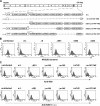
The NK cell-activating receptor NKG2D interacts with three different cellular ligands, all of which are regulated by mouse cytomegalovirus (MCMV). We set out to define the viral gene product regulating murine UL16-binding protein-like transcript (MULT)-1, a newly described NKG2D ligand. We show that MCMV infection strongly induces MULT-1 gene expression, but surface expression of this glycoprotein is nevertheless completely abolished by the virus. Screening a panel of MCMV deletion mutants defined the gene m145 as the viral regulator of MULT-1. The MCMV m145-encoded glycoprotein turned out to be necessary and sufficient to regulate MULT-1 by preventing plasma membrane residence of MULT-1. The importance of MULT-1 in NK cell regulation in vivo was confirmed by the attenuating effect of the m145 deletion that was lifted after NK cell depletion. Our findings underline the significance of escaping MULT-1/NKG2D signaling for viral survival and maintenance.
Figures


Similar articles
-
Effects of human cytomegalovirus infection on ligands for the activating NKG2D receptor of NK cells: up-regulation of UL16-binding protein (ULBP)1 and ULBP2 is counteracted by the viral UL16 protein.J Immunol. 2003 Jul 15;171(2):902-8. doi: 10.4049/jimmunol.171.2.902. J Immunol. 2003. PMID: 12847260
-
MCMV glycoprotein gp40 confers virus resistance to CD8+ T cells and NK cells in vivo.Nat Immunol. 2002 Jun;3(6):529-35. doi: 10.1038/ni799. Epub 2002 May 20. Nat Immunol. 2002. PMID: 12021778
-
Selective intracellular retention of virally induced NKG2D ligands by the human cytomegalovirus UL16 glycoprotein.Eur J Immunol. 2003 Jan;33(1):194-203. doi: 10.1002/immu.200390022. Eur J Immunol. 2003. PMID: 12594848
-
The UL16-binding proteins, a novel family of MHC class I-related ligands for NKG2D, activate natural killer cell functions.Immunol Rev. 2001 Jun;181:185-92. doi: 10.1034/j.1600-065x.2001.1810115.x. Immunol Rev. 2001. PMID: 11513139 Review.
-
UL16 binding proteins.Immunobiology. 2004;209(3):283-90. doi: 10.1016/j.imbio.2004.04.008. Immunobiology. 2004. PMID: 15518340 Review.
Cited by
-
The p36 isoform of murine cytomegalovirus m152 protein suffices for mediating innate and adaptive immune evasion.Viruses. 2013 Dec 16;5(12):3171-91. doi: 10.3390/v5123171. Viruses. 2013. PMID: 24351798 Free PMC article.
-
Human cytomegalovirus Fcγ binding proteins gp34 and gp68 antagonize Fcγ receptors I, II and III.PLoS Pathog. 2014 May 15;10(5):e1004131. doi: 10.1371/journal.ppat.1004131. eCollection 2014 May. PLoS Pathog. 2014. PMID: 24830376 Free PMC article.
-
Convergent Evolution by Cancer and Viruses in Evading the NKG2D Immune Response.Cancers (Basel). 2020 Dec 18;12(12):3827. doi: 10.3390/cancers12123827. Cancers (Basel). 2020. PMID: 33352921 Free PMC article. Review.
-
Can we build it better? Using BAC genetics to engineer more effective cytomegalovirus vaccines.J Clin Invest. 2010 Dec;120(12):4192-7. doi: 10.1172/JCI45250. Epub 2010 Nov 22. J Clin Invest. 2010. PMID: 21099107 Free PMC article.
-
Murine NKG2D ligands: "double, double toil and trouble".Mol Immunol. 2009 Mar;46(6):1011-9. doi: 10.1016/j.molimm.2008.09.035. Epub 2008 Dec 10. Mol Immunol. 2009. PMID: 19081632 Free PMC article. Review.
References
-
- Biron, C.A., K.B. Nguyen, G.C. Pien, L.P. Cousens, and T.P. Salazar-Mather. 1999. Natural killer cells in antiviral defense: function and regulation by innate cytokines. Annu. Rev. Immunol. 17:189–220. - PubMed
-
- Reddehase, M.J. 2002. Antigens and immunoevasins: opponents in cytomegalovirus immune surveillance. Nat. Rev. Immunol. 2:831–844. - PubMed
-
- Ljunggren, H.G., and K. Karre. 1990. In search of the ‘missing self’: MHC molecules and NK cell recognition. Immunol. Today. 11:237–244. - PubMed
Publication types
MeSH terms
Substances
LinkOut - more resources
Full Text Sources
Other Literature Sources
Miscellaneous

