Comparative Study
. 2004 Dec 20;167(6):1161-70.
doi: 10.1083/jcb.200410057.

Persistence of apoptotic cells without autoimmune disease or inflammation in CD14-/- mice

Affiliations
Comparative Study

Persistence of apoptotic cells without autoimmune disease or inflammation in CD14-/- mice

Andrew Devitt et al. J Cell Biol. .

Abstract

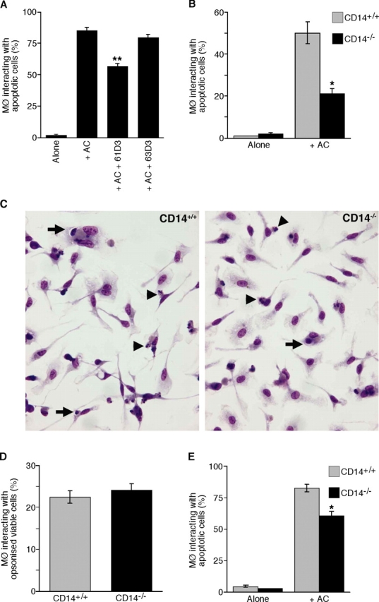
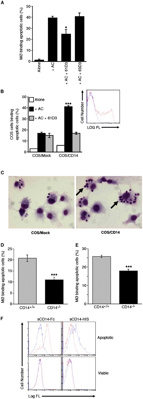
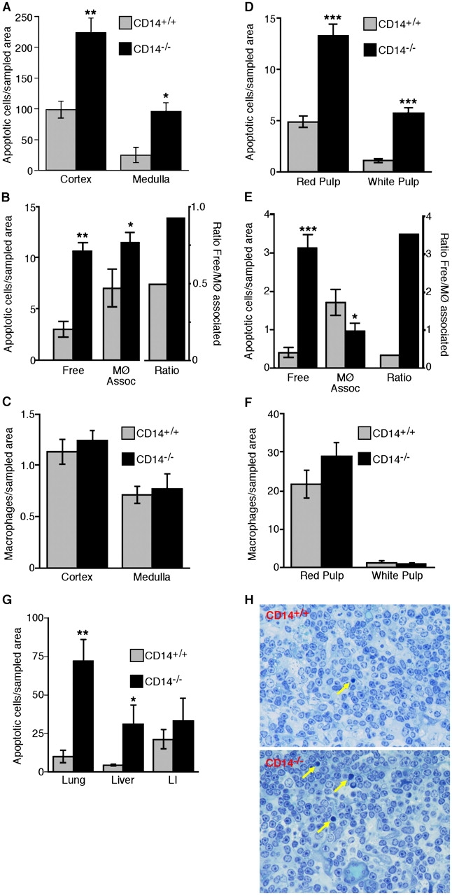
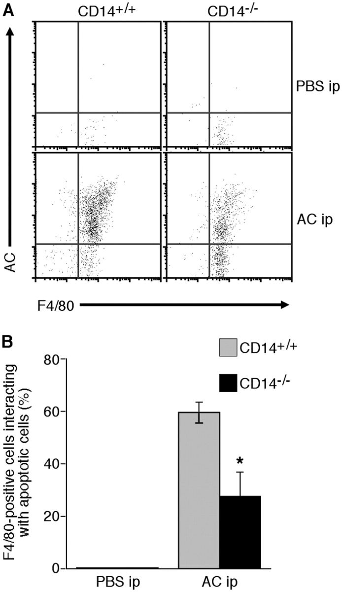
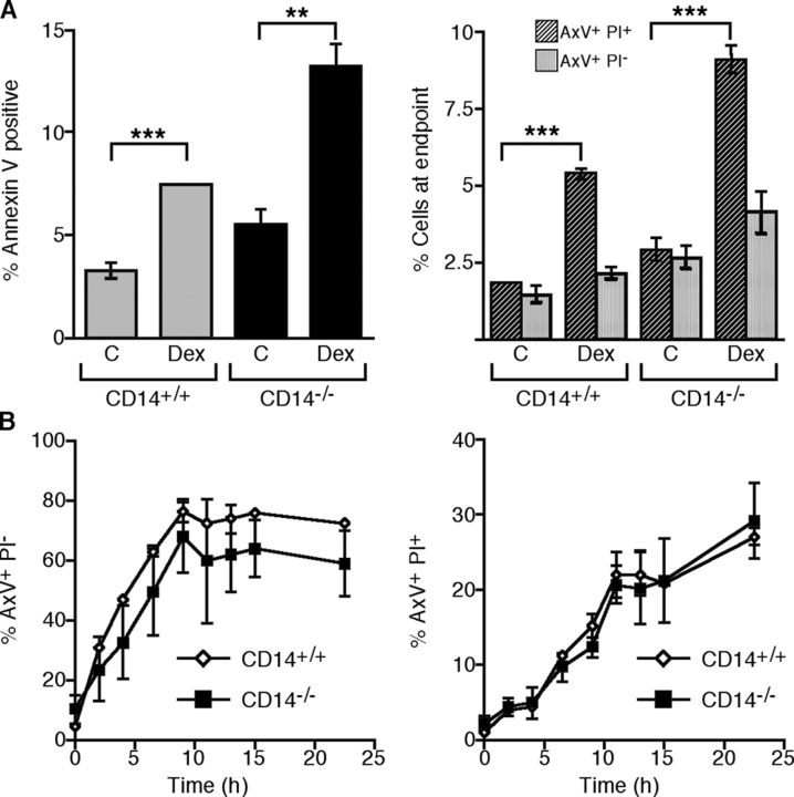
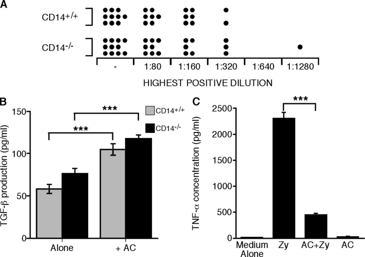
Interaction of macrophages with apoptotic cells involves multiple steps including recognition, tethering, phagocytosis, and anti-inflammatory macrophage responses. Defective apoptotic cell clearance is associated with pathogenesis of autoimmune disease. CD14 is a surface receptor that functions in vitro in the removal of apoptotic cells by human and murine macrophages, but its mechanism of action has not been defined. Here, we demonstrate that CD14 functions as a macrophage tethering receptor for apoptotic cells. Significantly, CD14(-/-) macrophages in vivo are defective in clearing apoptotic cells in multiple tissues, suggesting a broad role for CD14 in the clearance process. However, the resultant persistence of apoptotic cells does not lead to inflammation or increased autoantibody production, most likely because, as we show, CD14(-/-) macrophages retain the ability to generate anti-inflammatory signals in response to apoptotic cells. We conclude that CD14 plays a broad tethering role in apoptotic cell clearance in vivo and that apoptotic cells can persist in the absence of proinflammatory consequences.


Figures

Figure 1.
Figure 1.
CD14 dependence of interaction of apoptotic cells with human and murine macrophages. (A) Interaction (binding and phagocytosis) of 7-d HMDMs with apoptotic BL cells. Interaction between HMDMs and apoptotic BL cells (AC) was assessed after 60-min coculture at 37°C in the presence or absence of CD14 mAbs 61D3 or 63D3. Background interaction of apoptotic cells in the macrophage cultures without added BL cells is also shown (Alone). Results represent the percentage of macrophages interacting with apoptotic cells. Data shown are means ± SEM (n = 2). ANOVA: **, P < 0.01. (B) Interaction of 10-d BMDMs from CD14+/+ (gray bar) and CD14−/− (black bar) mice with apoptotic BL cells assessed after 30-min coculture at 37°C. Data shown are means ± SEM (n = 3). ANOVA: *, P < 0.05. Similar results were observed when syngeneic apoptotic thymocytes were tested in place of BL cells. (C) Photomicrographs showing 10-d BMDMs' interaction with apoptotic BL cells, comprising binding (arrowheads) and phagocytic (arrows) events. (D) Interaction of IgG-opsonized BL cells with the same macrophages as in B. Data are means ± SEM (n = 3). (E) Interaction of peritoneal macrophages from CD14+/+ (gray bar) and CD14−/− (black bar) mice with apoptotic BL cells assessed after 30-min coculture at 37°C. Data shown are means ± SEM (n = 3). ANOVA: *, P < 0.05.
Figure 2.
Figure 2.
Macrophage tethering of apoptotic cells by CD14. (A) Binding (in the absence of phagocytosis) of apoptotic BL cells (AC) by HMDMs assessed after coculture at 16–20°C for 60 min in the presence or absence of CD14 mAbs 61D3 or 63D3. Data shown are means ± SEM (n = 2). ANOVA: *, P < 0.05. (B) Binding of apoptotic BL cells (AC) by either mock-transfected or CD14-transfected COS-1 cells after coculture at 16–20°C for 60 min in the presence (gray bar) or absence (black bar) of CD14 mAb 61D3. (inset) Expression of CD14 by COS-1 cells as assessed by flow cytometry following immunofluorescence staining using mAb 63D3 and goat anti–mouse-FITC (shown in red) versus staining of mock transfectants (shown in blue). ANOVA: ***, P < 0.001. (C) Photomicrographs showing binding (but not phagocytosis) of apoptotic BL cells by COS cells. Binding is promoted by the expression of CD14 (arrow). (D) Binding of 10-d BMDMs from CD14+/+ (gray bar) and CD14−/− (black bar) mice with apoptotic BL cells assessed after 30-min coculture at 16–20°C. Data shown are means ± SEM (n = 3). ANOVA: ***, P < 0.001. (E) Binding of peritoneal macrophages from CD14+/+ (gray bar) and CD14−/− (black bar) mice with apoptotic BL cells assessed after 30-min coculture at 16–20°C. Data shown are means ± SEM (n = 3). ANOVA: ***, P < 0.001. (F) Binding of soluble recombinant human CD14 (sCD14) to apoptotic cells. BL cells undergoing spontaneous apoptosis were identified as “apoptotic” or “viable” according to light-scatter properties and confirmed by morphological analyses (Dive et al., 1992). Spontaneous apoptosis, a characteristic feature of group I BL cell lines (Gregory et al., 1991) was confirmed by morphological analysis as described previously (Devitt et al., 2003). Histograms of recombinant forms of CD14, sCD14-Fc, and sCD14-HIS binding to cells in these zones are shown. Control staining is shown in blue against staining with SCD14-Fc and SCD14-HIS in red.
Figure 3.
Figure 3.
Persistence of apoptotic cells in tissues of normal CD14 / mice. (A) Morphometric analysis of the frequency of ISEL+ nuclei present in the thymic cortex and medulla of CD14+/+ and CD14−/− mice. (B) Quantitative analyses of thymic sections showing the numbers of free and macrophage-associated apoptotic cells together with the ratios of free/macrophage-associated apoptotic cells for CD14+/+ and CD14−/− cortices. (C) Morphometric analyses of the distribution of macrophages (F4/80-reactive material) in the cortex and medulla of CD14+/+ and CD14−/− thymus. All thymus data shown are means ± SEM (n = 3 animals in each case). ANOVA: *, P < 0.05; **, P < 0.01. (D) Morphometric analysis of the frequency of ISEL+ nuclei present in the splenic red and white pulp of CD14+/+ and CD14−/− mice. ANOVA: ***, P < 0.001. (E) Quantitative analysis of the numbers of free and apoptotic cells together with the ratios of free/macrophage-associated apoptotic cells for CD14+/+ and CD14−/− splenic red pulp. (F) Morphometric analyses of the distribution of F4/80+ macrophages in the cortex and medulla of CD14+/+ and CD14−/− spleen. All spleen data shown are means ± SEM (n = 3 animals in each case). ANOVA: *, P < 0.05; ***, P < 0.001. (G) Persistence of apoptotic cells in non-lymphoid tissues of CD14−/− mice. Morphometric analyses of the frequency of ISEL+ nuclei present in the lung, liver, and large intestine (LI) of CD14+/+ and CD14−/− mice. Data are means ± SEM (n = 3 animals in each case). ANOVA: *, P < 0.1; **, P < 0.01. (H) Photomicrographs showing histological detail of CD14+/+ versus CD14−/− thymus. Note preponderance of free apoptotic cells in CD14−/− thymus (arrows) and absence of inflammatory cell infiltrate.
Figure 4.
Figure 4.
Inefficient interaction of apoptotic thymocytes with resident peritoneal macrophages in CD14 / mice. Fluorescent (green) autologous apoptotic thymocytes in PBS were introduced into the peritoneal cavity of CD14+/+ or CD14−/− mice by i.p. injection. Control animals received i.p. PBS alone. After 15 min, peritoneal cells were harvested, stained with F4/80, and analyzed by flow cytometry. Similar numbers of resident F4/80+ peritoneal macrophages were obtained from CD14+/+ and CD14−/− animals. (A) Representative flow cytometric dot plots of apoptotic (green) cells (AC) versus (red) F4/80+ events (macrophages). (B) Collated data from CD14+/+ and CD14−/− mice tested (means ± SEM; n = 3 animals in each case). ANOVA: *, P < 0.05.
Figure 5.
Figure 5.
Persistence of apoptotic cells in thymus of CD14 mice after dexamethasone treatment. (A) Flow cytometric analyses of thymocytes harvested from mock (C) or dexamethasone (DEX)-treated CD14+/+ or CD14−/− mice. Left, percentage of total AxV+ cells; right, percentage of apoptotic (AxV+PI) or secondarily necrotic (AxV+PI+) cells. ANOVA: **, P < 0.01; ***, P < 0.001. (B) Induction of apoptosis by dexamethasone treatment of thymocytes from CD14+/+ (open diamonds) or CD14−/− (closed squares) mice in vitro. Left, generation of apoptotic (AxV+PI) cells with time; right, generation of necrotic (AxV+PI+) cells with time. Data shown are means ± SEM (n = 3 animals per group).
Figure 6.
Figure 6.
Persistence of apoptotic cells in CD14 / mice does not lead to increased autoantibody production or inflammation. (A) Autoantibody production is not increased in CD14−/− mice. Titers of ANA antibodies in sera from aged CD14−/− and CD14+/+ mice were assessed by indirect immunofluorescence. See text for details. “−” denotes negative samples. (B) CD14−/− macrophages produce TGF-β effectively in response to apoptotic cells. TGF-β concentration in the supernatants of peritoneal macrophages following 18-h stimulation alone or with apoptotic thymocytes (AC). Data shown are means ± SEM, n = 5. ANOVA: ***, P < 0.001. (C) Apoptotic cells can inhibit proinflammatory responses of CD14-deficient macrophages. (left) TNF-α concentration in the supernatants of 10-d BMDMs following 18-h stimulation with both or either opsonized zymosan (Zy) and apoptotic thymocytes (AC). Data shown are means ± SEM, n = 3. ANOVA: ***, P < 0.001.

Similar articles

Cited by

References

    1. Albert, M.L. 2004. Death-defying immunity: do apoptotic cells influence antigen processing and presentation? Nat. Rev. Immunol. 4:223–231. - PubMed
    1. Ayala, A., C.D. Herdon, D.L. Lehman, C.A. Ayala, and I.H. Chaudry. 1996. Differential induction of apoptosis in lymphoid tissues during sepsis: variation in onset, frequency, and the nature of the mediators. Blood. 87:4261–4275. - PubMed
    1. Blander, J.M., and R. Medzhitov. 2004. Regulation of phagosome maturation by signals from toll-like receptors. Science. 304:1014–1018. - PubMed
    1. Botto, M., C. DellAgnola, A.E. Bygrave, E.M. Thompson, H.T. Cook, F. Petry, M. Loos, P.P. Pandolfi, and M.J. Walport. 1998. Homozygous C1q deficiency causes glomerulonephritis associated with multiple apoptotic bodies. Nat. Genet. 19:56–59. - PubMed
    1. Brown, S., I. Heinisch, E. Ross, K. Shaw, C.D. Buckley, and J. Savill. 2002. Apoptosis disables CD31-mediated cell detachment from phagocytes promoting binding and engulfment. Nature. 418:200–203. - PubMed

Publication types

MeSH terms