

. 2005 Jan 1;33(Database issue):D266-8.
doi: 10.1093/nar/gki001.
PDBsum more: new summaries and analyses of the known 3D structures of proteins and nucleic acids
Affiliations
- PMID: 15608193
- PMCID: PMC539955
- DOI: 10.1093/nar/gki001
Item in Clipboard
PDBsum more: new summaries and analyses of the known 3D structures of proteins and nucleic acids
Nucleic Acids Res.
.
Abstract
PDBsum is a database of mainly pictorial summaries of the 3D structures of proteins and nucleic acids in the Protein Data Bank. Its pages aim to provide an at-a-glance view of the contents of every 3D structure, plus detailed structural analyses of each protein chain, DNA-RNA chain and any bound ligands and metals. In the past year, the database has been significantly improved, in terms of both appearance and new content. Moreover, it has moved to its new address at http://www.ebi.ac.uk/thornton-srv/databases/pdbsum.
Figures
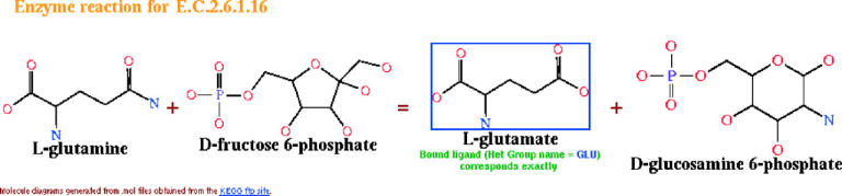
Diagram showing the reaction catalysed by enzymes of class E.C.2.6.1.16—the glucosamine 6-phosphate synthases. The diagram is taken from the PDBsum page for 1gdo, where the bound ligand—an l -glutamate—corresponds to one of the enzyme's products and is highlighted with a blue border in the diagram. Clicking on the highlighted molecule goes to the corresponding ligand page.
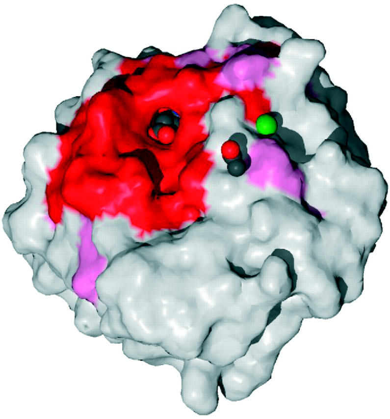
Surface of the glucosamine 6-phosphate synthase structure (PDB code 1gdo) coloured by residue conservation: red and pink for the most highly conserved regions, and blue for the most variable. The bound ligand—an l -glutamate—can be seen in spacefill representation within the highly conserved binding pocket. Also bound are an acetate ion and a sodium ion (green sphere).
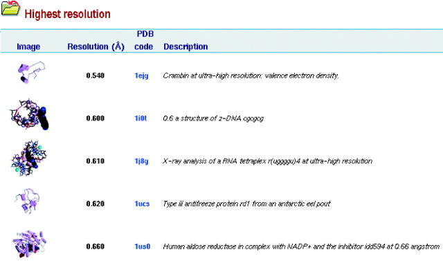
Example of one of the PDBsum highlights listings, here showing the top five structures in terms of the highest quoted resolution.
Similar articles
-
PDBsum: Structural summaries of PDB entries.Protein Sci. 2018 Jan;27(1):129-134. doi: 10.1002/pro.3289. Epub 2017 Oct 27. Protein Sci. 2018. PMID: 28875543 Free PMC article.
-
PDBsum1: A standalone program for generating PDBsum analyses.Protein Sci. 2022 Dec;31(12):e4473. doi: 10.1002/pro.4473. Protein Sci. 2022. PMID: 36251626 Free PMC article.
-
PDBsum extras: SARS-CoV-2 and AlphaFold models.Protein Sci. 2022 Jan;31(1):283-289. doi: 10.1002/pro.4238. Epub 2021 Nov 24. Protein Sci. 2022. PMID: 34779073 Free PMC article.
-
Computational modeling of RNA 3D structures and interactions.Curr Opin Struct Biol. 2016 Apr;37:22-8. doi: 10.1016/j.sbi.2015.11.007. Epub 2015 Dec 12. Curr Opin Struct Biol. 2016. PMID: 26689764 Review.
-
Inferring protein function from structure.Methods Biochem Anal. 2003;44:387-407. Methods Biochem Anal. 2003. PMID: 12647396 Review. No abstract available.
Cited by
-
Mycobacterium tuberculosis DosR regulon gene Rv0079 encodes a putative, 'dormancy associated translation inhibitor (DATIN)'.PLoS One. 2012;7(6):e38709. doi: 10.1371/journal.pone.0038709. Epub 2012 Jun 13. PLoS One. 2012. PMID: 22719925 Free PMC article.
-
IDBD: infectious disease biomarker database.Nucleic Acids Res. 2008 Jan;36(Database issue):D455-60. doi: 10.1093/nar/gkm925. Epub 2007 Nov 3. Nucleic Acids Res. 2008. PMID: 17982173 Free PMC article.
-
Structural evolution of the protein kinase-like superfamily.PLoS Comput Biol. 2005 Oct;1(5):e49. doi: 10.1371/journal.pcbi.0010049. Epub 2005 Oct 21. PLoS Comput Biol. 2005. PMID: 16244704 Free PMC article.
-
Comparative analysis of protein structure alignments.BMC Struct Biol. 2007 Jul 26;7:50. doi: 10.1186/1472-6807-7-50. BMC Struct Biol. 2007. PMID: 17672887 Free PMC article.
-
Crystal structure of the peptidase domain of Streptococcus ComA, a bifunctional ATP-binding cassette transporter involved in the quorum-sensing pathway.J Biol Chem. 2010 Apr 2;285(14):10777-85. doi: 10.1074/jbc.M109.093781. Epub 2010 Jan 25. J Biol Chem. 2010. PMID: 20100826 Free PMC article.
References
-
- Laskowski R.A., Hutchinson,E.G., Michie,A.D., Wallace,A.C., Jones,M.L., Thornton,J.M. (1997) PDBsum: a web-based database of summaries and analyses of all PDB structures. Trends Biochem. Sci., 22, 488–490. - PubMed
-
- Laskowski R.A., MacArthur,M.W., Moss,D.S., Thornton,J.M. (1993) PROCHECK—a program to check the stereochemical quality of protein structures. J. Appl. Crystallogr., 26, 283–291.
-
- Sayle R.A. and Milner-White,E.J. (1995) RASMOL: biomolecular graphics for all. Trends Biochem. Sci., 20, 374–376. - PubMed
Publication types
MeSH terms
Substances
LinkOut - more resources
Full Text Sources
Other Literature Sources
Miscellaneous
