. 2004 Dec;24(24):10636-49.
doi: 10.1128/MCB.24.24.10636-10649.2004.

The CRM1 nuclear export receptor controls pathological cardiac gene expression

Affiliations

The CRM1 nuclear export receptor controls pathological cardiac gene expression

Brooke C Harrison et al. Mol Cell Biol. 2004 Dec.

Abstract

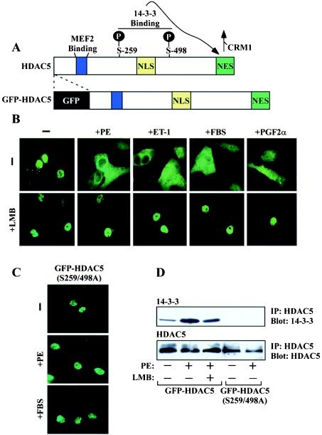
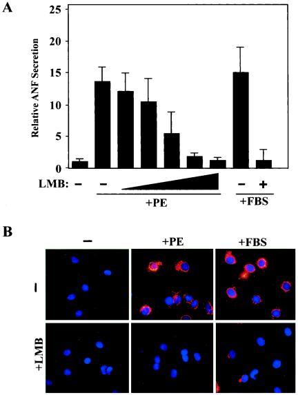
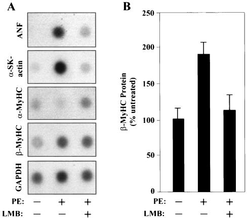
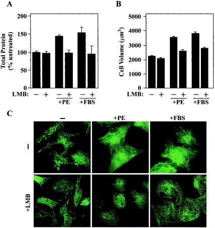
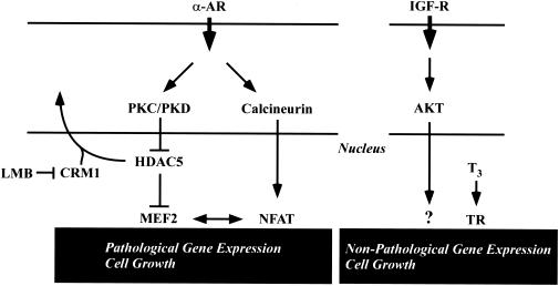
Diverse pathological insults trigger a cardiac remodeling process during which myocytes undergo hypertrophy, with consequent decline in cardiac function and eventual heart failure. Multiple transcriptional regulators of pathological cardiac hypertrophy are controlled at the level of subcellular distribution. For example, prohypertrophic transcription factors belonging to the nuclear factor of activated T cells (NFAT) and GATA families are subject to CRM1-dependent nuclear export but are rapidly relocalized to the nucleus in response to cues for hypertrophic growth. Here, we demonstrate that the antihypertrophic chromatin-modifying enzyme histone deacetylase 5 (HDAC5) is shuttled out of the cardiomyocyte nucleus via a CRM1-mediated pathway in response to diverse signals for hypertrophy. CRM1 antagonists block the agonist-mediated nuclear export of HDAC 5 and repress pathological gene expression and associated hypertrophy of cultured cardiomyocytes. Conversely, CRM1 activity is dispensable for nonpathological cardiac gene activation mediated by thyroid hormone and insulin-like growth factor 1, agonists that fail to trigger the nuclear export of HDAC5. These results suggest a selective role for CRM1 in derepression of pathological cardiac genes via its neutralizing effects on antihypertrophic factors such as HDAC5. Pharmacological approaches targeting CRM1-dependent nuclear export in heart muscle may have salutary effects on cardiac function by suppressing maladaptive changes in gene expression evoked by stress signals.


Figures

FIG. 1.
FIG. 1.
Signal-dependent nuclear export of HDAC5 in cardiomyocytes. (A) HDAC5 harbors an 18-amino-acid MEF2 binding domain (blue), a nuclear localization signal rich in basic residues (yellow), and a carboxy-terminal NES that is required for CRM1-mediated nucleo-cytoplasmic shuttling (green). Upon phosphorylation at two serine residues (S259 and S498 in human HDAC5), HDAC5 is bound by the intracellular chaperone protein 14-3-3, which results in exposure of the otherwise cryptic NES and the consequent nuclear export of the transcriptional repressor. Sequences encoding GFP were fused in frame to the amino terminus of HDAC5. (B) NRVMs were infected with adenovirus encodingGFP-HDAC5. Cells were treated for 1 h with PE (20 μM), ET-1 (50 nM), FBS (10%), or prostaglandin F2α (PGF2α; 10 μM) in the absence or presence of LMB (18.5 nM), and live-cell images were captured with an inverted fluorescence microscope. (C) NRVMs were infected with adenovirus encoding GFP fused to HDAC5 containing alanines in place of the 14-3-3 target sites [GFP-HDAC5 (S259/498A)] and treated with the indicated agonists, as described for panel B. (D) NRVMs were infected with adenovirus encoding GFP-HDAC5 or GFP-HDAC5 (S259/498A) and cultured overnight. Cells were washed and cultured in serum-free medium for 4 h prior to stimulation with PE (20 μM) for 1 h. HDAC5 was immunoprecipitated (IP) from cell lysates with HDAC5-specific antibody, and associated, endogenous 14-3-3 was detected by immunoblotting (top panel). Blots were reprobed with anti-GFP antibody to reveal total amounts of immunoprecipitated, ectopic HDAC5 (bottom panel).
FIG. 2.
FIG. 2.
Effects of a CRM1 inhibitor on ANF expression in cardiomyocytes. NRVMs were treated for 48 h with PE (20 μM) or FBS (5%) in the absence or presence of LMB (2.3, 4.6, 9.25, or 18.5 nM). ELISA was employed to measure concentrations of secreted ANF in culture supernatants. ANF values are presented relative to those of untreated controls (set at 1) ± standard deviations from eight independent samples. (B) NRVMs were treated with PE (20 μM) or FBS (10%) in the absence or presence of LMB (18.5 nM), and ANF protein was detected by indirect immunofluorescence with anti-ANF antibodies (red). Nuclei were stained with Hoechst dye (blue). Agonist-induced perinuclear expression of ANF was inhibited by LMB (lower panels).
FIG. 3.
FIG. 3.
A CRM1 inhibitor blocks the induction of the fetal cardiac gene program by PE. (A) NRVMs were treated for 48 h with PE (20 μM) in the absence or presence of LMB (18.5 nM), and the indicated transcripts were detected by RNA dot blot analysis. α-SK-actin, alpha-skeletal actin. (B) NRVMs were cultured for 48 h with LMB (18.5 nM) in the absence or presence of PE (20 μM). Levels of β-MyHC protein were measured by cytoblot analysis and are graphed as percentages of expression relative to the expression of untreated controls (set at 100%). Values are means ± standard deviations from eight independent samples.
FIG. 4.
FIG. 4.
A CRM1 inhibitor blocks agonist-induced increases in cardiomyocyte size and sarcomere organization. (A) NRVMs were treated with PE (20 μM) or FBS (5%) for 48 h in the absence or presence of LMB (18.5 nM). Total cellular protein was quantified by the Bradford assay and is presented as a percentage of that in untreated cells. Values are averages from eight independent samples ± standard deviations. (B) NRVMs were treated as described for panel A and trypsinized for Coulter Counter analysis. Average cell volumes (in cubic micrometers) were determined for 104 cells. Error bars represent standard deviations of results from three independent experiments. (C) NRVMs were treated for 48 h with PE (20 μM) or FBS (5%) in the absence or presence of LMB (18.5 nM). Cells were fixed and stained for α-actinin to reveal sarcomeres by indirect immunofluorescence. LMB prevents the enhanced organization of the sarcomeres in response to hypertrophic agonists.
FIG. 5.
FIG. 5.
CRM1 inhibition does not alter cardiomyocyte viability. (A) NRVMs were left untreated or stimulated with PE (20 μM) for 48 h in the absence or presence of the indicated concentrations of LMB. Cells were stained with calcein AM dye, which is retained by and fluoresces in viable cells (green), and ethidium bromide (red), which enters only cells with compromised membranes. (B) AK was detected in culture medium of NRVMs stimulated for 48 h with PE (20 μM) or FBS (5%) in the absence or presence of LMB (18.5 nM). Values represent the means ± standard deviations of results from eight independent samples. The higher values from FBS-treated cells are a consequence of the AK present in serum. (C) NRVMs were cultured in 96-well dishes and treated with the indicated concentrations of LMB for 48 h in the absence of hypertrophic stimulus. Intracellular ATP concentrations were determined for eight independent samples from each treatment group and are graphed relative to values for untreated cells (set at 100). Standard deviations are shown.
FIG. 6.
FIG. 6.
A newly discovered CRM1 inhibitor blocks cardiac hypertrophy. (A) NRVMs were infected with adenovirus encoding GFP-HDAC5 and treated for 2 h with ET-1 in the absence or presence of 5219668 (1.25 μM; 5219668 is the ChemBridge Corporation catalog number) or LMB (18.5 nM). (B) NRVMs were treated with PE (20 μM) for 48 h in the absence or presence of 5219668 (1.25 μM). Cells were fixed and analyzed by indirect immunofluorescence to reveal sarcomeres (green, top panels) and ANF (red, bottom panels). Nuclei were stained with Hoechst dye (blue, bottom panels). (C) NRVMs were treated for 48 h with PE (20 μM) in the absence or presence of 5219668 (1.25 μM). ELISA was employed to measure concentrations of secreted ANF in culture supernatants. ANF values are presented relative to those of untreated cells (set at 1) and were derived from eight independent samples for each condition. Standard deviations are shown. (D) The Bradford assay was employed to quantifytotal cellular protein from cells analyzed for the results shown in panel C. Values were averaged for eight independent samples for each condition and are presented as percentages of levels in untreated cells (100%) ± standard deviations. (E) NRVMs were treated for 48 h with PE (20 μM) in the absence or presence of 5219668 (1.25 μM). Average cell volumes for 104 cells per condition were measured using a Coulter Counter. Error bars represent standard deviations from three independent experiments.
FIG. 7.
FIG. 7.
Agonist-mediated induction of α-MyHC and SERCA expression are unaffected by CRM1 inhibition. (A) NRVMs were infected with adenovirus encoding GFP-HDAC5 and stimulated for 2 h with PE (20 μM), ET-1 (50 nM), T3 (3 nM), IGF-1 (100 nM), or T3 (3 nM) plus IGF-1 (100 nM). (B) NRVMs were treated for 48 h with T3 (3 nM) in the absence or presence of LMB (18.5 nM). Effects of LMB on T3-mediated regulation of α- and β-MyHC protein expression were assessed by cytoblot analysis. Average values were determined from at least eight independent wells per condition and are depicted relative to levels in untreated controls (set at 100%). Standard deviations are shown. (C) NRVMs were treated for 48 h with IGF-1 (100 nM) in the absence or presence of LMB (18.5 nM). Levels of α-MyHC protein expression were assessed by cytoblot analysis, as described for panel B. *, P < 0.0001 versus values for untreated cells. (D) NRVMs were cultured for 48 h with T3 (3 nM) or IGF-1 (100 nM) in the absence or presence of LMB (18.5 nM) or 5219668 (1.25 μM). Effects of LMB on T3- and IGF-1-induced expression of SERCA2 were determined by immunoblotting with anti-SERCA antibodies. The blot was reprobed with antibodies to the calnexin chaperone protein to control for protein loading.
FIG. 8.
FIG. 8.
Effects of a CRM1 inhibition on NFAT-dependent gene expression. (A) MCIP-1 functions in a negative-feedback loop to control calcineurin signaling. The Ca2+-calmodulin (CaM)-dependent phosphatase calcineurin is activated in response to stress signals that increase intracellular Ca2+. Calcineurin dephosphorylates NFAT, promoting its import into the nucleus. Calcineurin signaling stimulates the expression of a 28-kDa isoform of MCIP-1 by virtue of an alternative promoter harboring 15 NFAT binding sites. Newly expressed MCIP-1 protein binds to and negatively regulates calcineurin. (B) NRVMs were cultured in the absence or presence of LMB (18.5 nM), as indicated. Cells were stimulated with either T3 (3 nM) or PE (20 μM) for 48 h prior to preparation of protein lysates for immunoblot analysis with anti-MCIP1 antibodies. Induction of the calcineurin-responsive form of MCIP-1 (28 kDa) is blocked by LMB. Blots were reprobed with antibodies to the calnexin chaperone protein to control for protein loading (bottom panel).
FIG. 9.
FIG. 9.
Model for repression of pathological cardiac gene expression by CRM1. Pathological cardiac hypertrophy can be triggered by a number of stress stimuli, including agonists that stimulate the α-adrenergic receptor (α-AR). Hypertrophic stimuli activate the Ca2+-calmodulin-dependent phosphatase calcineurin. Calcineurin dephosphorylates the NFAT transcription factor, driving it to the nucleus, where it stimulates pathological gene expression. Stress signals also stimulate the PKC/PKD pathway, resulting in phosphorylation of HDAC5, which normally binds to repress the function of the MEF2 transcription factor. Phospho-HDAC5 is escorted from the nucleus via a CRM1-dependent pathway, freeing MEF2 to activate downstream target genes that promote pathological hypertrophy. CRM1 inhibitors such as LMB retain HDAC5 in the nucleus, thereby repressing MEF2-dependent transcription. Since MEF2 interacts with NFAT, nuclear retention of HDAC5 is also predicted to suppress NFAT target gene expression. Binding of the bioactive form of thyroid hormone (T3) to its receptor (TR) triggers expression of genes that are associated with increased cardiac contractility and nonpathological cardiac growth, including α-MyHC and SERCA. Nonpathological cardiac gene expression and growth can also be triggered by signaling via IGF receptor (IGF-R) and its downstream effectors, including the AKT kinase. These pathways for cardiac gene expression function independently of CRM1.

Similar articles

Cited by

References

    1. Antos, C. L., T. A. McKinsey, N. Frey, W. Kutschke, J. McAnally, J. M. Shelton, J. A. Richardson, J. A. Hill, and E. N. Olson. 2002. Activated glycogen synthase kinase-3β suppresses cardiac hypertrophy in vivo. Proc. Natl. Acad. Sci. USA 99:907-912. - PMC - PubMed
    1. Beals, C. R., N. A. Clipstone, S. N. Ho, and G. R. Crabtree. 1997. Nuclear localization of NF-ATc by a calcineurin-dependent, cyclosporin-sensitive intramolecular interaction. Genes Dev. 11:824-834. - PubMed
    1. Becker, T. C., R. J. Noel, W. S. Coats, A. M. Gomez-Foix, T. Alam, R. D. Gerard, and C. B. Newgard. 1994. Use of recombinant adenovirus for metabolic engineering of mammalian cells. Methods Cell Biol. 43:161-189. - PubMed
    1. Braunwald, E., and M. R. Bristow. 2000. Congestive heart failure: fifty years of progress. Circulation 102:IV14-IV23. - PubMed
    1. Bush, E., J. Fielitz, L. Melvin, M. Martinez-Arnold, T. A. McKinsey, R. Plichta, and E. N. Olson. 2004. A small molecular activator of cardiac hypertrophy uncovered in a chemical screen for modifiers of the calcineurin signaling pathway. Proc. Natl. Acad. Sci. USA 101:2870-2875. - PMC - PubMed

MeSH terms

LinkOut - more resources