

Prefrontal cortex stimulation induces 2-arachidonoyl-glycerol-mediated suppression of excitation in dopamine neurons
- PMID: 15564588
- PMCID: PMC6730123
- DOI: 10.1523/JNEUROSCI.3502-04.2004
Prefrontal cortex stimulation induces 2-arachidonoyl-glycerol-mediated suppression of excitation in dopamine neurons
Abstract
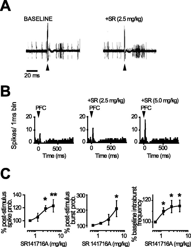
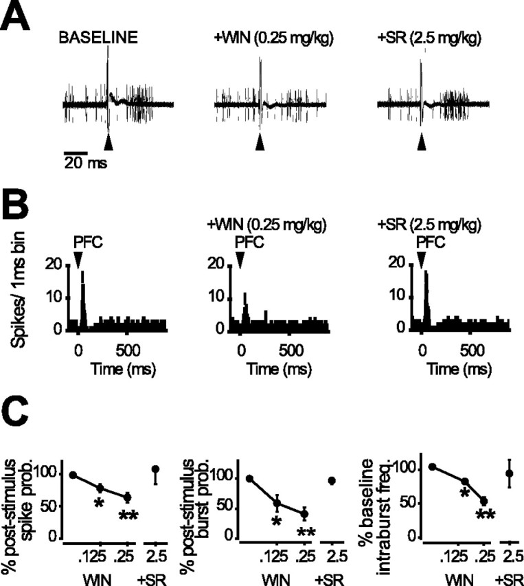
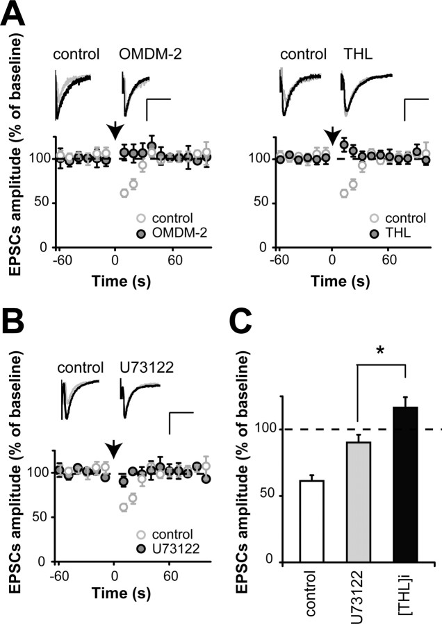
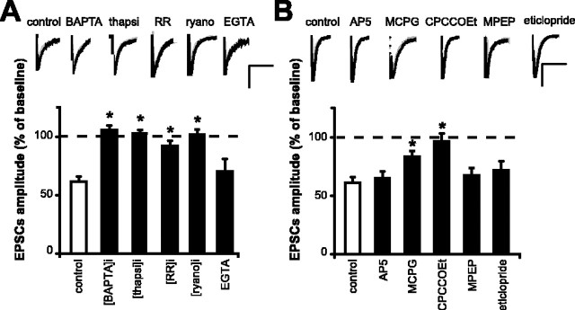
Endocannabinoids form a novel class of retrograde messengers that modulate short- and long-term synaptic plasticity. Depolarization-induced suppression of excitation (DSE) and inhibition (DSI) are the best characterized transient forms of endocannabinoid-mediated synaptic modulation. Stimulation protocols consisting of long-lasting voltage steps to the postsynaptic cell are routinely used to evoke DSE-DSI. Little is known, however, about more physiological conditions under which these molecules are released in vitro. Moreover, the occurrence in vivo of such forms of endocannabinoid-mediated modulation is still controversial. Here we show that physiologically relevant patterns of synaptic activity induce a transient suppression of excitatory transmission onto dopamine neurons in vitro. Accordingly, in vivo endocannabinoids depress the increase in firing and bursting activity evoked in dopamine neurons by prefrontal cortex stimulation. This phenomenon is selectively mediated by the endocannabinoid 2-arachidonoyl-glycerol (2-AG), which activates presynaptic cannabinoid type 1 receptors. 2-AG synthesis involves activation of metabotropic glutamate receptors and Ca2+ mobilization from intracellular stores. These findings indicate that dopamine neurons release 2-AG to shape afferent activity and ultimately their own firing pattern. This novel endocannabinoid-mediated self-regulatory role of dopamine neurons may bear relevance in the pathogenesis of neuropsychiatric disorders such as schizophrenia and addiction.
Figures

Similar articles
-
Regulation of plasticity of glutamate synapses by endocannabinoids and the cyclic-AMP/protein kinase A pathway in midbrain dopamine neurons.J Physiol. 2010 Jul 15;588(Pt 14):2589-604. doi: 10.1113/jphysiol.2010.190066. Epub 2010 May 24. J Physiol. 2010. PMID: 20498231 Free PMC article.
-
Postsynaptic diacylglycerol lipase mediates retrograde endocannabinoid suppression of inhibition in mouse prefrontal cortex.J Physiol. 2011 Oct 15;589(Pt 20):4857-84. doi: 10.1113/jphysiol.2011.212225. Epub 2011 Aug 1. J Physiol. 2011. PMID: 21807615 Free PMC article.
-
Independent presynaptic and postsynaptic mechanisms regulate endocannabinoid signaling at multiple synapses in the ventral tegmental area.J Neurosci. 2004 Dec 8;24(49):11070-8. doi: 10.1523/JNEUROSCI.3695-04.2004. J Neurosci. 2004. PMID: 15590923 Free PMC article.
-
Endocannabinoid-mediated short-term synaptic plasticity: depolarization-induced suppression of inhibition (DSI) and depolarization-induced suppression of excitation (DSE).Br J Pharmacol. 2004 May;142(1):9-19. doi: 10.1038/sj.bjp.0705726. Epub 2004 Apr 20. Br J Pharmacol. 2004. PMID: 15100161 Free PMC article. Review.
-
Endocannabinoid-mediated retrograde modulation of synaptic transmission.Curr Opin Neurobiol. 2014 Dec;29:1-8. doi: 10.1016/j.conb.2014.03.017. Epub 2014 Apr 18. Curr Opin Neurobiol. 2014. PMID: 24747340 Review.
Cited by
-
Involvement of the endogenous cannabinoid system in the effects of alcohol in the mesolimbic reward circuit: electrophysiological evidence in vivo.Psychopharmacology (Berl). 2005 Dec;183(3):368-77. doi: 10.1007/s00213-005-0195-0. Epub 2005 Oct 15. Psychopharmacology (Berl). 2005. PMID: 16228194
-
Dopaminergic regulation of dopamine D3 and D3nf receptor mRNA expression.Synapse. 2010 Aug;64(8):634-43. doi: 10.1002/syn.20770. Synapse. 2010. PMID: 20340170 Free PMC article.
-
Drug-induced psychosis: how to avoid star gazing in schizophrenia research by looking at more obvious sources of light.Front Behav Neurosci. 2011 Jan 17;5:1. doi: 10.3389/fnbeh.2011.00001. eCollection 2011. Front Behav Neurosci. 2011. PMID: 21267359 Free PMC article.
-
Cannabinoids and Vanilloids in Schizophrenia: Neurophysiological Evidence and Directions for Basic Research.Front Pharmacol. 2017 Jun 21;8:399. doi: 10.3389/fphar.2017.00399. eCollection 2017. Front Pharmacol. 2017. PMID: 28680405 Free PMC article. Review.
-
Analgesic and Anti-Inflammatory Effects of Perampanel in Acute and Chronic Pain Models in Mice: Interaction With the Cannabinergic System.Front Pharmacol. 2021 Feb 1;11:620221. doi: 10.3389/fphar.2020.620221. eCollection 2020. Front Pharmacol. 2021. PMID: 33597883 Free PMC article.
References
-
- Alger BE (2002) Retrograde signaling in the regulation of synaptic transmission: focus on endocannabinoids. Prog Neurobiol 68: 247-286. - PubMed
-
- Ameri A, Simmet T (2000) Effects of 2-arachidonylglycerol, an endogenous cannabinoid, on neuronal activity in rat hippocampal slices. Naunyn Schmiedebergs Arch Pharmacol 361: 265-272. - PubMed
-
- Batchelor AM, Knopfel T, Gasparini F, Garthwaite J (1997) Pharmacological characterization of synaptic transmission through mGluRs in rat cerebellar slices. Neuropharmacology 36: 401-403. - PubMed
-
- Beltramo M, Stella N, Calignano A, Lin SY, Makriyannis A, Piomelli D (1997) Functional role of high-affinity anandamide transport, as revealed by selective inhibition. Science 277: 1094-1097. - PubMed
-
- Bisogno T, Melck D, De Petrocellis L, Di Marzo V (1999a) Phosphatidic acid as the biosynthetic precursor of the endocannabinoid 2-arachidonoylglycerol in intact mouse neuroblastoma cells stimulated with ionomycin. J Neurochem 72: 2113-2119. - PubMed
Publication types
MeSH terms
Substances
LinkOut - more resources
Full Text Sources
Miscellaneous
