Replication-independent chromatin loading of Dnmt1 during G2 and M phases
- PMID: 15550930
- PMCID: PMC1299190
- DOI: 10.1038/sj.embor.7400295
Replication-independent chromatin loading of Dnmt1 during G2 and M phases
Abstract
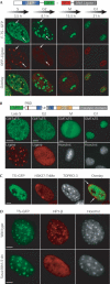
The major DNA methyltransferase, Dnmt1, associates with DNA replication sites in S phase maintaining the methylation pattern in the newly synthesized strand. In view of the slow kinetics of Dnmt1 in vitro versus the fast progression of the replication fork, we have tested whether Dnmt1 associates with chromatin beyond S phase. Using time-lapse microscopy of mammalian cells expressing green-fluorescent-protein-tagged Dnmt1 and DsRed-tagged DNA Ligase I as a cell cycle progression marker, we have found that Dnmt1 associates with chromatin during G2 and M. This association is mediated by a specific targeting sequence, shows strong preference for constitutive but not facultative heterochromatin and is independent of heterochromatin-specific histone H3 Lys 9 trimethylation, SUV39H and HP1. Moreover, photobleaching analyses showed that Dnmt1 is continuously loaded onto chromatin throughout G2 and M, indicating a replication-independent role of Dnmt1 that could represent a novel and separate pathway to maintain DNA methylation.
Figures


Similar articles
-
A role for LSH in facilitating DNA methylation by DNMT1 through enhancing UHRF1 chromatin association.Nucleic Acids Res. 2020 Dec 2;48(21):12116-12134. doi: 10.1093/nar/gkaa1003. Nucleic Acids Res. 2020. PMID: 33170271 Free PMC article.
-
Direct interaction between DNMT1 and G9a coordinates DNA and histone methylation during replication.Genes Dev. 2006 Nov 15;20(22):3089-103. doi: 10.1101/gad.1463706. Epub 2006 Nov 3. Genes Dev. 2006. PMID: 17085482 Free PMC article.
-
RFTS-dependent negative regulation of Dnmt1 by nucleosome structure and histone tails.FEBS J. 2017 Oct;284(20):3455-3469. doi: 10.1111/febs.14205. Epub 2017 Sep 11. FEBS J. 2017. PMID: 28834260
-
Mammalian cytosine DNA methyltransferase Dnmt1: enzymatic mechanism, novel mechanism-based inhibitors, and RNA-directed DNA methylation.Curr Med Chem. 2008;15(1):92-106. doi: 10.2174/092986708783330700. Curr Med Chem. 2008. PMID: 18220765 Review.
-
Coordinated Dialogue between UHRF1 and DNMT1 to Ensure Faithful Inheritance of Methylated DNA Patterns.Genes (Basel). 2019 Jan 18;10(1):65. doi: 10.3390/genes10010065. Genes (Basel). 2019. PMID: 30669400 Free PMC article. Review.
Cited by
-
Dynamics of Dnmt1 interaction with the replication machinery and its role in postreplicative maintenance of DNA methylation.Nucleic Acids Res. 2007;35(13):4301-12. doi: 10.1093/nar/gkm432. Epub 2007 Jun 18. Nucleic Acids Res. 2007. PMID: 17576694 Free PMC article.
-
Rapid and transient recruitment of DNMT1 to DNA double-strand breaks is mediated by its interaction with multiple components of the DNA damage response machinery.Hum Mol Genet. 2011 Jan 1;20(1):126-40. doi: 10.1093/hmg/ddq451. Epub 2010 Oct 11. Hum Mol Genet. 2011. PMID: 20940144 Free PMC article.
-
Epigenetic Enhancement of the Post-replicative DNA Mismatch Repair of Mammalian Genomes by a Hemi-mCpG-Np95-Dnmt1 Axis.Sci Rep. 2016 Nov 25;6:37490. doi: 10.1038/srep37490. Sci Rep. 2016. PMID: 27886214 Free PMC article.
-
Global DNA hypomethylation prevents consolidation of differentiation programs and allows reversion to the embryonic stem cell state.PLoS One. 2012;7(12):e52629. doi: 10.1371/journal.pone.0052629. Epub 2012 Dec 27. PLoS One. 2012. PMID: 23300728 Free PMC article.
-
Dissection of cell cycle-dependent dynamics of Dnmt1 by FRAP and diffusion-coupled modeling.Nucleic Acids Res. 2013 May;41(9):4860-76. doi: 10.1093/nar/gkt191. Epub 2013 Mar 27. Nucleic Acids Res. 2013. PMID: 23535145 Free PMC article.
References
-
- Bird AP, Wolffe AP (1999) Methylation-induced repression—belts, braces, and chromatin. Cell 99: 451–454 - PubMed
-
- Chuang LS, Ian HI, Koh TW, Ng HH, Xu G, Li BF (1997) Human DNA–(cytosine-5) methyltransferase–PCNA complex as a target for p21WAF1. Science 277: 1996–2000 - PubMed
-
- Doerfler W (1983) DNA methylation and gene activity. Annu Rev Biochem 52: 93–124 - PubMed
Publication types
MeSH terms
Substances
LinkOut - more resources
Full Text Sources
Other Literature Sources
Molecular Biology Databases
Research Materials

