

Direct measurement of transmural laminar architecture in the anterolateral wall of the ovine left ventricle: new implications for wall thickening mechanics
- PMID: 15550521
- PMCID: PMC2822837
- DOI: 10.1152/ajpheart.00813.2004
Direct measurement of transmural laminar architecture in the anterolateral wall of the ovine left ventricle: new implications for wall thickening mechanics
Abstract
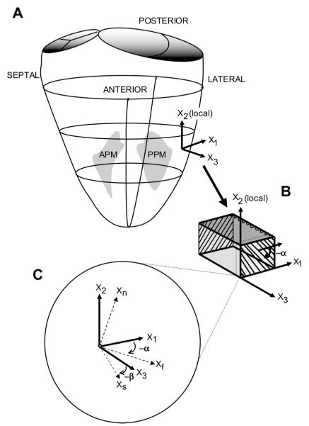
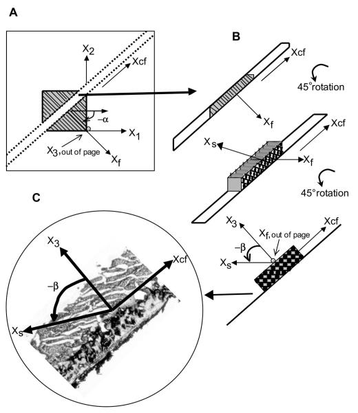
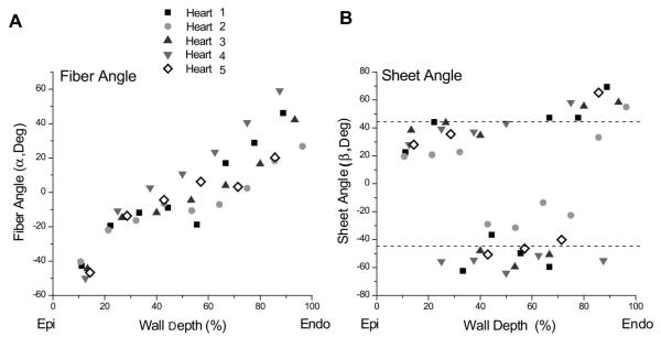
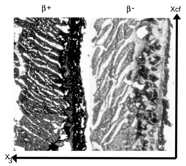
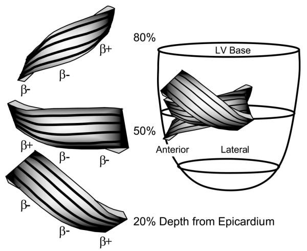
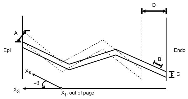
Laminar, or sheet, architecture of the left ventricle (LV) is a structural basis for normal systolic and diastolic LV dynamics, but transmural sheet orientations remain incompletely characterized. We directly measured the transmural distribution of sheet angles in the ovine anterolateral LV wall. Ten Dorsett-hybrid sheep hearts were perfusion fixed in situ with 5% buffered glutaraldehyde at end diastole and stored in 10% formalin. Transmural blocks of myocardial tissue were excised, with the edges cut parallel to local circumferential, longitudinal, and radial axes, and sliced into 1-mm-thick sections parallel to the epicardial tangent plane from epicardium to endocardium. Mean fiber directions were determined in each section from five measurements of fiber angles. Each section was then cut transverse to the fiber direction, and five sheet angles (beta) were measured and averaged. Mean fiber angles progressed nearly linearly from -41 degrees (SD 11) at the epicardium to +42 degrees (SD 16) at the endocardium. Two families of sheets were identified at approximately +45 degrees (beta(+)) and -45 degrees (beta(-)). In the lateral region (n = 5), near the epicardium, sheets belonged to the beta(+) family; in the midwall, to the beta(-) family; and near the endocardium, to the beta(+) family. This pattern was reversed in the basal anterior region (n = 4). Sheets were uniformly beta(-) over the anterior papillary muscle (n = 2). These direct measurements of sheet angles reveal, for the first time, alternating transmural families of predominant sheet angles. This may have important implications in understanding wall mechanics in the normal and the failing heart.
Figures
Similar articles
-
Heterogeneity of left ventricular wall thickening mechanisms.Circulation. 2008 Aug 12;118(7):713-21. doi: 10.1161/CIRCULATIONAHA.107.744623. Epub 2008 Jul 28. Circulation. 2008. PMID: 18663088 Free PMC article.
-
Relation of regional cross-fiber shortening to wall thickening in the intact heart. Three-dimensional strain analysis by NMR tagging.Circulation. 1994 Mar;89(3):1174-82. doi: 10.1161/01.cir.89.3.1174. Circulation. 1994. PMID: 8124804
-
Effects of undersized mitral annuloplasty on regional transmural left ventricular wall strains and wall thickening mechanisms.Circulation. 2006 Jul 4;114(1 Suppl):I600-9. doi: 10.1161/CIRCULATIONAHA.105.001529. Circulation. 2006. PMID: 16820645
-
The architecture of the ventricular mass and its functional implications for organ-preserving surgery.Eur J Cardiothorac Surg. 2005 Feb;27(2):183-90. doi: 10.1016/j.ejcts.2004.10.050. Eur J Cardiothorac Surg. 2005. PMID: 15691669 Review.
-
[Transmural heterogeneity of the left ventricular wall: subendocardial layer and subepicardial layer].J Cardiol. 2000 Mar;35(3):205-18. J Cardiol. 2000. PMID: 10808428 Review. Japanese.
Cited by
-
Myofiber angle distributions in the ovine left ventricle do not conform to computationally optimized predictions.J Biomech. 2008 Nov 14;41(15):3219-24. doi: 10.1016/j.jbiomech.2008.08.007. Epub 2008 Sep 20. J Biomech. 2008. PMID: 18805536 Free PMC article.
-
Assessment of Myocardial Microstructural Dynamics by In Vivo Diffusion Tensor Cardiac Magnetic Resonance.J Am Coll Cardiol. 2017 Feb 14;69(6):661-676. doi: 10.1016/j.jacc.2016.11.051. J Am Coll Cardiol. 2017. PMID: 28183509 Free PMC article.
-
[The antagonistic function of the heart muscle sustains the autoregulation according to Frank and Starling : Part I: Structure and function of heart muscle].Herz. 2020 Apr;45(2):170-177. doi: 10.1007/s00059-018-4734-y. Epub 2018 Jul 27. Herz. 2020. PMID: 30054713 Review. German.
-
Myocardial mesostructure and mesofunction.Am J Physiol Heart Circ Physiol. 2022 Aug 1;323(2):H257-H275. doi: 10.1152/ajpheart.00059.2022. Epub 2022 Jun 3. Am J Physiol Heart Circ Physiol. 2022. PMID: 35657613 Free PMC article. Review.
-
The presence of two local myocardial sheet populations confirmed by diffusion tensor MRI and histological validation.J Magn Reson Imaging. 2011 Nov;34(5):1080-91. doi: 10.1002/jmri.22725. Epub 2011 Sep 19. J Magn Reson Imaging. 2011. PMID: 21932362 Free PMC article.
References
-
- Arts T, Costa KD, Covell JW, McCulloch AD. Relating myocardial laminar architecture to shear strain and muscle fiber orientation. Am J Physiol Heart Circ Physiol. 2001;280:H2222–H2229. - PubMed
-
- Caulfield JB, Borg TK. The collagen network of the heart. Lab Invest. 1979;40:364–372. - PubMed
-
- Chen J, Song SK, Liu W, McLean M, Allen JS, Tan J, Wickline SA, Yu X. Remodeling of cardiac fiber structure after infarction in rats quantified with diffusion tensor MRI. Am J Physiol Heart Circ Physiol. 2003;285:H946–H954. - PubMed
Publication types
MeSH terms
Grants and funding
LinkOut - more resources
Full Text Sources
Other Literature Sources