Review
. 2004;6(6):248-64.
doi: 10.1186/ar1441. Epub 2004 Oct 8.

The tandem CCCH zinc finger protein tristetraprolin and its relevance to cytokine mRNA turnover and arthritis

Affiliations
Review

The tandem CCCH zinc finger protein tristetraprolin and its relevance to cytokine mRNA turnover and arthritis

Danielle M Carrick et al. Arthritis Res Ther. 2004.

Abstract

Tristetraprolin (TTP) is the best-studied member of a small family of three proteins in humans that is characterized by a tandem CCCH zinc finger (TZF) domain with highly conserved sequences and spacing. Although initially discovered as a gene that could be induced rapidly and transiently by the stimulation of fibroblasts with growth factors and mitogens, it is now known that TTP can bind to AU-rich elements in mRNA, leading to the removal of the poly(A) tail from that mRNA and increased rates of mRNA turnover. This activity was discovered after TTP-deficient mice were created and found to have a systemic inflammatory syndrome with severe polyarticular arthritis and autoimmunity, as well as medullary and extramedullary myeloid hyperplasia. The syndrome seemed to be due predominantly to excess circulating tumor necrosis factor-alpha (TNF-alpha), resulting from the increased stability of the TNF-alpha mRNA and subsequent higher rates of secretion of the cytokine. The myeloid hyperplasia might be due in part to increased stability of granulocyte-macrophage colony-stimulating factor (GM-CSF). This review highlights briefly the characteristics of the TTP-deficiency syndrome in mice and its possible genetic modifiers, as well as recent data on the characteristics of the TTP-binding site in the TNF-alpha and GM-CSF mRNAs. Recent structural data on the characteristics of the complex between RNA and one of the TTP-related proteins are reviewed, and used to model the TTP-RNA binding complex. We review the current knowledge of TTP sequence variants in humans and discuss the possible contributions of the TTP-related proteins in mouse physiology and in human monocytes. The TTP pathway of TNF-alpha and GM-CSF mRNA degradation is a possible novel target for anti-TNF-alpha therapies for rheumatoid arthritis, and also for other conditions proven to respond to anti-TNF-alpha therapy.


Figures
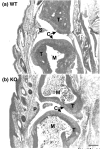
Figure 1
Figure 1
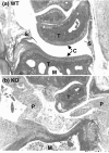
Interphalangeal joints in wild-type (a) and tristetraprolin knockout (b) mice. Shown are matching joints from littermate mice at about 7 months of age, stained with hematoxylin and eosin. C, articular cartilage; M, marrow; P, pannus; S, synovium; T, trabecular bone. Scale bar, 0.5 mm. Modified from [11].
Figure 2
Figure 2
Radial head histology in wild-type (a) and tristetraprolin knockout (b) mice. This low-power view is of radial head joints from littermate mice at about 7 months of age, with the radial head (RH) indicated. Other abbreviations are as in the legend to Fig. 1. Modified from [11].
Figure 3
Figure 3
Higher-power view of the radial head histology for the same littermate wild-type (a) and tristetraprolin knockout (b) mice as those shown in Fig. 2, with the radial head at the bottom of each panel. Abbreviations are as in the legend to Fig. 1. Modified from [11].
Figure 4
Figure 4
Proposed structure of the human tristetraprolin (TTP) tandem zinc finger domain in complex with the TTP-binding site 5'-UUAUUUAUU-3'. This proposed structure was modeled on the original nuclear magnetic resonance structure described by [32], using their pdb coordinates and the Swiss-Model program. The RNA oligonucleotide is shown in magenta, with the 5' and 3' ends indicated, along with the key residues A3, U5, and A7. The peptide is shown as a surface structure, with the buried zinc residues highlighted and the amino-terminal (N-term.) and carboxy-terminal (C-term.) ends of the peptide shown by arrows. The dark blue residues represent amino acids that are identical between human TTP and the ZFP36L2 (TIS11D) protein used in the original structure. The other colors represent progressively greater amino acid differences between the two proteins, ranging from minimally different (aquamarine, upper right), through green, yellow, and orange, with orange representing the most marked amino acid differences.
Figure 5
Figure 5
Mammalian tumour necrosis factor-α (TNF) and granulocyte/macrophage colony-stimulating factor (GM-CSF) AU-rich elements (AREs). (a) The central ARE region of the TNF mRNA 3' untranslated region from all mammalian species for which this region of the mRNA has been deposited in GenBank. In most cases these were derived from EST sequences; note that the horse sequence has not been completed at the 3' end. The overlines indicate the nine-base tristetraprolin (TTP)-binding site 5'-UUAUUUAUU-3'. Sequences from the various mammals are divided into groups based on the pattern of these nonamers, with the top group of 10 mammals being the most common group. (b) A similar approach was used to align the central ARE from the GM-CSF transcript, after alignment using the program ClustalW. The asterisks below the alignment represent base identity at that position; note that gaps were used to optimize the alignment. The overlines again represent the nonamer TTP-binding site. These data are modified from [37].
Figure 6
Figure 6
Schematic representation of the human tristetraprolin (TTP) gene (ZFP36) and its polymorphisms. The two exons of ZFP36 are shown as boxes, whereas the flanking regions and intron are indicated by a thin line. Open boxes represent untranslated regions, solid filled boxes represent protein-coding regions, and the hatched region represents the tandem zinc finger domain. The positions of the polymorphisms listed in Table 1 are indicated by arrowheads. Kb, kilobases. The data are modified from [52].
Figure 7
Figure 7
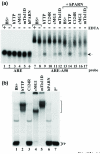
Effects of tristetraprolin (TTP)-related tandem CCCH zinc finger (TZF) proteins to bind AU-rich element (ARE)-containing probes and to promote their deadenylation. HEK-293 cells were maintained, and transient transfection of 1.2 × 10⁶ cells with expression plasmid constructs in calcium phosphate precipitates was performed, as described [22]. To each plate of HEK-293 cells was added 0.2 μg of the TZF protein expression constructs CMV.hTTP.tag (hTTP), a human TTP (hTTP) zinc finger mutant (C124R), CMV.cMG1.tag (cMG1), CMV.mTis11D.tag (mTis11D), 0.1 μg of human poly(A) exonuclease (hPARN) expression plasmid CMV.hPARN.flag (hPARN), or plasmid DNA alone (BS+). The zinc finger protein expression constructs were transfected either with vector alone or together with CMV.hPARN.flag; vector DNA (BS+) was added to each transfection to make the total amount of co-transfected DNA 5 μg per plate. Cytosolic extracts were prepared and used in deadenylation assays as described [23]. (a) Extracts (10 μg of protein per sample) were incubated with probes ARE or ARE-A50 at 37°C for 60 min in the presence (+) or absence (-) of 20 mM EDTA, as indicated. The samples were processed as described previously [23]. The arrow indicates the migration position of the ARE probe (lanes 1–6) and the deadenylated product of probe ARE-A50 (lanes 9, 11, 12, 14, 16 and 17). (b) The extracts used in lanes 7–13 of (a) were incubated with the ARE-A50 probe and used in a gel-shift assay. Lane 7 (P') was loaded with probe alone (digested with RNase T1). The migration positions of the zinc finger protein-RNA complexes are indicated by the bracket to the right of the gel, and the position of the free probe (FP) is also indicated. The bands present in the gel in lane 1 represent endogenous HEK-293 cell proteins shifting the probe; note that this pattern is identical in lane 3, representing a zinc finger mutant of TTP, and in lane 6, representing hPARN alone.
Figure 8
Figure 8
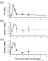
Tristetraprolin (TTP), ZFP36L1, and ZFP36L2 expression patterns in human monocytes stimulated with lipopolysaccharide (LPS). Purified monocytes from healthy human subjects (n = 5) were stimulated with LPS (or phosphate-buffered saline as control). Total cellular RNA from the monocytes was converted to cDNA and analyzed by real-time polymerase chain reaction for (a) TTP, (b) ZFP36L1, and (c) ZFP36L2 expression levels. Resulting Ct values were normalized to the geometric mean of four internal control transcripts and then to corresponding samples from PBS-treated cultures at the same time points, then converted to 2-ΔΔCt. The normalized values were then expressed as a fraction of the mean value at which maximum expression occurred (t = 1 hour for TTP and ZFP36L2; t = 1.5 hours for ZFP36L1). These were then expressed as means ± s.e.m.
Figure 9
Figure 9
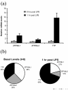
Relative basal and stimulated levels of tristetraprolin (TTP), ZFP36L1, and ZFP36L2 transcripts in cultured human monocytes. The real-time polymerase chain reaction (PCR) values were normalized to the geometric mean of four internal transcript controls and against PBS-treated cells at corresponding time points, then converted to 2-ΔΔCt to obtain expression level values for the TTP, ZFP36L1, and ZFP36L2 transcripts, shown in (a) as means ± s.e.m. from cultures derived from five individual subjects. These expression levels from the three transcripts can be compared with each other within subjects, because the primer/probe sets for each gene yielded equivalent PCR amplification efficiencies and fluorescence intensities, and the data were normalized to control for plate-to-plate variations. The basal levels are similar for each of the three genes in resting monocytes, shown as 0 hours after lipopolysaccharide (LPS) in (a) and t = 0 in (b). LPS treatment for 1 hour stimulated the expression of all three genes; however, after 1 hour of exposure to LPS, TTP levels were increased most markedly (a and b, right panel). In unstimulated cells, mean levels of all three transcripts representing TTP 'equivalents' were approximately equal (b, left panel). However, after 1 hour of stimulation with LPS, TTP represented about 69% of TTP equivalents, compared with 21% for ZFP36L1 and 10% for ZFP36L2.

Similar articles

Cited by

References

    1. Lai WS, Stumpo DJ, Blackshear PJ. Rapid insulin-stimulated accumulation of an mRNA encoding a proline-rich protein. J Biol Chem. 1990;265:16556–16563. - PubMed
    1. Ma Q, Herschman HR. A corrected sequence for the predicted protein from the mitogen-inducible TIS11 primary response gene. Oncogene. 1991;6:1277–1278. - PubMed
    1. Varnum BC, Lim RW, Sukhatme VP, Herschman HR. Nucleotide sequence of a cDNA encoding TIS11, a message induced in Swiss 3T3 cells by the tumor promoter tetradecanoyl phorbol acetate. Oncogene. 1989;4:119–120. - PubMed
    1. DuBois RN, McLane MW, Ryder K, Lau LF, Nathans D. A growth factor-inducible nuclear protein with a novel cysteine/histidine repetitive sequence. J Biol Chem. 1990;265:19185–19191. - PubMed
    1. Taylor GA, Thompson MJ, Lai WS, Blackshear PJ. Phosphorylation of tristetraprolin, a potential zinc finger transcription factor, by mitogen stimulation in intact cells and by mitogen-activated protein kinase in vitro. J Biol Chem. 1995;270:13341–13347. doi: 10.1074/jbc.270.22.13341. - DOI - PubMed

MeSH terms

Substances

LinkOut - more resources