Haploinsufficiency of delta-like 4 ligand results in embryonic lethality due to major defects in arterial and vascular development
- PMID: 15520367
- PMCID: PMC524697
- DOI: 10.1073/pnas.0407290101
Haploinsufficiency of delta-like 4 ligand results in embryonic lethality due to major defects in arterial and vascular development
Abstract
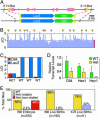
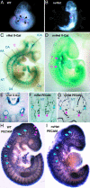
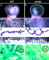
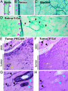
Vascular development depends on the highly coordinated actions of a variety of angiogenic regulators, most of which apparently act downstream of vascular endothelial growth factor (VEGF). One potential such regulator is delta-like 4 ligand (Dll4), a recently identified partner for the Notch receptors. We generated mice in which the Dll4 gene was replaced with a reporter gene, and found that Dll4 expression is initially restricted to large arteries in the embryo, whereas in adult mice and tumor models, Dll4 is specifically expressed in smaller arteries and microvessels, with a striking break in expression just as capillaries merge into venules. Consistent with these arterial-specific expression patterns, heterozygous deletion of Dll4 resulted in prominent albeit variable defects in arterial development (reminiscent of those in Notch knockouts), including abnormal stenosis and atresia of the aorta, defective arterial branching from the aorta, and even arterial regression, with occasional extension of the defects to the venous circulation; also noted was gross enlargement of the pericardial sac and failure to remodel the yolk sac vasculature. These striking phenotypes resulting from heterozygous deletion of Dll4 indicate that vascular development may be as sensitive to subtle changes in Dll4 dosage as it is to subtle changes in VEGF dosage, because VEGF accounts for the only other example of haploid insufficiency, resulting in obvious vascular abnormalities. In summary, Dll4 appears to be a major trigger of Notch receptor activities previously implicated in arterial and vascular development, and it may represent a new opportunity for pro- and anti-angiogenic therapies.
Figures


Similar articles
-
Delta-like ligand 4 (Dll4) is induced by VEGF as a negative regulator of angiogenic sprouting.Proc Natl Acad Sci U S A. 2007 Feb 27;104(9):3219-24. doi: 10.1073/pnas.0611206104. Epub 2007 Feb 12. Proc Natl Acad Sci U S A. 2007. PMID: 17296940 Free PMC article.
-
The Notch ligand Delta-like 4 negatively regulates endothelial tip cell formation and vessel branching.Proc Natl Acad Sci U S A. 2007 Feb 27;104(9):3225-30. doi: 10.1073/pnas.0611177104. Epub 2007 Feb 12. Proc Natl Acad Sci U S A. 2007. PMID: 17296941 Free PMC article.
-
The role of the vascular endothelial growth factor-Delta-like 4 ligand/Notch4-ephrin B2 cascade in tumor vessel remodeling and endothelial cell functions.Cancer Res. 2006 Sep 1;66(17):8501-10. doi: 10.1158/0008-5472.CAN-05-4226. Cancer Res. 2006. PMID: 16951162
-
Role of the vascular endothelial growth factor isoforms in retinal angiogenesis and DiGeorge syndrome.Verh K Acad Geneeskd Belg. 2005;67(4):229-76. Verh K Acad Geneeskd Belg. 2005. PMID: 16334858 Review.
-
New pathways and mechanisms regulating and responding to Delta-like ligand 4-Notch signalling in tumour angiogenesis.Biochem Soc Trans. 2011 Dec;39(6):1612-8. doi: 10.1042/BST20110721. Biochem Soc Trans. 2011. PMID: 22103496 Review.
Cited by
-
Endothelial-Specific Molecule 1 Inhibition Lessens Productive Angiogenesis and Tumor Metastasis to Overcome Bevacizumab Resistance.Cancers (Basel). 2022 Nov 18;14(22):5681. doi: 10.3390/cancers14225681. Cancers (Basel). 2022. PMID: 36428773 Free PMC article.
-
Cis inhibition of NOTCH1 through JAGGED1 sustains embryonic hematopoietic stem cell fate.Nat Commun. 2024 Feb 21;15(1):1604. doi: 10.1038/s41467-024-45716-y. Nat Commun. 2024. PMID: 38383534 Free PMC article.
-
Vascular endothelial growth factor signaling regulates the segregation of artery and vein via ERK activity during vascular development.Biochem Biophys Res Commun. 2013 Jan 25;430(4):1212-6. doi: 10.1016/j.bbrc.2012.12.076. Epub 2012 Dec 22. Biochem Biophys Res Commun. 2013. PMID: 23266606 Free PMC article.
-
Biochemical characterization and cellular effects of CADASIL mutants of NOTCH3.PLoS One. 2012;7(9):e44964. doi: 10.1371/journal.pone.0044964. Epub 2012 Sep 18. PLoS One. 2012. PMID: 23028706 Free PMC article.
-
Cerebral cavernous malformation protein CCM1 inhibits sprouting angiogenesis by activating DELTA-NOTCH signaling.Proc Natl Acad Sci U S A. 2010 Jul 13;107(28):12640-5. doi: 10.1073/pnas.1000132107. Epub 2010 Jun 24. Proc Natl Acad Sci U S A. 2010. PMID: 20616044 Free PMC article.
References
-
- Ferrara, N., Carver-Moore, K., Chen, H., Dowd, M., Lu, L., O'Shea, K. S., Powell-Braxton, L., Hillan, K. J. & Moore, M. W. (1996) Nature 380, 439–442. - PubMed
-
- Carmeliet, P., Ferreira, V., Breier, G., Pollefeyt, S., Kieckens, L., Gertsenstein, M., Fahrig, M., Vandenhoeck, A., Harpal, K., Eberhardt, C., et al. (1996) Nature 380, 435–439. - PubMed
-
- Suri, C., Jones, P. F., Patan, S., Bartunkova, S., Maisonpierre, P. C., Davis, S., Sato, T. N. & Yancopoulos, G. D. (1996) Cell 87, 1171–1180. - PubMed
-
- Wang, H. U., Chen, Z. F. & Anderson, D. J. (1998) Cell 93, 741–753. - PubMed
-
- Gerety, S. S., Wang, H. U., Chen, Z. F. & Anderson, D. J. (1999) Mol. Cell 4, 403–414. - PubMed
MeSH terms
Substances
LinkOut - more resources
Full Text Sources
Other Literature Sources
Molecular Biology Databases

