Comparative Study
. 2004 Nov 10;23(22):4506-16.
doi: 10.1038/sj.emboj.7600451. Epub 2004 Oct 28.

alpha-Synuclein produces a long-lasting increase in neurotransmitter release

Affiliations
Comparative Study

alpha-Synuclein produces a long-lasting increase in neurotransmitter release

Shumin Liu et al. EMBO J. .

Abstract

Wild-type alpha-synuclein, a protein of unknown function, has received much attention because of its involvement in a series of diseases that are known as synucleinopathies. We find that long-lasting potentiation of synaptic transmission between cultured hippocampal neurons is accompanied by an increase in the number of alpha-synuclein clusters. Conversely, suppression of alpha-synuclein expression through antisense nucleotide and knockout techniques blocks the potentiation, as well as the glutamate-induced increase in presynaptic functional bouton number. Consistent with these findings, alpha-synuclein introduction into the presynaptic neuron of a pair of monosynaptically connected cells causes a rapid and long-lasting enhancement of synaptic transmission, and rescues the block of potentiation in alpha-synuclein null mouse cultures. Also, we report that the application of nitric oxide (NO) increases the number of alpha-synuclein clusters, and inhibitors of NO-synthase block this increase, supporting the hypothesis that NO is involved in the enhancement of the number of alpha-synuclein clusters. Thus, alpha-synuclein is involved in synaptic plasticity by augmenting transmitter release from the presynaptic terminal.


Figures

Figure 1
Figure 1
Glutamate produces rapid increases in α-Syn and Sys I IR puncta number, and sites where α-Syn and Sys I are colocalized. (A1) Immunoreactivity for α-Syn (green) is present in puncta along the processes and is also present in the cell bodies in a 10-day in vitro neuron. Scale bar=20 μm. (A2) After 14 days in vitro, α-Syn immunoreactivity is predominantly localized to the presynaptic terminal. (B) Examples of α-Syn (green) and Sys I IR puncta (red), and colocalization (yellow) in a control dish (left), a dish fixed 5 min after application of 200 μM glutamate (middle) and a dish fixed after glutamate in the presence of D-APV (200 μM) (right). Scale bar=5 μm. (C) Average results from experiments like the one shown in (B, middle panel) (n=9 dishes per group). After 1 min glutamate, the number of α-Syn-IR puncta increased significantly compared to controls, as well as the number of Sys I-IR puncta, and the number of sites where α-Syn and Sys I IR puncta were colocalized. **P<0.01 compared to control in this and subsequent figures. The data have been normalized to the average puncta number in a representative field in control dishes from the same culture batch. (D) Average of experiments like the one shown in (B, right panel) (n=6 dishes per group). D-APV blocks the glutamate-induced increase in α-Syn and Sys I IR puncta number, and colocalized puncta; #P<0.05 compared to glutamate alone in this and subsequent figures.
Figure 2
Figure 2
α-Syn AS treatment blocks glutamate-induced increase in α-Syn and Sys I IR puncta number, and sites where α-Syn and Sys I are colocalized. (A) Examples of α-Syn and Sys I IR puncta, and colocalization in a control dish (left), a dish fixed after 4 days treatment with AS (middle) and a dish fixed after glutamate (200 μM) for 1 min and AS (5 μM) for 4 days (right). Scale bar=5 μm. (B) Example of Western blot demonstrating that 4-day AS treatment decreases α-Syn protein expression in cultured hippocampal neurons, whereas NSE levels remain constant. AS also slightly reduced Sys I expression. (C) Average results from experiments like the one shown in (A, middle panel) (n=6 dishes). After 4 days of AS, the number of α-Syn-IR puncta was significantly reduced compared to control dishes. The number of Sys I-IR puncta was also reduced, and also the number of sites where α-Syn and Sys I were colocalized. (D) Average of experiments like the one shown in (A, right panel) (n=6). α-Syn AS blocked the glutamate-induced increase in the number of α-Syn and Sys I IR puncta, and colocalized puncta.
Figure 3
Figure 3
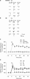
α-Syn AS treatment blocks glutamate-induced mEPSC frequency increase. (A, B) Examples of spontaneous mEPSCs before (Pre) and 2.5 and 45 min after brief application of 200 μM glutamate (Glu) to a control dish (A) or a dish treated with AS for 4 days (B). (C) Average increase in mEPSC frequency in experiments like the one shown in (A). Glutamate produced a rapid and long-lasting increase in mEPSC frequency (filled squares). This increase was blocked by D-APV (50 μM) (open squares). Bath application alone did not enhance the mEPSC frequency (filled diamonds). Data were normalized to the average baseline value during the 10 min before glutamate in each experiment. (D) Average increase in mEPSC frequency in experiments like the one shown in (B). Cultured hippocampal neurons were either treated with AS (filled diamonds), scrambled (Scr) (filled circles), sense (S) (open diamonds) or serum-free (Sf) solution (filled triangles) for 4 days.
Figure 4
Figure 4
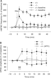
Hippocampal neurons in culture derived from α-Syn KO mice do not show potentiation of spontaneous and evoked responses. (A) Average increase in mEPSC frequency in experiments from α-Syn KO mice (−/−) and their control wt (+/+) littermates. This increase was blocked by D-APV (50 μM) (average frequency at 45 min 98.1±20.1 of baseline values before glutamate, n=7, data not shown). (B) A tetanus (filled circles) in an Mg2+-free medium caused an immediate and long-lasting EPSC amplitude increase in cultures from wt mice, whereas it did not increase EPSC amplitude in cultures from α-Syn KO mice (filled squares). Application of APV blocked the EPSC amplitude increase by tetanus in wt cultures (open circles). Data were normalized to the average baseline value during the 10 min before tetanus. The three vertical arrows indicate the tetanus application.
Figure 5
Figure 5
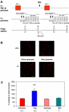
Hippocampal neurons in culture derived from α-Syn KO mice do not show glutamate-induced increase in active synaptic bouton number. (A) Experimental protocol for FM1-43 staining and destaining of synaptic vesicles. Loading of FM1-43 was induced by changing the perfusion medium from normal saline solution to hyperkalemic solution. The excess dye was washed in normal bath solution for 10 min with ADVASEP-7 introduced for 60 s in the washing bath solution at 1 and 6 min of washing. The culture was then exposed to multiple applications of hyperkalemic bath solution (without FM1-43) to facilitate release of the dye from vesicles. The difference between the images before and after destaining gave the measure of FM1-43-stained vesicles. The same procedure was repeated 30 min after glutamate. The percentage increase in active boutons after glutamate was calculated (Supplementary Materials). (B) Examples of activity-dependent FM1-43 staining before and after glutamate in −/− and +/+ hippocampal cultures. Glutamate increased presynaptic bouton number in +/+ cultures but not in −/− cultures. Scale bar=10 μm. (C) Percentage increase in presynaptic active boutons 30 min after glutamate in 0 Mg2+ in +/+ and −/− cultures. Glutamate increased the number of active boutons in +/+ (n=10) but not in −/− (n=8) cultures (**P<0.01). Saline solution did not increase the active bouton number in +/+ (n=6) and −/− (n=8) cultures.
Figure 6
Figure 6
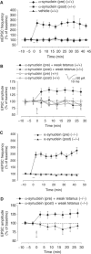
α-Syn introduction into the presynaptic neuron induces a long-lasting transmitter release increase. (A) Presynaptic injection of unmodified, murine α-Syn (filled diamonds) produced an immediate and long-lasting mEPSC frequency increase in microcultured neurons. Postsynaptic unmodified α-Syn (filled squares) and presynaptic vehicle (filled triangles) did not change the mEPSC frequency. Data were normalized to the average baseline value during the 10 min before glutamate in each experiment. (B) Presynaptic unmodified α-Syn paired with weak tetanus (filled diamonds) produced an immediate and long-lasting EPSC amplitude increase in cultured neurons. Presynaptic α-Syn alone (open diamonds), postsynaptic α-Syn paired with weak tetanus (filled triangles) or postsynaptic α-Syn alone (open triangles) did not change the EPSC amplitude. Data were normalized to the average baseline value during the 10 min before α-Syn paired with weak tetanus or alone in each experiment. The vertical arrow indicates the weak tetanus application. (Inset) Examples of EPSCs recorded before (dotted line) and after (solid line) presynaptic α-Syn. (C) Presynaptic unmodified α-Syn (filled diamonds) produced an immediate and long-lasting mEPSC frequency increase in cultures from α-Syn KO mice. Postsynaptic α-Syn (filled triangles) did not change the mEPSC frequency. (D) Presynaptic unmodified α-Syn paired with weak tetanus (filled diamonds) produced an immediate and long-lasting EPSC amplitude increase in cultures from α-Syn KO mice. Postsynaptic α-Syn paired with weak tetanus (filled triangles) did not enhance EPSC amplitude.
Figure 7
Figure 7
NO increased α-Syn-IR puncta number. (A) After a brief NO application (20 nM), the number of α-Syn-IR puncta increased significantly compared to control dishes. **P<0.01 compared to control (n=8 dishes per group). (B) The NO-synthase inhibitor L-NMA) (100 μM) blocks the glutamate-induced increase in α-Syn-IR puncta number. **P<0.01 compared to glutamate alone (n=7 dishes per group).

Similar articles

Cited by

References

    1. Abeliovich A, Schmitz Y, Farinas I, Choi-Lundberg D, Ho WH, Castillo PE, Shinsky N, Verdugo JM, Armanini M, Ryan A, Hynes M, Phillips H, Sulzer D, Rosenthal A (2000) Mice lacking alpha-synuclein display functional deficits in the nigrostriatal dopamine system. Neuron 25: 239–252 - PubMed
    1. Antonova I, Arancio O, Trillat AC, Wang HG, Zablow L, Udo H, Kandel ER, Hawkins RD (2001) Rapid increase in immunoreactive presynaptic terminals during long-lasting potentiation. Science 294: 1547–1550 - PubMed
    1. Antonova I, Trillat AC, Arancio O, De Vente J, Hawkins RD (1999) Glutamate and nitric oxide produce increases in cGMP and synaptophysin immunofluorescence in cultured hippocampal neurons. Soc Neurosci Abstr 25: 733
    1. Arancio O, Antonova I, Gambaryan S, Lohmann SM, Wood JS, Lawrence DS, Hawkins RD (2001) Presynaptic role of cGMP-dependent protein kinase during long-lasting potentiation. J Neurosci 21: 143–149 - PMC - PubMed
    1. Arancio O, Kandel ER, Hawkins RD (1995) Activity-dependent long-term enhancement of transmitter release by presynaptic 3′, 5′ -cyclic GMP in cultured hippocampal neurons. Nature 376: 74–80 - PubMed

Publication types

MeSH terms