. 2004 Nov;24(22):9848-62.
doi: 10.1128/MCB.24.22.9848-9862.2004.

Neuroprotective role of the Reaper-related serine protease HtrA2/Omi revealed by targeted deletion in mice

Affiliations

Neuroprotective role of the Reaper-related serine protease HtrA2/Omi revealed by targeted deletion in mice

L Miguel Martins et al. Mol Cell Biol. 2004 Nov.

Abstract

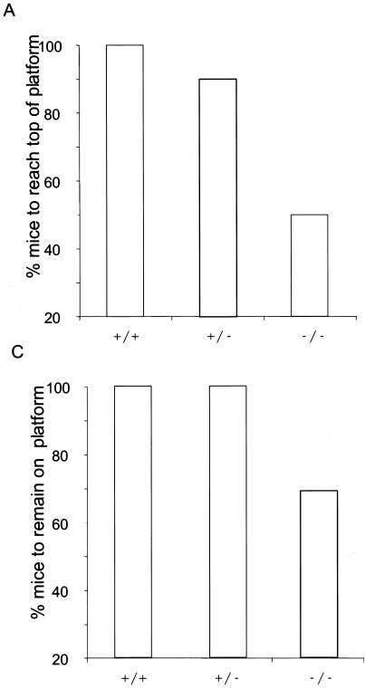
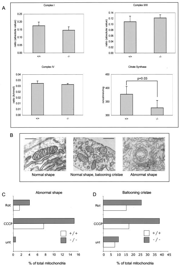
The serine protease HtrA2/Omi is released from the mitochondrial intermembrane space following apoptotic stimuli. Once in the cytosol, HtrA2/Omi has been implicated in promoting cell death by binding to inhibitor of apoptosis proteins (IAPs) via its amino-terminal Reaper-related motif, thus inducing caspase activity, and also in mediating caspase-independent death through its own protease activity. We report here the phenotype of mice entirely lacking expression of HtrA2/Omi due to targeted deletion of its gene, Prss25. These animals, or cells derived from them, show no evidence of reduced rates of cell death but on the contrary suffer loss of a population of neurons in the striatum, resulting in a neurodegenerative disorder with a parkinsonian phenotype that leads to death of the mice around 30 days after birth. The phenotype of these mice suggests that it is the protease function of this protein and not its IAP binding motif that is critical. This conclusion is reinforced by the finding that simultaneous deletion of the other major IAP binding protein, Smac/DIABLO, does not obviously alter the phenotype of HtrA2/Omi knockout mice or cells derived from them. Mammalian HtrA2/Omi is therefore likely to function in vivo in a manner similar to that of its bacterial homologues DegS and DegP, which are involved in protection against cell stress, and not like the proapoptotic Reaper family proteins in Drosophila melanogaster.


Figures

FIG. 1.
FIG. 1.
Targeting the HtrA2/Omi gene by homologous recombination. (A) Schematic representations of the wild-type mouse HtrA2/Omi (WT mHtrA2) locus, the targeting construct used in this study, and the targeted HtrA2/Omi allele. (B) Genotyping of the wild-type and mutant HtrA2/Omi loci by PCR. (C) Western blot analysis of protein lysates obtained from wild-type, HtrA2/Omi heterozygous, and HtrA2/Omi knockout MEFs.
FIG. 2.
FIG. 2.
Phenotypic alterations of HtrA2/Omi knockout mice. (A) Body weights of wild-type (HtrA2/Omi+/+), heterozygous (HtrA2/Omi+/−), and knockout (HtrA2/Omi−/−) littermates. Data shown are means ± standard deviations of results for at least six animals for each point. (B) Appearance of day 30 HtrA2/Omi knockout and littermate control wild-type animals. (C) HtrA2/Omi knockout mice show a general decrease in organ size. This decrease is more dramatic with the spleen, thymus, and heart; the brain is approximately 75% of the weight of wild-type control brains.
FIG. 3.
FIG. 3.
Sensorimotor tests on knockout mice. HtrA2+/+, HtrA2+/−, and HtrA2−/− mice were placed on a 60o-inclined platform. (A) Percentages of mice that climbed to the top of the platform. Wild-type (+/+; n = 8); heterozygote (+/−; n = 20), and knockout (−/−; n = 16) littermates were compared. (B) Average elapsed times for mice to climb to the top of the platform. Data are means ± standard deviations of results from a minimum of eight trials. (C) Percentages of mice that remained on the platform for the full 60-s trial period. Wild-type (+/+; n = 8), heterozygote (+/−; n = 20), and knockout (−/−; n = 16) littermates were compared.
FIG. 3.
FIG. 3.
Sensorimotor tests on knockout mice. HtrA2+/+, HtrA2+/−, and HtrA2−/− mice were placed on a 60o-inclined platform. (A) Percentages of mice that climbed to the top of the platform. Wild-type (+/+; n = 8); heterozygote (+/−; n = 20), and knockout (−/−; n = 16) littermates were compared. (B) Average elapsed times for mice to climb to the top of the platform. Data are means ± standard deviations of results from a minimum of eight trials. (C) Percentages of mice that remained on the platform for the full 60-s trial period. Wild-type (+/+; n = 8), heterozygote (+/−; n = 20), and knockout (−/−; n = 16) littermates were compared.
FIG. 4.
FIG. 4.
Morphological features of localized striatal degeneration in HtrA2/Omi knockout mice. (A) Hematoxylin and eosin staining showing focal loss of neurons and occasional ballooning in the striata of HtrA2/Omi knockout animals (right) compared to controls (+/+) (left). (B) GFAP immunohistochemistry revealing the reaction of astrocytes in knockout animals (right). (C) MAP2 immunohistochemistry revealing the loss of this protein in sections from knockout animals (right). (D) Diagram of a mouse brain in sagital orientation indicating the area of striatal degeneration (red box) in the striatum. (E and F) Density of NeuN-positive neurons in the striata (Str) of wild-type and HtrA2/Omi knockout mice scored for both the whole striatum (total basal ganglia) and the area where localized degeneration was observed (posterior portion of the basal ganglia). Analysis was performed with both P20 and P30 animals. (G and H) Average optical density of TH staining in both the total basal ganglia and the area where localized degeneration was observed (posterior portion of the basal ganglia). (I) Average number of TH-positive neurons in the substantia nigra pars compacta of wild-type and HtrA2/Omi knockout animals.
FIG. 4.
FIG. 4.
Morphological features of localized striatal degeneration in HtrA2/Omi knockout mice. (A) Hematoxylin and eosin staining showing focal loss of neurons and occasional ballooning in the striata of HtrA2/Omi knockout animals (right) compared to controls (+/+) (left). (B) GFAP immunohistochemistry revealing the reaction of astrocytes in knockout animals (right). (C) MAP2 immunohistochemistry revealing the loss of this protein in sections from knockout animals (right). (D) Diagram of a mouse brain in sagital orientation indicating the area of striatal degeneration (red box) in the striatum. (E and F) Density of NeuN-positive neurons in the striata (Str) of wild-type and HtrA2/Omi knockout mice scored for both the whole striatum (total basal ganglia) and the area where localized degeneration was observed (posterior portion of the basal ganglia). Analysis was performed with both P20 and P30 animals. (G and H) Average optical density of TH staining in both the total basal ganglia and the area where localized degeneration was observed (posterior portion of the basal ganglia). (I) Average number of TH-positive neurons in the substantia nigra pars compacta of wild-type and HtrA2/Omi knockout animals.
FIG. 5.
FIG. 5.
Mitochondrial function in wild-type and HtrA2/Omi knockout animals. (A) Activities of mitochondrial complexes I, II and III, and IV corrected for mitochondrial enrichment by normalizing the activity against the mitochondrial matrix enzyme citrate synthase; values for detected citrate synthase activities are also shown. Extracts were prepared from striata of P30 control (+/+) and HtrA2/Omi knockout (−/−) animals. A statistically significant (t test analysis) decrease in the amount of citrate synthase activity was detected in samples from HtrA2/Omi−/− animals. (B) Ultrastructural features of mitochondria present in cells from wild-type and HtrA2/Omi knockout animals. Scale bars correspond to 500 nm. (C and D) Quantification of mitochondrial morphological abnormalities detected in cells before and after treatment with mitochondrial stress stimuli. Results are representative of three independent experiments performed on three to five different cell lines isolated from different animals. Rot, rotenone; unt, untreated.
FIG. 6.
FIG. 6.
Deletion of HtrA2/Omi results in increased sensitivity to mitochondrion-damaging agents. Thymocytes were isolated from control and HtrA2/Omi knockout animals and incubated with increasing concentrations of anti-Fas antibody (A), etoposide (B), or the mitochondrion-damaging agents CCCP (C) and rotenone (D). Viability was determined 16 h after treatment by flow cytometry using propidium iodide. Results are representative of three independent experiments. (E) Viability of simian virus 40 large-T-antigen-immortalized MEFs derived from wild-type (+/+) and HtrA2/Omi knockout animals, determined by measurement of sub-G1 cell populations by flow cytometry. Cells were incubated in the presence of CCCP (25 μM for 27 h), rotenone (25 μM for 27 h), tunicamycin (2.5 μg/ml for 27 h), or hydrogen peroxide (3 μM for 4.5 h) and compared to untreated control cells. Results show the means ± standard deviations of results of three independent experiments. (F) The neuronalresponse to glutamate-induced cytotoxicity was determined in primary neurons isolated from E14.5 embryos cultured in vitro for 10 days. Following incubation with the indicated concentrations of glutamate for 4 h, cells were stained with Hoechst 33342 and nuclear morphology was determined. Results show the means ± standard deviations of results from two independent experiments where six fields with at least 40 cells were scored for each data point.
FIG. 7.
FIG. 7.
Immunoprecipitation of Smac/DIABLO from wild-type and HtrA2/Omi knockout cells effectively depletes endogenous XIAP. Lysates were prepared from MEFs and incubated with an anti-Smac/DIABLO monoclonal antibody. Following separation by sodium dodecyl sulfate-polyacrylamide gel electrophoresis, Western blot analysis was performed using antibodies to detect endogenous XIAP, Smac/DIABLO, and HtrA2/Omi. “I” indicates the input lysates (10% of the total immunoprecipitated proteins loaded), “ID” indicates the equivalent amount of lysate following immunodepletion, and “IP” indicates the immunopurified eluate (50% of the total protein equivalent).
FIG. 8.
FIG. 8.
Analysis of HtrA2/Omi Smac/DIABLO double-knockout animals. (A) Detection of the wild-type and mutant HtrA2/Omi and Smac/DIABLO loci in the offspring of animals with a heterozygous deletion of the genes for both HtrA2/Omi and Smac/DIABLO. PCR was performed on genomic tail DNA templates. (B) Average body weights of Smac/DIABLO+/+ HtrA2/Omi+/+ (wild-type [wt]) and Smac/DIABLO−/− HtrA2/Omi−/− (double-knockout [KO]) mice. (C and D) The sensitivities of wild-type and double-knockout MEFs to TNF-α (C), and etoposide (D) are shown. Viability was determined 16 h following treatment by flow cytometry using propidium iodide. Results show the means ± standard deviations of results from three independent experiments.

Similar articles

Cited by

References

    1. Darzynkiewicz, Z., and E. Bedner. 2000. Analysis of apoptotic cells by flow and laser scanning cytometry. Methods Enzymol. 322:18-39. - PubMed
    1. Du, C., M. Fang, Y. Li, L. Li, and X. Wang. 2000. Smac, a mitochondrial protein that promotes cytochrome c-dependent caspase activation by eliminating IAP inhibition. Cell 102:33-42. - PubMed
    1. Faccio, L., C. Fusco, A. Chen, S. Martinotti, J. V. Bonventre, and A. S. Zervos. 2000. Characterization of a novel human serine protease that has extensive homology to bacterial heat shock endoprotease HtrA and is regulated by kidney ischemia. J. Biol. Chem. 275:2581-2588. - PubMed
    1. Ghadially, F. N. 1982. Ultrastructural pathology of the cell and matrix, 2nd ed. Butterworths, London, United Kingdom.
    1. Gray, C. W., R. V. Ward, E. Karran, S. Turconi, A. Rowles, D. Viglienghi, C. Southan, A. Barton, K. G. Fantom, A. West, J. Savopoulos, N. J. Hassan, H. Clinkenbeard, C. Hanning, B. Amegadzie, J. B. Davis, C. Dingwall, G. P. Livi, and C. L. Creasy. 2000. Characterization of human HtrA2, a novel serine protease involved in the mammalian cellular stress response. Eur. J. Biochem. 267:5699-5710. - PubMed

Publication types

MeSH terms

LinkOut - more resources