. 2004 Nov;24(21):9339-50.
doi: 10.1128/MCB.24.21.9339-9350.2004.

BRUCE, a giant E2/E3 ubiquitin ligase and inhibitor of apoptosis protein of the trans-Golgi network, is required for normal placenta development and mouse survival

Affiliations

BRUCE, a giant E2/E3 ubiquitin ligase and inhibitor of apoptosis protein of the trans-Golgi network, is required for normal placenta development and mouse survival

Kristina Lotz et al. Mol Cell Biol. 2004 Nov.

Abstract

BRUCE is a highly conserved 528-kDa peripheral membrane protein of the trans-Golgi network. Owing to the presence of an N-terminal single baculovirus inhibitor repeat, BRUCE functions as an inhibitor of apoptosis protein and blocks apoptosis when overexpressed. In addition, due to the presence of a C-terminal ubiquitin-conjugating domain, BRUCE can covalently attach ubiquitin to substrates. Here we report the generation and characterization of BRUCE-deficient mice. Complete inactivation of the BRUCE gene resulted in perinatal lethality and growth retardation discernible after embryonic day 14. The growth defect is linked to impaired placental development and may be caused by insufficient oxygen and nutrient transfer across the placenta. Chorioallantoic placentation initiated normally, but the mutant placenta showed an impaired maturation of the labyrinth layer and a significant reduction of the spongiotrophoblast. No evidence for an elevated apoptosis rate was detectable in embryonic and extraembryonic tissues and in knockout fibroblasts. Thus, although BRUCE is broadly expressed in embryonic, extraembryonic, and adult mouse tissues, this bifunctional protein might play a unique role in normal trophoblast differentiation and embryonic survival.


Figures

FIG. 1.
FIG. 1.
Targeted disruption of the BRUCE gene by homologous recombination. (A) Schematic representation of the targeting strategy for BRUCE, showing the partial BRUCE locus with exons (Ex) 3 to 6, the targeting construct, and the targeted allele. Exon 5 is deleted and replaced with a neoR cassette. The probe used for Southern hybridization of neomycin-resistant ES cell clones and genomic DNA, which recognizes 2.9-kb WT and 2.3-kb mutant (ko) PvuII fragments, is indicated. Arrowheads represent primers used for screening ES cell DNA for homologous recombination events and for genotyping. (B) PCR analysis of yolk sac DNA from embryos derived from heterozygous matings (top panel). The positions of primer pairs for amplification of the 556-bp WT and the 624-bp mutant PCR fragments are shown in panel (A). Immunoblot analysis of BRUCE protein in E10 whole-embryo lysates from WT, heterozygous, and BRUCE null embryos by using a monoclonal antibody against the N terminus of the murine BRUCE protein (middle panel). Equal protein shown by an antiactin immunoblot (bottom panel).
FIG. 2.
FIG. 2.
Loss of BRUCE results in increasing growth retardation during embryonic development. (A) Growth-retarded knockout neonates within the offspring of a heterozygous mating (BRUCE+/− × BRUCE+/−). (B) Growth curves of WT (+/+), heterozygous (+/−), and mutant (−/−) BRUCE embryos and their placentas from mid- to late gestation. The average body and placental weights were determined over time; error bars indicate standard deviations. Males and females were pooled. The number of monitored pups was at least five. In most cases, 10 or more embryos per genotype were examined.
FIG. 3.
FIG. 3.
External appearance of WT (+/+) and mutant (−/−) BRUCE embryos and extraembryonic tissues at different embryonic and fetal stages. Knockout embryos are retarded in growth compared to WT littermates after E13.5. They appear paler, and major blood vessels are often not clearly visible. In 34% of the BRUCE null mutants, temporary bleedings in the ventricle system of the brain and in the neural tube could be observed between E12 and E14 (arrowheads). In the extraembryonic tissues, defects already appear at E9.5. Mutant placenta is paler and smaller than WT, and the major blood vessels in the yolk sac are less pronounced than in the WT (arrowheads).
FIG. 4.
FIG. 4.
In situ hybridization analysis of BRUCE expression. Radiolabled antisense probe was reacted to E13.5 placenta of WT (BRUCE +/+) and mutant (BRUCE −/−) embryos to detect BRUCE mRNA levels in extraembryonic tissue. BRUCE is expressed predominantly in the diploid trophoblast of the placenta, including the labyrinth layer. Increased levels of BRUCE mRNA are found in the spongiotrophoblast layer, whereas in trophoblast giant cells BRUCE expression is reduced. The specificity of the BRUCE probe was demonstrated by parallel hybridization of WT tissue with a sense probe and by hybridization of knockout tissue with the antisense probe, which shows absence of BRUCE mRNA. Bars, 1 mm.
FIG. 5.
FIG. 5.
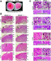
Morphology and histology of WT and BRUCE mutant placentas. (A) Ventral view of a WT placenta (+/+) and a BRUCE mutant placenta (−/−) at E17.5, showing apparent decreased vascularization of the mutant placenta, as shown by its paler color. Bar, 0.5 cm. (B) Hematoxylin- and eosin-stained radial sections of WT and mutant placentas at different developmental ages. All characteristic layers are present in the mutant placenta (de, maternal decidual tissue; gi, trophoblast giant cells; sp, spongiotrophoblast layer; la, labyrinthine layer; ch, chorionic plate). Chorioallantoic fusion occurs normally, and decidua and trophoblast giant cell development appear to be normal. The spongiotrophoblast of the mutant placenta is significantly decreased in thickness, whereas the mutant labyrinth layer is not markedly reduced in size. Bars, 0.5 mm. (C) Higher magnification of the labyrinthine and spongiotrophoblast layers of WT and mutant placentas. Fetal blood vessels (arrowheads) and maternal blood sinuses (asterisks) are present in normal number and come into close contact at early developmental stages (e.g., E11.5). Later, the labyrinthine trophoblast of the mutant placenta appears less compact and fails to mature over time. The number of trophoblast cells within the labyrinth seems to be reduced. The spongiotrophoblast of the mutant placenta consists of a few cell layers only (in contrast to the WT), and the trophoblast cells are enlarged. Bars, 10 μm.
FIG. 6.
FIG. 6.
Placental marker analysis of WT and BRUCE mutants at E11.5, E13.5, E15.5, and E17.5. (A) Specific antisense riboprobes were reacted with serial placental sections of WT (BRUCE +/+) and mutant (BRUCE −/−) embryos to discriminate the different placental layers. Placental lactogen (Pl-1) is highly expressed in trophoblast giant cells, and the fms-like tyrosine kinase Flt-1 was used to label spongiotrophoblast cells. The fetal liver kinase Flk-1 is specifically expressed in fetal endothelial cells and was therefore used as a marker for the labyrinth layer. Expression of the transcription factor eHand is detectable in diploid trophoblast cells of the labyrinth as well as in the spongiotrophoblast layer and at higher level in trophoblast giant cells. All layers are present in WT as well as in mutant placenta. Note the reduction of the spongiotrophoblast layer at all developmental stages examined and the increasing number of Pl-1-expressing cells in BRUCE-deficient placenta. Bars, 1 mm. (B) Development of the placental vascular network (from top to bottom). Immunohistochemical staining of basal lamina of fetal capillaries in the labyrinth layer by using an antibody to laminin shows the delay in maturation of the mutant placental vascular network. Thus, the fetal blood vessels in the labyrinth layer appear to be less branched and dilated, even at the end of gestation. Bars, 50 μm.
FIG. 7.
FIG. 7.
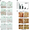
Apoptosis in extraembryonic tissues. (A) Similar rates of apoptotic cell death in the placentas of WT and BRUCE null mutants, as shown by immunohistochemical detection of caspase 3 activity and TUNEL staining (positive cells are marked by arrowheads). Staining methods were performed on sections of at least three different placentas per indicated genotype. The stained labyrinth layer of E15.5 WT and mutant placentas is shown as representative area. (B) For quantification, TUNEL-positive cells were counted in five randomly chosen labyrinth areas (200-fold magnification) of at least three different placentas of WT and knockout genotypes at the indicated developmental stages. Each bar represents the average ± standard deviation of apoptotic cells per area. (C) Levels of IAP proteins in WT (+/+) and BRUCE-deficient (−/−) placental tissue of E17.5 embryos. Expression levels of proteins were determined by Western blotting of placental lysates (excluding maternal tissue). Equal protein loading was shown by antiactin staining.
FIG. 8.
FIG. 8.
Cell proliferation in placenta. (A) Proliferating cells were visualized by immunohistochemical detection of BrdU incorporation (labeling of placental layers as for Fig. 5). The proliferation pattern is changed in placenta of BRUCE null mutants. There are almost no proliferating cells detectable in the strongly reduced spongiotrophoblast layer. Proliferation is also impaired in the labyrinthine layer. There are clearly fewer proliferating cells at early stages (e.g., E11.5 and E 13.5), whereas at later stages (e.g., E15.5 and E17.5), when cells in the mature WT placenta stop proliferating, cells still proliferate in the labyrinth layer of the mutant placenta. BrdU incorporation was monitored in three different WT and three mutant placentas. A representative sample of each phenotype is shown. Bars, 1 mm. (B) Higher magnification (×400) of the spongiotrophoblasts, the labyrinthine layers, and the chorionic plates of WT and mutant placentas. Note that the proliferating activity of endothelial cells of fetal blood vessels within the chorionic plate of mutant placenta does not seem to be altered. Bars, 50 μm. (C) For quantification, BrdU-positive cells were counted in five randomly chosen labyrinth areas (400-fold magnification) of at least three different placentas of WT and knockout genotype at the indicated developmental stages. Each bar represents the average ± standard deviation of cells in S phase per area.
FIG. 9.
FIG. 9.
Characterization of BRUCE-deficient fibroblast cultures. (A) Growth curves of WT (BRUCE +/+), heterozygous (BRUCE +/−), and BRUCE-deficient (BRUCE −/−) primary fibroblasts. Cells were inoculated at suboptimal high (9 × 10⁵ cells per 6-cm-diameter dish) and low (1 × 10⁵ cells per 6-cm-diameter dish) densities and at optimal growth density (3 × 10⁵ cells per 6-cm-diameter dish) and counted after the indicated period of growth. The average number of isolated cells (± standard deviation) from four individual littermates of each genotype, determined in duplicate, is shown. (B) Proliferation (left) and apoptosis (right) in asynchronous cultures of primary WT (+/+) and knockout (−/−) fibroblasts. The ordinate shows the percentage of cells that had incorporated BrdU (left) or the number of TUNEL-positive cells (right) in a randomly chosen area. Each point represents the average ± standard deviation from at least two cell lines from individual littermates of the indicated genotype, each determined in quadruplicate. (C) Effect of different apoptosis-inducing stimuli on fibroblasts from WT (MEF +/+) and mutant (MEF −/−) littermates. Cells were treated for the indicated time with the different apoptotic stimuli (actinomycin D, UV light, H2O2 [oxidative stress], and TNF-α in combination with 1.0 μg of cycloheximide [CHX] per ml). Cell death is shown as a function of concentration of the inducers of apoptosis and was assessed by measuring crystal violet staining of surviving cells (optical density [OD] at 595 nm). Primary fibroblasts isolated from three individual littermates of the indicated genotypes were analyzed in the experiment, and data represent the means ± standard deviations. (D) Levels of IAP proteins and caspase 3 in WT (+/+) and BRUCE-deficient (−/−) fibroblasts. Either cells were untreated or apoptosis was induced by treatment with 100 ng of human TNF-α per ml for 16 h. Expression levels of proteins were determined by Western blotting of whole-cell lysates. Equal protein loading was shown by antiactin staining.

Similar articles

Cited by

References

    1. Arama, E., J. Agapite, and H. Steller. 2003. Caspase activity and a specific cytochrome C are required for sperm differentiation in Drosophila. Dev. Cell 4:687-697. - PubMed
    1. Bartke, T., Pohl, C., G. Pyrowolakis, and S. Jentsch. 2004. Dual role of BRUCE as an antiapoptotic IAP and a chimeric E2/E3 ubiquitin ligase. Mol. Cell 14:801-811. - PubMed
    1. Chen, Z., M. Naito, S. Hori, T. Mashima, T. Yamori, and T. Tsuruo. 1999. A human IAP-family gene, apollon, expressed in human brain cancer cells. Biochem. Biophys. Res. Commun. 264:847-854. - PubMed
    1. Colosi, P., F. Talamantes, and D. I. Linzer. 1987. Molecular cloning and expression of mouse placental lactogen I complementary deoxyribonucleic acid. Mol. Endocrinol. 1:767-776. - PubMed
    1. Cross, J. C. 2000. Genetic insights into trophoblast differentiation and placental morphogenesis. Semin. Cell Dev. Biol. 11:105-113. - PubMed

Publication types

MeSH terms