. 2004 Oct 27;23(21):4297-306.
doi: 10.1038/sj.emboj.7600435. Epub 2004 Oct 14.

HSF4 is required for normal cell growth and differentiation during mouse lens development

Affiliations

HSF4 is required for normal cell growth and differentiation during mouse lens development

Mitsuaki Fujimoto et al. EMBO J. .

Abstract

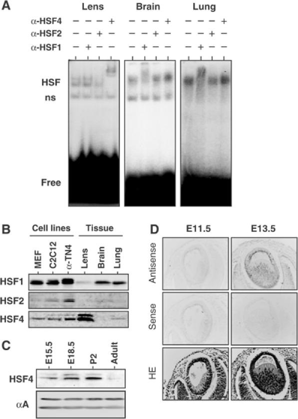
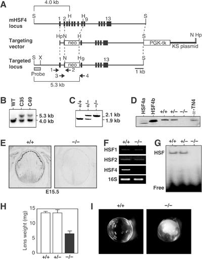
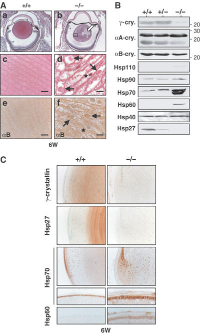
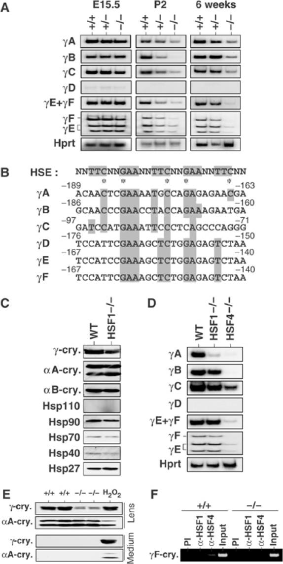
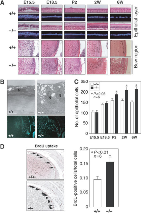
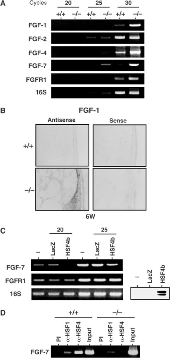
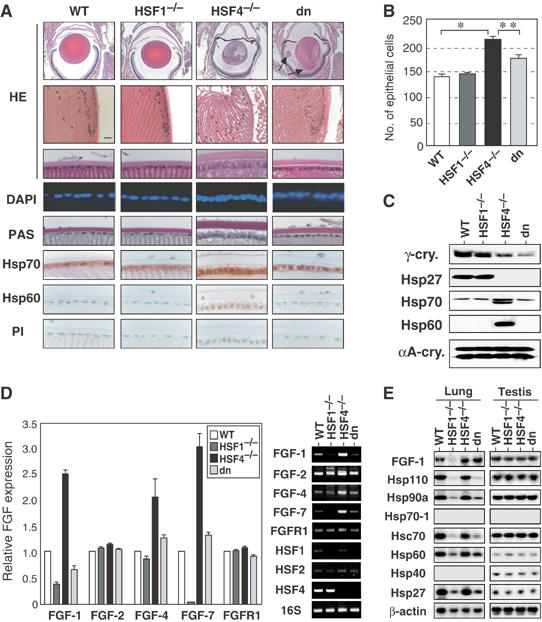
The heat shock transcription factor (HSF) family consists of three members in mammals and regulates expression of heat shock genes via a heat shock element. HSF1 and HSF2 are required for some developmental processes, but it is unclear how they regulate these processes. To elucidate the mechanisms of developmental regulation by HSFs, we generated mice in which the HSF4 gene is mutated. HSF4-null mice had cataract with abnormal lens fiber cells containing inclusion-like structures, probably due to decreased expression of gamma-crystallin, which maintains protein stability. Furthermore, we found increased proliferation and premature differentiation of the mutant lens epithelial cells, which is associated with increased expression of growth factors, FGF-1, FGF-4, and FGF-7. Unexpectedly, HSF1 competed with HSF4 for the expression of FGFs not only in the lens but also in other tissues. These findings reveal the lens-specific role of HSF4, which activates gamma-crystallin genes, and also indicate that HSF1 and HSF4 are involved in regulating expression of growth factor genes, which are essential for cell growth and differentiation.


Figures

Figure 1
Figure 1
HSF4 consists of a major HSE-binding activity in the mouse lens. (A) Gel shift assay in the presence or absence of antibody for HSF1, HSF2, or HSF4. Whole tissue extracts were prepared from the lens, brain, and lung in 6-week-old mice, and were mixed with the binding reaction containing a 32P-labeled HSE oligonucleotide and antibody. HSF indicates the complexes of HSFs and an HSE probe, ns indicates nonspecific binding activity, and free indicates an unbound HSE probe. (B) Protein levels of HSF1, HSF2, and HSF4 in cells and tissues. Cell extracts (40 μg) prepared from MEF, C2C12, and α-TN4, and tissue extracts (80 μg) prepared from the lens, brain, and lung in 2-day-old mice were subjected to Western blot analysis using each specific antibody. (C) Protein levels of HSF4 in the lens at E15.5, E18.5, p2, and 6-week-old (adult) were analyzed by Western blot. Level of αA-crystallin was constant during development. (D) In situ hybridization was performed on the eye sections of E11.5 and E13.5 embryos using sense and antisense probes specific for HSF4. The sections were also stained with hematoxylin and eosin (HE). Bar, 50 μm.
Figure 2
Figure 2
Cataract formation in HSF4-null mice. (A) Schematic representation of wild-type and mutant HSF4 loci together with targeting vector. The targeting vector was constructed to replace a part of exon 2 and exons 3–8 with a neomycin-resistant gene cassette. Locations of an external probe used to confirm correct targeting, and locations of PCR primers used to screen targeted ES clones (primers 1 and 2) and to identify genotype (primers 3 and 4) are shown. S, SalI; H, HindIII; Hp, HpaI; N, NotI; X, XbaI. (B) Southern blot of SalI/HindIII-digested genomic DNAs isolated from wild-type and targeted two ES clones (C39 and C49) using 32P-labeled probes described in (A). (C) PCR genotypic analysis for targeted locus. Mouse tail genomic DNA was isolated and was used to amplify DNA fragments by PCR using primers 3 and 4. (D) Western blot analysis of extracts of the lens of 6-week-old mice (+/+, +/−, −/−), 293 cells ectopically expressing HSF4a and HSF4b, and mouse lens epithelial α-TN4 cells. (E) In situ hybridization on the eye sections of E15.5 wild-type (+/+) and HSF4-null (−/−) mice using an antisense probe specific for HSF4. (F) mRNA levels of HSFs in wild-type(+/+) and HSF4-null (−/−) lens were examined by RT–PCR. (G) Whole-cell extracts were prepared from the lenses, and gel shift assay was performed using a 32P-labeled HSE oligonucleotide. Positions of free probe and probe bound by HSF are shown. (H) Lens weights in 6-week-old wild-type and mutant mice. Means and standard deviations were estimated by analyzing each of the three mice. (I) Lens opacity in 6-week-old wild-type and HSF4-null mice.
Figure 3
Figure 3
The lens fiber cells contain inclusion-like structures in HSF4-null mice. (A) Histological examination of the lens sections of the 6-week-old wild-type (a, c) and HSF4-null (b, d) mice stained with HE. Immunostaining of αB-crystallin was performed using the lens sections of wild-type (e) and HSF4-null (f) mice. The open arrow in (b) indicates the lesion where the structure of the fiber cells was not recognized. The legion indicated by a square in (b) was enlarged into (d). Arrows in (d, f) indicate cytoplasmic inclusion-like structures and stars indicate the nucleus. Magnification: (a, b) × 25; (c–f) × 1000. Bars, 10 μm. (B) Expression of Hsps and αA-, αB-, and γ-crystallins. Proteins isolated from the lens in 6-week-old wild-type (+/+) and mutant (+/− and −/−) mice were analyzed by Western blot analysis using each specific antibody. In the Hsp70 column, an upper band represents Hsc70 and a lower band represents an inducible Hsp70. (C) Immunohistochemistry of 6-week-old mice using antiserum for each specific antiserum. The bow regions (upper columns) and the epithelial layers (lower columns) are shown.
Figure 4
Figure 4
Expression of the γ-crystallin genes is markedly reduced in HSF4-null lens. (A) RT–PCR analysis of mRNAs of the γ-crystallin genes using specific primers. Total RNAs were isolated from lenses in 6-week-old mice, 2-day-old mice (P2), and 15.5 dpc embryos (E15.5). Images of autoradiography are shown. To identify γE and γF, the PCR products amplified with the same set of primers were digested with BglII. (B) Promoter sequence alignment of the six mouse γ-crystallin genes. Sequences identical to the consensus HSE sequences are shown in gray boxes. The asterisks indicate key nucleotides essential for HSF1 binding. (C) Expression of crystallins and Hsps in 6-week-old wild-type (WT) and HSF1-null (HSF1−/−) mice examined by Western blot analysis. (D) RT–PCR analysis of mRNAs of the γ-crystallin genes using total RNAs isolated from lenses in 6-week-old mice. Images of autoradiography are shown. (E) Lenses were removed from 2-week-old wild-type (+/+) and HSF4-null (−/−) mice, and were incubated in medium at 37°C for 24 h. To damage lens cells, the lenses from wild-type mice were incubated in the presence of 1 mM H2O2. Proteins in the lens and medium were analyzed by Western blot analysis using antiserum specific for αA- or γ-crystallin. (F) ChIP-enriched DNAs from 2-week-old wild-type (+/+) and HSF4-null (−/−) lenses using preimmune serum (PI), anti-HSF1 serum (α-HSF1), and anti-HSF4 serum (α-HSF4) as well as an input DNA were amplified using primers specific for the γF-crystallin gene by PCR analysis. The DNA fragment (−349 to +6) was amplified.
Figure 5
Figure 5
Increased proliferation and premature differentiation of the lens epithelial cells in HSF4-null mice. (A) Histological examination of the lens sections of 6-week-old, 2-week-old, 2-day-old, E18.5, and E15.5 wild-type (+/+) and HSF4-null (−/−) mice. Sections were stained with HE and DAPI. The epithelial layer (upper columns) is only found in the anterior of the lens. The bow regions, where epithelial cells differentiate into fiber cells, are also shown in the lower columns. (B) Transmission electron microscopic analysis (upper columns) and DAPI staining (lower columns) of 2-week-old wild-type and HSF4-null lens. The nuclei of the epithelial cells are indicated as N. Bar, 2 μm. (C) Numbers of total epithelial cells per section in six lenses were counted. The stars indicate P<0.05. (D) BrdU incorporation in the lens epithelial cells of E18.5 mice. The arrows indicate cells incorporated with BrdU. Percentages of BrdU-positive cells are shown. The stars indicate P<0.01.
Figure 6
Figure 6
Expression of FGFs is high in HSF4-null lenses. (A) RT–PCR analysis of mRNAs of the FGF-related genes using specific primers. RT–PCR was performed using total RNAs isolated from lenses of 6-week-old wild-type (+/+) and HSF4-null (−/−) mice, and DNA bands were stained with ethidium bromide. Representative data are shown. (B) In situ hybridization was performed on 6-week-old wild-type (+/+) and HSF4-null (−/−) lenses using sense and antisense probes specific for FGF-1. (C) RT–PCR analysis of mRNAs of FGF-related genes in MEF cells overexpressing HSF4b or LacZ as a control. MEF cells were transfected with adenovirus expressing HSF4b or LacZ. At 48 h after transfection, mRNA levels of FGF-7 and FGFR1 were estimated by RT–PCR. Western blot analysis of HSF4 protein is shown. (D) ChIP-enriched DNAs from 2-week-old wild-type (+/+) and HSF4-null (−/−) lenses using preimmune serum (PI), anti-HSF1 serum (α-HSF1), and anti-HSF4 serum (α-HSF4) as well as an input DNA were amplified using primers specific for the FGF7 gene by PCR analysis. The DNA fragment (−615 to +10) was amplified.
Figure 7
Figure 7
HSF1 competes with HSF4 for the expression of FGFs. (A) Histological examination of the lens sections of 6-week-old wild-type, HSF1-null, HSF4-null, and double-null (dn) mice. Sections were stained with HE and DAPI, PAS, or immunostained using a preimmune serum (PI) or an antiserum specific for Hsp70 or Hsp60. Lens extrusion was heavily accumulated in double-null mice (arrows). (B) Numbers of total epithelial cells per section in six lenses were shown on the right. The stars indicate P<0.01. (B) In situ hybridization was performed on 6-week-old wild-type (+/+), and HSF4-null (−/−) lenses using sense and antisense probes specific for FGF-1. (C) Proteins isolated from the lenses in 6-week-old mice were analyzed by Western blot analysis using each specific antibody. (D) RT–PCR analysis of mRNAs of the FGF-related genes in wild-type, HSF1-null, HSF4-null, and double-null (dn) mice. RT–PCR analysis was performed using total RNAs isolated from lenses in 6-week-old mice. Relative FGF expression levels are estimated from three experiments. Representative data are shown on the right. (E) Northern blot analysis of FGF-1 and Hsp mRNAs using total RNAs isolated from the lung and testis.

Similar articles

Cited by

References

    1. Bhat SP (2003) Crystallins, genes and cataract. Prog Drug Res 60: 205–262 - PubMed
    1. Bu L, Jin Y, Shi Y, Chu R, Ban A, Eiberg H, Andres L, Jiang H, Zheng G, Qian M, Cui B, Xia Y, Liu J, Hu L, Zhao G, Hayden MR, Kong X (2002) Mutant DNA-binding domain of HSF4 is associated with autosomal dominant lamellar and Marner cataract. Nat Genet 31: 276–278 - PubMed
    1. Burgess WH, Maciag T (1989) The heparin-binding (fibroblast) growth factor family of proteins. Annu Rev Biochem 58: 575–606 - PubMed
    1. Christians E, Davis AA, Thomas SD, Benjamin IJ (2000) Maternal effect of Hsf1 on reproductive success. Nature 407: 693–694 - PubMed
    1. Fagerholm PP, Philipson BT, Lindstrom B (1981) Normal human lens—the distribution of protein. Exp Eye Res 33: 615–620 - PubMed

Publication types

MeSH terms