DC-SIGN and DC-SIGNR interact with the glycoprotein of Marburg virus and the S protein of severe acute respiratory syndrome coronavirus
- PMID: 15479853
- PMCID: PMC523257
- DOI: 10.1128/JVI.78.21.12090-12095.2004
DC-SIGN and DC-SIGNR interact with the glycoprotein of Marburg virus and the S protein of severe acute respiratory syndrome coronavirus
Abstract
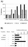
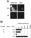
The lectins DC-SIGN and DC-SIGNR can augment viral infection; however, the range of pathogens interacting with these attachment factors is incompletely defined. Here we show that DC-SIGN and DC-SIGNR enhance infection mediated by the glycoprotein (GP) of Marburg virus (MARV) and the S protein of severe acute respiratory syndrome coronavirus and might promote viral dissemination. SIGNR1, a murine DC-SIGN homologue, also enhanced infection driven by MARV and Ebola virus GP and could be targeted to assess the role of attachment factors in filovirus infection in vivo.
Figures


Similar articles
-
pH-dependent entry of severe acute respiratory syndrome coronavirus is mediated by the spike glycoprotein and enhanced by dendritic cell transfer through DC-SIGN.J Virol. 2004 Jun;78(11):5642-50. doi: 10.1128/JVI.78.11.5642-5650.2004. J Virol. 2004. PMID: 15140961 Free PMC article.
-
West Nile virus discriminates between DC-SIGN and DC-SIGNR for cellular attachment and infection.J Virol. 2006 Feb;80(3):1290-301. doi: 10.1128/JVI.80.3.1290-1301.2006. J Virol. 2006. PMID: 16415006 Free PMC article.
-
Analysis of the interaction of Ebola virus glycoprotein with DC-SIGN (dendritic cell-specific intercellular adhesion molecule 3-grabbing nonintegrin) and its homologue DC-SIGNR.J Infect Dis. 2007 Nov 15;196 Suppl 2(Suppl 2):S237-46. doi: 10.1086/520607. J Infect Dis. 2007. PMID: 17940955 Free PMC article.
-
The role of DC-SIGN and DC-SIGNR in HIV and Ebola virus infection: can potential therapeutics block virus transmission and dissemination?Expert Opin Ther Targets. 2002 Aug;6(4):423-31. doi: 10.1517/14728222.6.4.423. Expert Opin Ther Targets. 2002. PMID: 12223058 Review.
-
DC-SIGN: binding receptors for hepatitis C virus.Chin Med J (Engl). 2004 Sep;117(9):1395-400. Chin Med J (Engl). 2004. PMID: 15377434 Review.
Cited by
-
An improved Fuzzy based GWO algorithm for predicting the potential host receptor of COVID-19 infection.Comput Biol Med. 2022 Dec;151(Pt A):106050. doi: 10.1016/j.compbiomed.2022.106050. Epub 2022 Aug 25. Comput Biol Med. 2022. PMID: 36334362 Free PMC article.
-
Angiodiversity and organotypic functions of sinusoidal endothelial cells.Angiogenesis. 2021 May;24(2):289-310. doi: 10.1007/s10456-021-09780-y. Epub 2021 Mar 21. Angiogenesis. 2021. PMID: 33745018 Free PMC article. Review.
-
C-type lectins and extracellular vesicles in virus-induced NETosis.J Biomed Sci. 2021 Jun 11;28(1):46. doi: 10.1186/s12929-021-00741-7. J Biomed Sci. 2021. PMID: 34116654 Free PMC article. Review.
-
Virus entry: old viruses, new receptors.Curr Opin Virol. 2012 Feb;2(1):4-13. doi: 10.1016/j.coviro.2011.12.005. Epub 2012 Jan 2. Curr Opin Virol. 2012. PMID: 22440960 Free PMC article. Review.
-
Primary severe acute respiratory syndrome coronavirus infection limits replication but not lung inflammation upon homologous rechallenge.J Virol. 2012 Apr;86(8):4234-44. doi: 10.1128/JVI.06791-11. Epub 2012 Feb 15. J Virol. 2012. PMID: 22345460 Free PMC article.
References
-
- Appelmelk, B. J., I. van Die, S. J. van Vliet, C. M. J. E. Vandenbroucke-Grauls, T. B. H. Geijtenbeek, and Y. van Kooyk. 2003. Cutting edge: carbohydrate profiling identifies new pathogens that interact with dendritic cell-specific ICAM-3-grabbing nonintegrin on dendritic cells. J. Immunol. 170:1635-1639. - PubMed
-
- Baribaud, F., S. Pöhlmann, T. Sparwasser, M. T. Kimata, Y. K. Choi, B. S. Haggarty, N. Ahmad, T. Macfarlan, T. G. Edwards, G. J. Leslie, J. Arnason, T. A. Reinhart, J. T. Kimata, D. R. Littman, J. A. Hoxie, and R. W. Doms. 2001. Functional and antigenic characterization of human, rhesus macaque, pigtailed macaque, and murine DC-SIGN. J. Virol. 75:10281-10289. - PMC - PubMed
-
- Bashirova, A. A., T. B. Geijtenbeek, G. C. van Duijnhoven, S. J. Van Vliet, J. B. Eilering, M. P. Martin, L. Wu, T. D. Martin, N. Viebig, P. A. Knolle, V. N. KewalRamani, Y. Van Kooyk, and M. Carrington. 2001. A dendritic cell-specific intercellular adhesion molecule 3-grabbing nonintegrin (DC-SIGN)-related protein is highly expressed on human liver sinusoidal endothelial cells and promotes HIV-1 infection. J. Exp. Med. 193:671-678. - PMC - PubMed
Publication types
MeSH terms
Substances
Grants and funding
LinkOut - more resources
Full Text Sources
Other Literature Sources
Molecular Biology Databases

