. 2004 Oct 27;23(21):4166-76.
doi: 10.1038/sj.emboj.7600427. Epub 2004 Oct 7.

TI-VAMP/VAMP7 is required for optimal phagocytosis of opsonised particles in macrophages

Affiliations

TI-VAMP/VAMP7 is required for optimal phagocytosis of opsonised particles in macrophages

Virginie Braun et al. EMBO J. .

Abstract

Phagocytosis relies on extension of plasmalemmal pseudopods generated by focal actin polymerisation and delivery of membranes from intracellular pools. Here we show that compartments of the late endocytic pathway, bearing the tetanus neurotoxin-insensitive vesicle-associated membrane protein (TI-VAMP/VAMP7), are recruited upon particle binding and undergo exocytosis before phagosome sealing in macrophages during Fc receptor (FcR)-mediated phagocytosis. Expression of the dominant-negative amino-terminal domain of TI-VAMP or depletion of TI-VAMP with small interfering RNAs inhibited phagocytosis mediated by Fc or complement receptors. In addition, inhibition of TI-VAMP activity led to a reduced exocytosis of late endocytic vesicles and this resulted in an early blockade of pseudopod extension, as observed by scanning electron microscopy. Therefore, TI-VAMP defines a new pathway of membrane delivery required for optimal FcR-mediated phagocytosis.


Figures
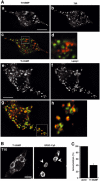
Figure 1
Figure 1
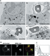
Endogenous TI-VAMP/VAMP7, colocalised with the lysosomal marker Lamp1, is recruited early and accumulates at the site of phagocytosis. (A) RAW264.7 cells were fixed, permeabilised and stained with anti-TI-VAMP and Cy3-anti-mouse IgG (a, c, e, g) and either anti-TfR (b, d) or anti-Lamp1 (f, h) followed by Cy2-anti-rat IgG. (c, g) Combined images; (d, h) insets in (c, g). Cells were analysed by wide-field fluorescence microscopy with deconvolution. Medial optical sections are shown. Bar, 5 μm. An average of 3.7±0.5 (n=10) TI-VAMP-positive and TfR-positive structures were colocalised per cell representing 8.7±1.1% of total TfR-positive structures while an average of 12±1.6 (n=10) TI-VAMP-positive structures were found to overlap with Lamp1-positive structures per cell which corresponded to 41±5.8% of total Lamp1-positive structures. (B) RAW264.7 cells were incubated for 10 min at 37°C with IgG-SRBCs, and then fixed and stained with Cy3-anti-rabbit IgG (right panel). This staining reveals particles that are still accessible to antibodies and therefore in nonclosed phagosomes, whereas internal particles were observed by phase contrast. The cells were then permeabilised, labelled with anti-TI-VAMP followed by Cy2-anti-mouse IgG (left panel) and analysed by confocal microscopy. One optical section is shown. The arrowheads point to external particles positive for TI-VAMP. Bar, 5 μm. (C) GFP-TI-VAMP is recruited early to phagosomes. RAW264.7 cells transiently transfected to express GFP-TI-VAMP were incubated for 10 min at 37°C with IgG-SRBCs, then placed on ice and, without fixation, stained with Cy3-anti-rabbit IgG to detect external particles. Then, cells were fixed, permeabilised and polymerised actin was labelled with phalloïdine-Alexa350. The number of accumulations of polymerised actin and GFP-TI-VAMP were scored for 50 GFP-TI-VAMP-expressing cells and expressed as an index of accumulation per cell. Then, the index obtained for TI-VAMP recruitment was expressed as a percentage of the index of actin cups. Data are the mean±s.e.m. of three independent experiments.
Figure 2
Figure 2
TI-VAMP/VAMP7-positive compartments are recruited early during phagocytosis. RAW264.7 cells transfected to express GFP-TI-VAMP were put into contact with IgG-SRBCs and recorded at 37°C using 4D deconvolution video microscopy. A time-stack was built with maximum intensity projections of deconvolved image stacks (see Movie 1, Supplementary data). Selected images are shown (top left corner, time in s).
Figure 3
Figure 3
TI-VAMP/Lamp1-positive compartments are exocytosed early during phagocytosis. (A) RAW264.7 cells were allowed to internalise HRP (50 mg/ml) for 30 min at 37°C. After a 1 h chase, phagocytosis was performed for 10 min. The cells were then fixed and processed for DAB cytochemistry and conventional electron microscopy. The arrowheads point to HRP-loaded vesicles recruited to sites of red blood cell attachment and the arrows to exocytosed DAB-positive membranes. The white stars label external SRBCs. Bar, 2 μm. (B) RAW264.7 cells were incubated with IgG-SRBCs for 10 min at 37°C, then placed on ice and, without fixation, stained with anti-Lamp1 antibodies followed by Cy3-anti-rat IgG. External red blood cells were detected with Cy2-anti-rabbit IgG antibodies. Finally, the cells were fixed and analysed by confocal microscopy. One medial optical section is shown. Bar, 5 μm. (C) RAW264.7 cells were treated as in (B), except that secondary antibodies were RPE-coupled. Live cells were then analysed by flow cytometry (bold line). As a control, the cells were incubated with IgG-SRBCs on ice before labelling (dotted histogram).
Figure 4
Figure 4
Exocytosis of Lamp1- and TI-VAMP-positive compartments. (A) RAW264.7 macrophages were allowed to phagocytose IgG-SRBCs for 10 min at 37°C and then fixed and processed for ultrathin cryosectioning. Cryosections were immunogold labelled with anti-Lamp1 antibody followed by Protein A-Gold. Bar, 1 μm. Staining of SRBCs was due to a nonspecific reaction. The asterisks indicate Lamp1-positive compartments. The arrows show Lamp1 that was found on membrane ruffles under particles, facing the extracellular medium. (B) Cells were treated as described in (A). Cryosections were immunogold labelled with anti-TI-VAMP antibody followed by Protein A-Gold. Bar, 1 μm. A TI-VAMP-positive compartment is indicated by an asterisk and the arrows show TI-VAMP facing the extracellular medium.
Figure 5
Figure 5
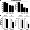
The amino-terminal domain of TI-VAMP inhibits FcR and CR-mediated phagocytosis. (A) RAW264.7 macrophages transiently expressing GFP-TI-VAMP, GFP-Longin or GFP-Cdc42N17 were incubated with IgG-SRBCs for 60 min at 37°C. The cells were then fixed and external SRBCs stained with Cy3-anti-rabbit IgG antibodies. The efficiencies of association (left) and phagocytosis (right) were calculated on 50 transfected and 50 control cells. The mean±s.e.m. of nine independent experiments is plotted. Control, GFP-negative cells. (B) RAW264.7 macrophages transiently expressing GFP-TI-VAMP (grey bars) or GFP-Longin (black bars) were allowed to phagocytose zymosan or C3bi-SRBCs for 60 min at 37°C. The samples were processed as in (A). The mean±s.e.m. of three independent experiments is plotted. Control, GFP-negative cells.
Figure 6
Figure 6
TI-VAMP siRNA treatment inhibits phagocytosis. RAW264.7 macrophages were transfected with siRNA directed against TI-VAMP (RNA.1 or RNA.2). After 24 and 48 h of treatment, cells were analysed by immunofluorescence (A), Western blot (B) or allowed to phagocytose IgG-SRBCs (C). (A) Cells were fixed, permeabilised, then labelled with anti-TI-VAMP followed by Cy3-anti-mouse IgG and analysed by wild-field fluorescence microscopy. Control cells were mock transfected. Bar, 10 μm. (B) Lysates were prepared and Western blotting was performed with anti-TI-VAMP (lower panel) and, after stripping of the membrane, with anti-clathrin HC (upper panel). Control cells were mock-transfected cells. (C) Transfected cells were incubated for 60 min at 37°C with IgG-SRBCs, then fixed and stained with Cy2-anti-rabbit IgG. The cells were then permeabilised and labelled with anti-TI-VAMP followed by Cy3-anti-mouse IgG. The efficiencies of association (left) and phagocytosis (right) were calculated on 50 cells that presented a decrease of TI-VAMP signal and 50 control cells (mock-transfected cells or cells transfected with siRNA GFP). The mean±s.e.m. of three independent experiments is plotted.
Figure 7
Figure 7
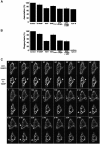
VAMP3- and TI-VAMP-mediated membrane delivery. (A) RAW264.7 macrophages transiently expressing GFP-TI-VAMP, GFP-Longin, TeNTE234Q, TeNT or both GFP-Longin and TeNT, or nontransfected cells pretreated for 30 min at 37°C with cytochalasin D (Cyto D) were processed for phagocytosis as described in Figure 5. Controls for transfection were negative cells and for Cytochalasin D, DMSO-treated cells. The mean efficiency of association±s.e.m. of four independent transfection experiments is plotted. (B) RAW264.7 cells were treated as in (A). The mean efficiency of phagocytosis±s.e.m. of four independent transfection experiments is represented. (C) RAW264.7 cells cotransfected to express YFP-TI-VAMP and CFP-VAMP3 were put into contact with IgG-SRBCs and followed at 37°C under a Zeiss LSM510 Meta confocal microscope. Images were recorded simultaneously in the two channels with a line average of eight every 3 s (see Movie 2, Supplementary data). Selected images are shown (top left corner, time in s). The arrowheads point to accumulation of YFP-TI-VAMP and the arrows to CFP-VAMP3.
Figure 8
Figure 8
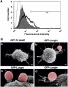
TI-VAMP controls exocytosis and membrane extension during phagocytosis. (A) RAW264.7 cells transfected to express GFP-TI-VAMP or GFP-Longin were incubated with IgG-SRBCs for 10 min at 37°C, then placed on ice and, without fixation, stained with anti-Lamp1 antibodies followed by RPE-anti-rat IgG. At least 5000 live, GFP-positive cells were gated and analysed by flow cytometry. Control cells incubated with secondary antibodies alone (dotted histogram), GFP-Longin-expressing cells (filled histogram), GFP-TI-VAMP-expressing cells (bold line) are shown. M1: Lamp1-positive cells, 18% for GFP-TI-VAMP- and 6% for GFP-Longin-expressing cells. (B) RAW264.7 cells expressing GFP-TI-VAMP (a) or GFP-Longin (b–d) were grown on CeLLocate coverslips, incubated with IgG-SRBCs for 60 min at 37°C, fixed, located on coverslips by fluorescence microscopy and then imaged by SEM. Red blood cells were artificially coloured under Adobe Photoshop 7.0. Bar, 1 μm.

Similar articles

Cited by

References

    1. Ackerman AL, Kyritsis C, Tampe R, Cresswell P (2003) Early phagosomes in dendritic cells form a cellular compartment sufficient for cross presentation of exogenous antigens. Proc Natl Acad Sci USA 100: 12889–12894 - PMC - PubMed
    1. Aderem A, Underhill DM (1999) Mechanisms of phagocytosis in macrophages. Annu Rev Immunol 17: 593–623 - PubMed
    1. Advani RJ, Bae HR, Bock JB, Chao DS, Doung YC, Prekeris R, Yoo JS, Scheller RH (1998) Seven novel mammalian SNARE proteins localize to distinct membrane compartments. J Biol Chem 273: 10317–10324 - PubMed
    1. Advani RJ, Yang B, Prekeris R, Lee KC, Klumperman J, Scheller RH (1999) VAMP-7 mediates vesicular transport from endosomes to lysosomes. J Cell Biol 146: 765–776 - PMC - PubMed
    1. Allen LA, Yang C, Pessin JE (2002) Rate and extent of phagocytosis in macrophages lacking VAMP3. J Leukoc Biol 72: 217–221 - PMC - PubMed

Publication types

MeSH terms