. 2004;2004(4):185-194.
doi: 10.1155/S1110724304403131.

Retrotransposition-Competent Human LINE-1 Induces Apoptosis in Cancer Cells With Intact p53

Retrotransposition-Competent Human LINE-1 Induces Apoptosis in Cancer Cells With Intact p53

Abdelali Haoudi et al. J Biomed Biotechnol. 2004.

Abstract

Retrotransposition of human LINE-1 (L1) element, a major representative non-LTR retrotransposon in the human genome, is known to be a source of insertional mutagenesis. However, nothing is known about effects of L1 retrotransposition on cell growth and differentiation. To investigate the potential for such biological effects and the impact that human L1 retrotransposition has upon cancer cell growth, we examined a panel of human L1 transformed cell lines following a complete retrotransposition process. The results demonstrated that transposition of L1 leads to the activation of the p53-mediated apoptotic pathway in human cancer cells that possess a wild-type p53. In addition, we found that inactivation of p53 in cells, where L1 was undergoing retrotransposition, inhibited the induction of apoptosis. This suggests an association between active retrotransposition and a competent p53 response in which induction of apoptosis is a major outcome. These data are consistent with a model in which human retrotransposition is sensed by the cell as a "genetic damaging event" and that massive retrotransposition triggers signaling pathways resulting in apoptosis.


Figures

Figure 1
Figure 1
Retrotransposition rationale. L1 plasmid is tagged with an antisense copy of the indicator gene NEO, which is disrupted by an intron, (I), in the sense orientation of L1. The NEO gene is flanked by a heterologous promoter (P2) and a polyadenylation signal (An). This ensures that G418R cells will only appear when an L1 transcript (P1: L1 promoter) is spliced (SD: splicing donor, SA: splicing acceptor), reverse transcribed, and reintegrated into chromosomal DNA, thus allowing expression of the uninterrupted NEO gene (NEO active). L1 tagged construct is subcloned into pCEP4 expression vector, which contains hygromycin selectable marker gene.
Figure 2
Figure 2
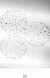
Human L1 retrotransposition in different cancer cell lines. (a) Colony forming ability of HCT-116 and SW480 cell lines. 103 cells either untransfected or transfected with L1 and selected for either hygromycin or G418 resistance were plated in triplicate until colonies were measurable, then stained and counted. Both cell lines exhibit similar colony forming ability when mock-treated or selected only for stable transformation with the plasmid (HygR). However, a dramatic difference in their survival ability was observed in G418-resistant clones bearing a retrotransposition-competent L1. (b) Confirmation of L1 retrotransposition by PCR in HCT-116 and SW480 cell lines. Lane M: DNA marker λ/HindIII; lane 1: HCT-116 mock-transfected cells; lane 2: HCT-116 HygR cells did not show the spliced form in either cell line; lane 3: HCT-116 G418R cells showing the correctly spliced form of NEO at 468 bp; lane 4: SW480 mock-transfected cells; lane 5: SW480 HygR cells; lane 6: SW480 G418R cells; lane 7: no DNA; and lane 8: the DNA plasmid alone with the unspliced form shown at 1361 bp.
Figure 2
Figure 2
Human L1 retrotransposition in different cancer cell lines. (a) Colony forming ability of HCT-116 and SW480 cell lines. 103 cells either untransfected or transfected with L1 and selected for either hygromycin or G418 resistance were plated in triplicate until colonies were measurable, then stained and counted. Both cell lines exhibit similar colony forming ability when mock-treated or selected only for stable transformation with the plasmid (HygR). However, a dramatic difference in their survival ability was observed in G418-resistant clones bearing a retrotransposition-competent L1. (b) Confirmation of L1 retrotransposition by PCR in HCT-116 and SW480 cell lines. Lane M: DNA marker λ/HindIII; lane 1: HCT-116 mock-transfected cells; lane 2: HCT-116 HygR cells did not show the spliced form in either cell line; lane 3: HCT-116 G418R cells showing the correctly spliced form of NEO at 468 bp; lane 4: SW480 mock-transfected cells; lane 5: SW480 HygR cells; lane 6: SW480 G418R cells; lane 7: no DNA; and lane 8: the DNA plasmid alone with the unspliced form shown at 1361 bp.
Figure 3
Figure 3
Analysis of apoptosis induction in the presence of RC-L1. (a) Apoptosis was highly induced in HCT-116 G418R cells with a wild-type p53 (G418R L1). No significant apoptosis induction was detected in SW480 cells with a mutant p53. Both HCT-116 and SW480 transfected with the vector alone (G418R NEO) did not show any apoptosis induction. Nuclease treatment was used as positive control. (b) Specific induction of Bax expression following L1 retrotransposition. HCT-116 cells transfected with either the L1 plasmid (JM101) or the vector alone (pBRV1). Bax induction was observed only in G418R L1-transfected cells (lane 3). No Bax expression was observed in mock-transfected cells (lane 1), HygR cells transfected with L1 (Lane 2), or G418R cells transfected with a plasmid containing a NEO gene alone (lane 4). α-tubulin is shown as loading control.
Figure 3
Figure 3
Analysis of apoptosis induction in the presence of RC-L1. (a) Apoptosis was highly induced in HCT-116 G418R cells with a wild-type p53 (G418R L1). No significant apoptosis induction was detected in SW480 cells with a mutant p53. Both HCT-116 and SW480 transfected with the vector alone (G418R NEO) did not show any apoptosis induction. Nuclease treatment was used as positive control. (b) Specific induction of Bax expression following L1 retrotransposition. HCT-116 cells transfected with either the L1 plasmid (JM101) or the vector alone (pBRV1). Bax induction was observed only in G418R L1-transfected cells (lane 3). No Bax expression was observed in mock-transfected cells (lane 1), HygR cells transfected with L1 (Lane 2), or G418R cells transfected with a plasmid containing a NEO gene alone (lane 4). α-tubulin is shown as loading control.
Figure 4
Figure 4
p53 inactivation by human papillomavirus E6 gene. HCT-116 cells transformed with L1 and selected for hygromycin resistance HygR were either (a) subjected to G418 selection directly (L1) or (b) exposed to either a deleted form of E6, ΔE6 (L1 + ΔE6), or (c) a complete form of E6 (L1 + E6). A histogram showing the effects of L1 retrotransposition on the number of clones in the presence and absence of E6 is shown in (d).
Figure 4
Figure 4
p53 inactivation by human papillomavirus E6 gene. HCT-116 cells transformed with L1 and selected for hygromycin resistance HygR were either (a) subjected to G418 selection directly (L1) or (b) exposed to either a deleted form of E6, ΔE6 (L1 + ΔE6), or (c) a complete form of E6 (L1 + E6). A histogram showing the effects of L1 retrotransposition on the number of clones in the presence and absence of E6 is shown in (d).
Figure 5
Figure 5
L1 retrotransposition in isogenic HT1080 cells. (a) Confirmation of L1 retrotransposition in HT1080 wt and HT1080 mut cells: a 468 bp DNA fragment was generated by PCR in HT1080 wt and HT1080 mut G418R clones demonstrating a correct splicing of the NEO marker gene. (b) Western blot analysis: whole cell extracts from HT1080 wt and HT1080 mut cells showing an increase of Bax expression following transfection with human L1 (1) and selection for G418R clones (2) harboring L1-retrotransposition elements predominantly in HT1080 wt. (c) Densitometric analysis of Bax expression levels in HT1080 wt and HT1080 mut before and after generation of G418R clones. More pronounced increase of Bax expression in HT1080 wt in comparison with HT1080 mut. Shown are relative arbitrary numbers. (d) Western blot analysis of HT10807thinsp;wt cells: Bcl2 expression before transfection (1) and after selection of G418R clones following L1 retrotransposition (2). (e) Western blot analysis of HT1080 mut cells: Bcl2 expression before transfection (1) and after selection of G418R (2). α-tubulin is shown as loading control.
Figure 5
Figure 5
L1 retrotransposition in isogenic HT1080 cells. (a) Confirmation of L1 retrotransposition in HT1080 wt and HT1080 mut cells: a 468 bp DNA fragment was generated by PCR in HT1080 wt and HT1080 mut G418R clones demonstrating a correct splicing of the NEO marker gene. (b) Western blot analysis: whole cell extracts from HT1080 wt and HT1080 mut cells showing an increase of Bax expression following transfection with human L1 (1) and selection for G418R clones (2) harboring L1-retrotransposition elements predominantly in HT1080 wt. (c) Densitometric analysis of Bax expression levels in HT1080 wt and HT1080 mut before and after generation of G418R clones. More pronounced increase of Bax expression in HT1080 wt in comparison with HT1080 mut. Shown are relative arbitrary numbers. (d) Western blot analysis of HT10807thinsp;wt cells: Bcl2 expression before transfection (1) and after selection of G418R clones following L1 retrotransposition (2). (e) Western blot analysis of HT1080 mut cells: Bcl2 expression before transfection (1) and after selection of G418R (2). α-tubulin is shown as loading control.

Similar articles

Cited by

References

    1. Hutchison C.A, Hardies S.C, Loeb D.D, Shehee W.R, Edgell M.H. LINEs and related retroposons: long interspersed repeated sequences in the eucaryotic genome. In: Berg D.E, Howe M.M, editors. Mobile DNA. Washington, DC, Wash: ASM Press; 1989. pp. 593–617.
    1. Boeke J.D, Stoye J.P. Retrotransposons, endogenous retroviruses, and the evolution of retroelements. In: Coffin J.M, Hughes S.H, Varmus H.E, editors. Retroviruses. Cold Spring Harbor, NY: Cold Spring Harbor Laboratory Press; 1997. pp. 343–435. - PubMed
    1. Fanning T.G, Singer M.F. LINE-1: a mammalian transposable element. Biochim Biophys Acta. 1987;910(3):203–212. - PubMed
    1. Moran J.V, Gilbert N. Mammalian LINE-1 retrotransposons and related elements. In: Craig N, Craigie R, Gellert M, et al., editors. Mobile DNA II. Washington, DC, Wash: ASM Press; 2002. pp. 836–869.
    1. Smit A.F, Toth G, Riggs A.D, Jurka J. Ancestral, mammalian-wide subfamilies of LINE-1 repetitive sequences. J Mol Biol. 1995;246(3):401–417. - PubMed