The PWWP domain of Dnmt3a and Dnmt3b is required for directing DNA methylation to the major satellite repeats at pericentric heterochromatin
- PMID: 15456878
- PMCID: PMC517890
- DOI: 10.1128/MCB.24.20.9048-9058.2004
The PWWP domain of Dnmt3a and Dnmt3b is required for directing DNA methylation to the major satellite repeats at pericentric heterochromatin
Abstract
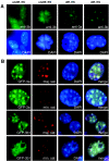
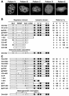
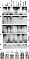
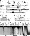
Dnmt3a and Dnmt3b are responsible for the establishment of DNA methylation patterns during development. These proteins contain, in addition to a C-terminal catalytic domain, a unique N-terminal regulatory region that harbors conserved domains, including a PWWP domain. The PWWP domain, characterized by the presence of a highly conserved proline-tryptophan-tryptophan-proline motif, is a module of 100 to 150 amino acids found in many chromatin-associated proteins. However, the function of the PWWP domain remains largely unknown. In this study, we provide evidence that the PWWP domains of Dnmt3a and Dnmt3b are involved in functional specialization of these enzymes. We show that both endogenous and green fluorescent protein-tagged Dnmt3a and Dnmt3b are particularly concentrated in pericentric heterochromatin. Mutagenesis analysis indicates that their PWWP domains are required for their association with pericentric heterochromatin. Disruption of the PWWP domain abolishes the ability of Dnmt3a and Dnmt3b to methylate the major satellite repeats at pericentric heterochromatin. Furthermore, we demonstrate that the Dnmt3a PWWP domain has little DNA-binding ability, in contrast to the Dnmt3b PWWP domain, which binds DNA nonspecifically. Collectively, our results suggest that the PWWP domains of Dnmt3a and Dnmt3b are essential for targeting these enzymes to pericentric heterochromatin, probably via a mechanism other than protein-DNA interactions.
Figures


Similar articles
-
DNMT3B PWWP mutations cause hypermethylation of heterochromatin.EMBO Rep. 2024 Mar;25(3):1130-1155. doi: 10.1038/s44319-024-00061-5. Epub 2024 Jan 30. EMBO Rep. 2024. PMID: 38291337 Free PMC article.
-
Chromatin targeting of de novo DNA methyltransferases by the PWWP domain.J Biol Chem. 2004 Jun 11;279(24):25447-54. doi: 10.1074/jbc.M312296200. Epub 2004 Mar 3. J Biol Chem. 2004. PMID: 14998998
-
The PWWP domain of mammalian DNA methyltransferase Dnmt3b defines a new family of DNA-binding folds.Nat Struct Biol. 2002 Mar;9(3):217-24. doi: 10.1038/nsb759. Nat Struct Biol. 2002. PMID: 11836534 Free PMC article.
-
Using human disease mutations to understand de novo DNA methyltransferase function.Biochem Soc Trans. 2024 Oct 30;52(5):2059-2075. doi: 10.1042/BST20231017. Biochem Soc Trans. 2024. PMID: 39446312 Free PMC article. Review.
-
Enzymology of Mammalian DNA Methyltransferases.Adv Exp Med Biol. 2016;945:87-122. doi: 10.1007/978-3-319-43624-1_5. Adv Exp Med Biol. 2016. PMID: 27826836 Review.
Cited by
-
Role of DNA methyltransferases in regulation of human ribosomal RNA gene transcription.J Biol Chem. 2006 Aug 4;281(31):22062-22072a. doi: 10.1074/jbc.M601155200. Epub 2006 May 30. J Biol Chem. 2006. Retraction in: J Biol Chem. 2018 Mar 9;293(10):3591. doi: 10.1074/jbc.W118.002433. PMID: 16735507 Free PMC article. Retracted.
-
Catalytically inactive Dnmt3b rescues mouse embryonic development by accessory and repressive functions.Nat Commun. 2019 Sep 26;10(1):4374. doi: 10.1038/s41467-019-12355-7. Nat Commun. 2019. PMID: 31558711 Free PMC article.
-
The Dnmt3a PWWP domain reads histone 3 lysine 36 trimethylation and guides DNA methylation.J Biol Chem. 2010 Aug 20;285(34):26114-20. doi: 10.1074/jbc.M109.089433. Epub 2010 Jun 11. J Biol Chem. 2010. PMID: 20547484 Free PMC article.
-
Disruption of Ledgf/Psip1 results in perinatal mortality and homeotic skeletal transformations.Mol Cell Biol. 2006 Oct;26(19):7201-10. doi: 10.1128/MCB.00459-06. Mol Cell Biol. 2006. PMID: 16980622 Free PMC article.
-
Effect of Disease-Associated Germline Mutations on Structure Function Relationship of DNA Methyltransferases.Genes (Basel). 2019 May 14;10(5):369. doi: 10.3390/genes10050369. Genes (Basel). 2019. PMID: 31091831 Free PMC article. Review.
References
-
- Bachman, K. E., M. R. Rountree, and S. B. Baylin. 2001. Dnmt3a and Dnmt3b are transcriptional repressors that exhibit unique localization properties to heterochromatin. J. Biol. Chem. 276:32282-32287. - PubMed
-
- Bird, A. 2002. DNA methylation patterns and epigenetic memory. Genes Dev. 16:6-21. - PubMed
-
- Chen, T., and E. Li. 2004. Structure and function of eukaryotic DNA methyltransferases. Curr. Top. Dev. Biol. 60:55-89. - PubMed
Publication types
MeSH terms
Substances
Grants and funding
LinkOut - more resources
Full Text Sources
Other Literature Sources
Molecular Biology Databases
