Identification of proteins associated with murine cytomegalovirus virions
- PMID: 15452238
- PMCID: PMC521832
- DOI: 10.1128/JVI.78.20.11187-11197.2004
Identification of proteins associated with murine cytomegalovirus virions
Abstract
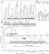
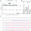
Proteins associated with the murine cytomegalovirus (MCMV) viral particle were identified by a combined approach of proteomic and genomic methods. Purified MCMV virions were dissociated by complete denaturation and subjected to either separation by sodium dodecyl sulfate-polyacrylamide gel electrophoresis and in-gel digestion or treated directly by in-solution tryptic digestion. Peptides were separated by nanoflow liquid chromatography and analyzed by tandem mass spectrometry (LC-MS/MS). The MS/MS spectra obtained were searched against a database of MCMV open reading frames (ORFs) predicted to be protein coding by an MCMV-specific version of the gene prediction algorithm GeneMarkS. We identified 38 proteins from the capsid, tegument, glycoprotein, replication, and immunomodulatory protein families, as well as 20 genes of unknown function. Observed irregularities in coding potential suggested possible sequence errors in the 3'-proximal ends of m20 and M31. These errors were experimentally confirmed by sequencing analysis. The MS data further indicated the presence of peptides derived from the unannotated ORFs ORF(c225441-226898) (m166.5) and ORF(105932-106072). Immunoblot experiments confirmed expression of m166.5 during viral infection.
Figures


Similar articles
-
Pox proteomics: mass spectrometry analysis and identification of Vaccinia virion proteins.Virol J. 2006 Mar 1;3:10. doi: 10.1186/1743-422X-3-10. Virol J. 2006. PMID: 16509968 Free PMC article.
-
Identification, analysis, and evolutionary relationships of the putative murine cytomegalovirus homologs of the human cytomegalovirus UL82 (pp71) and UL83 (pp65) matrix phosphoproteins.J Virol. 1996 Nov;70(11):7929-39. doi: 10.1128/JVI.70.11.7929-7939.1996. J Virol. 1996. PMID: 8892916 Free PMC article.
-
Cloning, characterization, and expression of the murine cytomegalovirus homologue of the human cytomegalovirus 28-kDa matrix phosphoprotein (UL99).Virology. 1994 Dec;205(2):417-29. doi: 10.1006/viro.1994.1662. Virology. 1994. PMID: 7975245
-
DNA sequence and transcriptional analysis of the glycoprotein M gene of murine cytomegalovirus.J Gen Virol. 1995 Nov;76 ( Pt 11):2895-901. doi: 10.1099/0022-1317-76-11-2895. J Gen Virol. 1995. PMID: 7595401
-
The multiple origins of proteins present in tupanvirus particles.Curr Opin Virol. 2019 Jun;36:25-31. doi: 10.1016/j.coviro.2019.02.007. Epub 2019 Mar 16. Curr Opin Virol. 2019. PMID: 30889472 Review.
Cited by
-
Mutations in the M112/M113-coding region facilitate murine cytomegalovirus replication in human cells.J Virol. 2010 Aug;84(16):7994-8006. doi: 10.1128/JVI.02624-09. Epub 2010 Jun 2. J Virol. 2010. PMID: 20519391 Free PMC article.
-
Cytomegalovirus downregulates IRE1 to repress the unfolded protein response.PLoS Pathog. 2013;9(8):e1003544. doi: 10.1371/journal.ppat.1003544. Epub 2013 Aug 8. PLoS Pathog. 2013. PMID: 23950715 Free PMC article.
-
Laboratory strains of murine cytomegalovirus are genetically similar to but phenotypically distinct from wild strains of virus.J Virol. 2008 Jul;82(13):6689-96. doi: 10.1128/JVI.00160-08. Epub 2008 Apr 16. J Virol. 2008. PMID: 18417589 Free PMC article.
-
The Impact of Mass Spectrometry-Based Proteomics on Fundamental Discoveries in Virology.Annu Rev Virol. 2014 Nov;1(1):581-604. doi: 10.1146/annurev-virology-031413-085527. Epub 2014 Jul 14. Annu Rev Virol. 2014. PMID: 26958735 Free PMC article.
-
Proteomic analyses of human cytomegalovirus strain AD169 derivatives reveal highly conserved patterns of viral and cellular proteins in infected fibroblasts.Viruses. 2014 Jan 7;6(1):172-88. doi: 10.3390/v6010172. Viruses. 2014. PMID: 24402306 Free PMC article.
References
-
- Bahr, U., and G. Darai. 2004. Re-evaluation and in silico annotation of the Tupaia herpesvirus proteins. Virus Genes 28:99-120. - PubMed
Publication types
MeSH terms
Substances
Grants and funding
LinkOut - more resources
Full Text Sources
Other Literature Sources

