. 2004 Oct 1;23(19):3721-34.
doi: 10.1038/sj.emboj.7600396. Epub 2004 Sep 23.

PKC controls HGF-dependent c-Met traffic, signalling and cell migration

Affiliations

PKC controls HGF-dependent c-Met traffic, signalling and cell migration

Stéphanie Kermorgant et al. EMBO J. .

Abstract

The growth factor/receptor pair HGF/c-Met exerts control on proliferation, morphogenesis and motility, and through overexpression and mutation is implicated in cancer. Here we have investigated the relationship between receptor signalling and traffic, and its control by specific PKC isotypes. It is shown that c-Met signalling to the ERK cascade occurs within endosomal compartments and that it is in this compartment that PKCepsilon specifically exerts its control on the pathway with the consequent accumulation of ERK in focal complexes. These events are clearly separated from the subsequent microtubule-dependent sorting of c-Met to its perinuclear destination, which is shown to be under the control of PKCalpha. Thus while it is shown that traffic to endosomes is essential for HGF/c-Met to trigger an ERK response, the subsequent traffic and signalling of c-Met controlled by these two PKC isotypes are unconnected events. The dynamic properties conferred by the PKCepsilon control are shown to be essential for a normal HGF-dependent migratory response. Thus PKCs are shown to control both receptor traffic and signal traffic to relay HGF/c-Met responses.


Figures
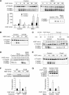
Figure 1
Figure 1
BIM-I sustains dynamic activation of ERK1/2. (A–E) Representative P-ERK1/2 and α-tubulin Western blots. (A) HeLa cells were stimulated with HGF in the absence (control) or presence of BIM-I (1 μM) or TPA (400 nM) or BIM-I plus TPA. Densitometric results are in arbitrary units. (B, C) Cells were stimulated with HGF in the absence (control) or presence of U0126 (10 μM) added 20 min before HGF. In (C) only, after 15 min of HGF exposure, the cells were washed, some fresh medium was replaced and cells were harvested at the indicated time. The last lane, ‘20*', is a control: cells pretreated with U0126 were harvested after 20 min of HGF treatment without washing. (D) Cells pretreated or not (control) for 20 min with BIM-I (1 μM) were treated with HGF. After 110 min, U0126 was added. Cells were harvested 5 or 10 min later (at 115 or 120 min). (E) Cells were pretreated or not (control) with concanavalin A (ConA) (250 μg/ml) before HGF stimulation. (F) Cells cotransfected with GFP-ERK2 and dynamin WT or dominant-negative (K44A) were stimulated with HGF for 0 or 120 min. Representative P-ERK and GFP Western blots are shown. (G) Cells transfected with an RNAi specific to clathrin heavy chain (HCC) were stimulated with HGF for 0 or 120 min. Representative HCC, α-tubulin and P-ERK Western blots are shown. (F, G) Densitometric results are in arbitrary units.
Figure 2
Figure 2
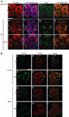
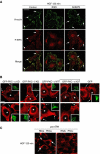
BIM-I inhibits c-Met-dependent ERK translocation to focal complexes. Representative confocal images for (A) pan ERK (red), phalloidin (blue) and c-Met (green), and (B) vinculin (green) and P-ERK (red). The arrows show examples of colocalisation. Bar, 20 μm. HeLa cells were stimulated for 0 or 120 min with HGF alone or with BIM-I.
Figure 3
Figure 3
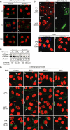
BIM-I does not modify STAT3 phosphorylation but sustains dynamic activation of JNK and c-Jun. (A) Representative P-STAT3 (PY705), α-tubulin and P-ERK1/2 Western blots from cells stimulated for 0 or 120 min with HGF alone or with BIM-I. Densitometric results are in fold increase. (B–D) Representative P-JNK and α-tubulin Western blots. (B) HeLa cells were stimulated with HGF in the absence (control) or presence of BIM-I (1 μM). The densitometric results are in arbitrary units. (C, D) Cells were pretreated or not (control) with concanavalin A (ConA) (250 μg/ml) (C) or U0126 (10 μM) (D) before HGF stimulation. (E) Representative Western blot and confocal medial sections for P-c-Jun. Bar, 20 μm. HeLa cells were UV treated (100 μJ) (positive control) or stimulated with HGF alone (control) or in the presence of BIM-I (1 μM). For each experiment, the ratio of P-c-Jun-positive nuclei was counted in 10 random fields of 10 cells (*P<0.05). The graph is in arbitrary units and is the mean of three independent experiments.
Figure 4
Figure 4
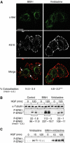
BIM-I sustains endocytic signalling from c-Met independent of trans-cytosolic traffic. (A, B) HeLa cells were stimulated by HGF in the presence of BIM-I (1 μM) or vinblastine (1 μM). (A) Representative confocal projections of five Z-sections for c-Met (green) and 4G10 (white or red). Bar, 20 μm. The numbers represent the statistical analysis of the colocalisation between c-Met and 4G10; *P<0.0001 (see Materials and methods). (B, C) Representative P-ERK1/2 and α-tubulin Western blots. Densitometric results are in arbitrary units (*P<0.01). Cells were stimulated by HGF in the presence or not (control) of BIM-1 (1 μM) or vinblastine (1 μM) alone or vinblastine and BIM-I (1 μM).
Figure 5
Figure 5
PKCα controls traffic of c-Met. (A–C) HeLa cells were pretreated or not (control) with Gö6976 (1 μM) for 10 min and stimulated with HGF. (A) Representative basomedial confocal images for c-Met (green) and propidium iodide (red). Bar, 20 μm. (B) Representative c-Met and α-tubulin Western blots. Densitometric results are in arbitrary units. (C) Representative medial confocal pictures for c-Met (red for (a, c); green for (e, f)). Bar, 20 μm. HeLa cells transfected with a kinase-inactive GFP-PKCα mutant (GFP-PKCα KD) were treated for 10 min with TPA plus 15 min with HGF (a, b) or with HGF alone for 240 min (c, d). * Indicates transfected cells. (e, f) Mouse embryonic fibroblasts knocked out for PKCα (PKCα KO) or re-expressing PKCα were treated for 10 min with TPA plus 15 min with HGF. (D) Representative confocal images for c-Met (green) and propidium iodide (red). Bar, 20 μm. HeLa cells transfected with RNAi control (a, e, i, m) or RNAis specific to PKCα (b, f, j, n), PKCδ (c, g, k, o) or PKCɛ (d, h, l, p) were stimulated with HGF alone for 0, 15 or 120 min or for 10 min with TPA plus 15 min with HGF.
Figure 6
Figure 6
PKCɛ but not PKCα controls c-Met-dependent ERK activation. (A) HeLa cells transfected with RNAi control or RNAis specific to PKCα, δ or ɛ were stimulated with HGF for 0 or 120 min. Western bots were performed for each PKC, α-tubulin and P-ERK. The densitometric results for P-ERK2 are in arbitrary units (*P<0.05). (B) HeLa cells were pretreated or not (control) with BIM-I (1 μM) or Gö6976 (1 μM) for 10 min and then stimulated with HGF for 120 min. Representative P-ERK1/2 Western blots and the densitometric results in fold increase are shown.
Figure 7
Figure 7
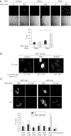
PKCɛ controls c-Met-dependent activation of c-Jun. (A) Representative confocal images for the same fields show P-Jun and phase. Bar, 20 μm. HeLa cells transfected with RNAi control or RNAis specific to PKCα or ɛ were stimulated with HGF alone for 0 or 120 min, fixed and stained for P-Jun. The graph represents the intensity of P-Jun in arbitrary units measured on 100 cells for each condition. (B) Representative basomedial confocal images for P-c-Jun. Bar, 20 μm. HeLa cells were pretreated or not (control) with BIM-I (1 μM) or Gö6976 (1 μM) for 10 min and stimulated with HGF for 120 min. The number of positive nuclei was determined as in Figure 3. (C) HeLa cells were transfected with GFP-PKCα, δ or ɛ constructs, WT or kinase-inactive (KD) or an empty EGFP vector. After 24 h in 0.1% FBS medium, the cells were stimulated or not with HGF, fixed and stained for P-c-Jun. A total of 10 confocal medial sections (10 cells/section) were randomly acquired. The ratio of transfected cells positive for P-c-Jun was obtained. The value obtained for each PKC construct was divided by the value obtained with empty GFP vector. Representative confocal sections for GFP-PKCɛ KD and GFP-PKCα KD are shown. Bar, 20 μm. The graph represents the P-Jun activation for each GFP-PKC at 0- and 120-min HGF stimulation; data are derived from three independent experiments (*P<0.001). For each coverslip, the proportions of nontransfected cells positive for P-c-Jun have been counted and represent an internal control. The last column represents this control for GFP-PKCɛ KD.
Figure 8
Figure 8
PKCɛ controls c-Met-dependent ERK translocation to focal complexes. (A) Representative confocal images for vinculin (green) and P-ERK (red). The arrows indicate examples of colocalisation. Bar, 20 μm. HeLa cells were pretreated or not (control) with BIM-I (1 μM) or Gö6976 (1 μM) for 10 min and stimulated with HGF for 120 min. (B) HeLa cells were transfected and HGF treated as indicated in Figure 7C and stained for pan ERK (red). Representative medial confocal sections are shown for PKCα and δ KD, PKɛ WT and KD and GFP alone. Bar, 10 μm. The arrows indicate examples of pan ERK localisation at the plasma membrane. * Indicates transfected cells. (C) Confocal images for pan ERK of HeLa cells transfected with RNAis control or specific to PKCɛ and stimulated with HGF for 120 min. Bar, 20 μm.
Figure 9
Figure 9
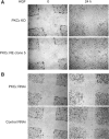
Confluent cells in 0.5% FBS medium for 24 h were wounded with a tip, washed and replaced with 0.5% FBS medium containing HGF. The same wound was photographed at 0 and 24 h. (A) Mouse embryo PKCɛ knockout (PKCɛ KO) or re-expressing (PKCɛ RE, clone 5). (B) HeLa cells transfected with RNAis specific to PKCɛ or control.
Figure 10
Figure 10
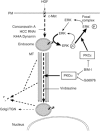
Scheme of PKC, c-Met controls. The figure illustrates the elements in the HGF-induced c-Met pathway under the control of PKC (see Discussion).

Similar articles

Cited by

References

    1. Alves dos Santos CM, van Kerkhof P, Strous GJ (2001) The signal transduction of the growth hormone receptor is regulated ubiquitin/proteasome system and continues after endocytosis. J Biol Chem 276: 10839–10846 - PubMed
    1. Awasthi V, King RJ (2000) PKC, p42/p44 MAPK, and p38 MAPK are required for HGF-induced of H441 cells. Am J Physiol Lung Cell Mol Physiol 279: L942–L949 - PubMed
    1. Burke P, Schooler K, Wiley HS (2001) Regulation of epidermal growth factor receptor signalling by endocytosis and intracellular trafficking. Mol Biol Cell 12: 1897–1910 - PMC - PubMed
    1. Ceresa BP, Kao AW, Santeler SR, Pessin JE (1998) Inhibition of clathrin-mediated endocytosis selectively attenuates specific insulin receptor signal transduction pathways. Mol Cell Biol 18: 3862–3870 - PMC - PubMed
    1. Chapell R, Bueno OF, Alvarez-Hernandez X, Robinson LC, Leidenheimer NJ (1998) Activation of protein kinase C induces gamma-aminobutyric acid type A receptor internalization in Xenopus oocytes. J Biol Chem 273: 32595–32601 - PubMed

MeSH terms