. 2004 Sep 20;200(6):701-12.
doi: 10.1084/jem.20041270.

Loss of HIV-1-specific CD8+ T cell proliferation after acute HIV-1 infection and restoration by vaccine-induced HIV-1-specific CD4+ T cells

Affiliations

Loss of HIV-1-specific CD8+ T cell proliferation after acute HIV-1 infection and restoration by vaccine-induced HIV-1-specific CD4+ T cells

Mathias Lichterfeld et al. J Exp Med. .

Abstract

Virus-specific CD8(+) T cells are associated with declining viremia in acute human immunodeficiency virus (HIV)1 infection, but do not correlate with control of viremia in chronic infection, suggesting a progressive functional defect not measured by interferon gamma assays presently used. Here, we demonstrate that HIV-1-specific CD8(+) T cells proliferate rapidly upon encounter with cognate antigen in acute infection, but lose this capacity with ongoing viral replication. This functional defect can be induced in vitro by depletion of CD4(+) T cells or addition of interleukin 2-neutralizing antibodies, and can be corrected in chronic infection in vitro by addition of autologous CD4(+) T cells isolated during acute infection and in vivo by vaccine-mediated induction of HIV-1-specific CD4(+) T helper cell responses. These data demonstrate a loss of HIV-1-specific CD8(+) T cell function that not only correlates with progressive infection, but also can be restored in chronic infection by augmentation of HIV-1-specific T helper cell function. This identification of a reversible defect in cell-mediated immunity in chronic HIV-1 infection has important implications for immunotherapeutic interventions.


Figures
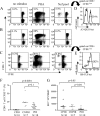
Figure 1.
Figure 1.
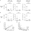
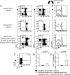
Cross-sectional assessment of CD8+ T cell proliferation after stimulation with HIV-1 peptide pools in individuals with primary, chronic progressive, and chronic long-term nonprogressive HIV-1 infection. (A–C) Dot plots showing the flow cytometric analysis of HIV-1–specific CD8+ T cell proliferation after stimulation of PBMCs with no stimulus, phytohemagglutinin (PHA), or a pool of overlapping peptides spanning the entire HIV-1 Nef protein in subjects with long-term nonprogressive (A), chronic progressive (B), or primary (C) HIV-1 infection. Values in top left corner of dot plots indicate the proportion of CFSElow CD8+ T cells. (D and E) Corresponding antigen specificity of proliferation cells. 34% of proliferating cells in the study individual in A were binding to the HLA-A3-QVPLRPMTYK (QK10) tetramer (D), whereas 82% of the CD8+ T cells proliferating in the study person in C were specific for the HLA-B8-FLKEKGGL (FL8) tetramer (E). (F and G) Comparative analysis of proliferation and interferon γ secretion by CD8+ T cells in response to stimulation with overlapping peptides spanning the entire HIV-1 proteome. Data from study subjects with chronic progressive HIV-1 infection (CPHI; n = 10), chronic long-term nonprogressive HIV-1 infection (CNPHI; n = 7), and primary HIV-1 infection (PHI; n = 18) are shown.
Figure 2.
Figure 2.
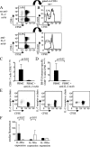
Longitudinal evolution of HIV-1–specific CD8+ T cell proliferative responses after primary HIV-1 infection. (A and B) Simultaneous assessment of antigen-specific proliferation and interferon γ secretion of CD8+ and CD4+ T cells after stimulation with overlapping peptides spanning the entire HIV-1 proteome at baseline and after 1 yr of follow-up in study persons with untreated (A) and treated (B) primary HIV-1 infection. (C and D) Correlation between HIV-1–specific CD8+ and CD4+ T cell lymphoproliferative responses. Proportions of CD8+ cells proliferating after exposure to overlapping peptides spanning the HIV-1 proteome were plotted against the corresponding proportion of CD4+ T cells (C) and against the corresponding magnitude of CD8+ T cell–mediated SFCs/106 PBMCs using an interferon γ ELISPOT assay (D). Data from the cross-sectional and longitudinal analysis were included. Dashed lines indicate the 95% confidence interval of the regression line.
Figure 3.
Figure 3.
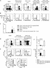
Antigen-specific ex vivo proliferation of HIV-1–specific CD8+ T cells critically depends on IL-2. (A and B) Dot plots (A) or histograms (B) showing the flow cytometric analysis of the proportion of CD8+ T cells (A) or HLA-B8-FLKEKGGL (FL8) tetramer–specific CD8+ T cells (B) proliferating in response to stimulation with a pool of overlapping peptides spanning HIV-1 Nef in the presence or absence of anti–IL-2 mAb. Values in top left corner of dot plots indicate the proportion of CFSElow CD8+ T cells. (C and D) Proportion of CD8+ T cells (C) or B8-FL8 tetramer–specific CD8+ T cells (D) proliferating after exposure to a pool of overlapping Nef peptides in the presence or absence of IL-2 antibodies. Mean and standard deviation of four experiments in four different study persons are shown. (E) Flow cytometric analysis of the surface expression of the α chain of the IL-2, IL-7, and IL-15 receptor in CD8+ T cells proliferating after stimulation with overlapping HIV-1 Nef peptides. (F) Median fluorescence of antibodies directed against the α chain of the IL-2, IL-7, and IL-15 receptor in CD8+ T cells proliferating (black bars) or nonproliferating (white bars) after stimulation with HIV-1 Nef peptides. Data reflect the mean and standard deviation of five independent experiments in four different study subjects.
Figure 4.
Figure 4.
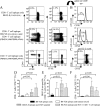
Ex vivo proliferation of HIV-1–specific CD8+ T cells is supported by CD4+ T cells. (A–C) Dot plots (A) and histograms (B) indicating the flow cytometric assessment of CD8+ T cells (A) or B8-FL8–specific CD8+ T cells (B) proliferating after stimulation with a Nef peptide pool in the presence or absence of indicated leukocellular subsets. Cells were gated according to forward scatter (FSC)/side scatter (SSC) characteristics of the lymphocyte population in A. In B, lymphocytes were additionally gated according to CD8+ expression and tetramer binding. Values in top left corner of dot plots indicate the proportion of CFSElow CD8+ T cells. A and B show one representative experiment and C indicates the mean and standard deviation of the proportion of CFSElow CD8+ T cells in four independent experiments (*, P < 0.05). (D–G) Dot plots (D) and histograms (E) showing the flow cytometric analysis of the proliferation of CD8+ T cells (D) or B8-FL8–specific CD8+ T cells (E) in responses to stimulation with a Nef peptide pool in whole PBMC samples, CD4+ cell–depleted PBMC samples, and in CD4+ cell–depleted PBMC samples that were supplemented with exogenous IL-2. Gating was performed as described for A and B. D and E show one representative experiment and F and G give the mean and standard deviation from ten independent experiments for bulk CD8+ T cells (F) and three different experiments for tetramer-specific cells (G), respectively. (F and G) Left black bars represent proliferating cells in whole PBMC samples, middle bars show PBMC samples depleted of CD4+ cells, and right white bars indicate PBMC samples depleted of CD4+ cells, but supplemented with exogenous IL-2. (H and I) Rescue of HIV-1–specific CD8+ T cell proliferation by exogenous IL-2 in chronic replicative HIV-1 infection. (H) CD8+ T cell proliferation in the presence of exogenous IL-2 after stimulation of PBMC samples from chronic HIV-1 infection with Nef pool peptides (solid line) or no antigenic stimulation (dashed line). Gating was performed according to FSC/SSC characteristics and CD8+ expression. (I) Proliferation of HLA-A3-QK10 tetramer–specific CD8+ T cells in the presence of IL-2 after stimulation of PBMC samples from chronic HIV-1 infection with Nef pool peptides (solid line) or no antigenic stimulation (dashed line). Cells were gated according to FSC/SSC characteristics, as well as CD8 expression and HLA-A3 QK10 tetramer binding. (H and I) Percentages indicate the proportion of CFSElow CD8+ T cells. One representative example of four different experiments is shown.
Figure 5.
Figure 5.
Ex vivo proliferative activities of HIV-1–specific CD8+ T cells are dramatically enhanced by simultaneous stimulation of antigen-specific CD4+ T cells. (A) CD8+ and CD4+ T cell proliferation after stimulation with the HIV-1 Nef CD8+ T cell epitope B8-FL8 or with the overlapping HIV-1 Nef peptide PEKEVLVWKFDSRLAFHH, or both peptides together. Dot plots of one representative flow cytometric experiment are shown. Cells were gated according to FSC/SSC characteristics of the lymphocyte population. (B) B8-FL8 tetramer–specific CD8+ T cells proliferating after stimulation with B8-FL8 peptide only (top) or in conjunction with the overlapping Nef peptide PEKEVLVWKFDSRLAFHH (bottom). (C) CD8+ T cell proliferation after simultaneous stimulation of PBMC samples with the CD8+ T cell epitope B8-FL8 and the HIV-1 Nef peptide PEKEVLVWKFDSRLAFHH in the presence of IL-2 antibodies. Cells were gated according to FSC/SSC characteristics of the lymphocyte population. (D and E) Proportion of CD8+ T cells (D), B8-FL8 tetramer–specific CD8+ T cells (E), or CD4+ T cells (F) proliferating after stimulation with the B8-FL8 epitopic peptide in the presence or absence of concomitant stimulation with tetanus toxoid, a CMV peptide, or the overlapping HIV-1 Nef peptide PEKEVLVWKFDSRLAFHH. Mean and standard deviation from three independent experiments in three different study subjects are shown.
Figure 6.
Figure 6.
Autologous CD4+ T cells harvested during primary HIV-1 infection can restore the ex vivo proliferative activity of HIV-1–specific CD8+ T cells in chronic HIV-1 infection. (A) Flow cytometric analysis of CD8+ and CD4+ T cell proliferation after stimulation with a pool of overlapping Nef peptides in study subject AC-98 during primary and chronic HIV-1 infection and in chronic HIV-1 infection after the addition of isolated autologous CD4+ T cells harvested during acute HIV-1 infection. Values in top left corner of dot plots indicate the proportion of CFSElow CD8+ or CD4+ T cells, respectively. (B) Corresponding histograms indicating the proportion of proliferating A3-QK10–specific CD8+ T cells. (C) Intracellular cytokine staining of CD4+ T cells from acute HIV infection in study subject AC-98 after stimulation with Nef pool peptides. Cells were gated according to FSC/SSC characteristics. Value in top right corner indicates proportion of IL-2+ CD4+ T cells. (D and E) Proportion of CD8+ T cells (D) and HIV-1 tetramer–specific CD8+ T cells (E) proliferating in chronic HIV-1 infection after stimulation with a pool of HIV-1–specific Nef peptides in the presence (white bars) or absence (black bars) of added autologous CD4+ T cells from acute HIV-1 infection. Data indicate the mean and standard deviation from three study individuals described in Results.
Figure 7.
Figure 7.
In vivo reconstitution of HIV-1–specific CD8+ T cell lymphoproliferative activities by vaccine-mediated induction of IL-2–secreting, HIV-1–specific CD4+ T cells. (A and B) Dot plots reflecting the lymphoproliferative activity of HIV-1–specific CD4+ (A) and CD8+ T (B) cells after stimulation with a pool of overlapping Gag peptides before and after five consecutive administrations of an Env-depleted immunogen or placebo. Percentages indicate the proportion of CFSElow CD8+ and CD4+ T cells. (C and D) Proportions of CD4+ (C) and CD8+ (D) T cells proliferating in response to stimulation with HIV-1 peptides spanning the entire HIV-1 proteome in five recipients of placebo and the vaccine. Data from baseline and after five consecutive administrations of the immunogen/placebo are shown.

Similar articles

Cited by

References

    1. Kahn, J.O., and B.D. Walker. 1998. Acute human immunodeficiency virus type 1 infection. N. Engl. J. Med. 339:33–39. - PubMed
    1. Borrow, P., H. Lewicki, B.H. Hahn, G.M. Shaw, and M.B. Oldstone. 1994. Virus-specific CD8+ cytotoxic T-lymphocyte activity associated with control of viremia in primary human immunodeficiency virus type 1 infection. J. Virol. 68:6103–6110. - PMC - PubMed
    1. Koup, R.A., J.T. Safrit, Y. Cao, C.A. Andrews, G. McLeod, W. Borkowsky, C. Farthing, and D.D. Ho. 1994. Temporal association of cellular immune responses with the initial control of viremia in primary human immunodeficiency virus type 1 syndrome. J. Virol. 68:4650–4655. - PMC - PubMed
    1. Altfeld, M., E.S. Rosenberg, R. Shankarappa, J.S. Mukherjee, F.M. Hecht, R.L. Eldridge, M.M. Addo, S.H. Poon, M.N. Phillips, G.K. Robbins, et al. 2001. Cellular immune responses and viral diversity in individuals treated during acute and early HIV-1 infection. J. Exp. Med. 193:169–180. - PMC - PubMed
    1. Dalod, M., M. Dupuis, J.C. Deschemin, C. Goujard, C. Deveau, L. Meyer, N. Ngo, C. Rouzioux, J.G. Guillet, J.F. Delfraissy, et al. 1999. Weak anti-HIV CD8(+) T-cell effector activity in HIV primary infection. J. Clin. Invest. 104:1431–1439. - PMC - PubMed

Publication types