. 2004 Oct 1;23(19):3793-802.
doi: 10.1038/sj.emboj.7600397. Epub 2004 Sep 16.

Tissue plasminogen activator is a potent activator of PDGF-CC

Affiliations

Tissue plasminogen activator is a potent activator of PDGF-CC

Linda Fredriksson et al. EMBO J. .

Abstract

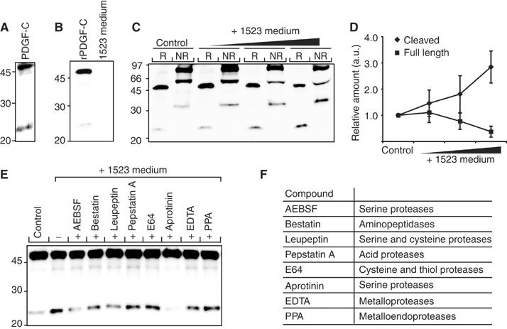
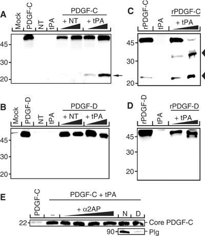
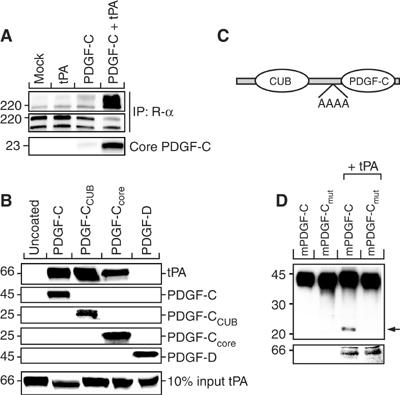
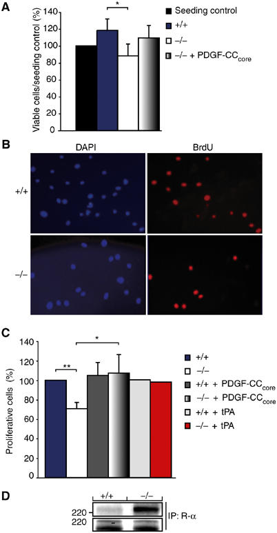
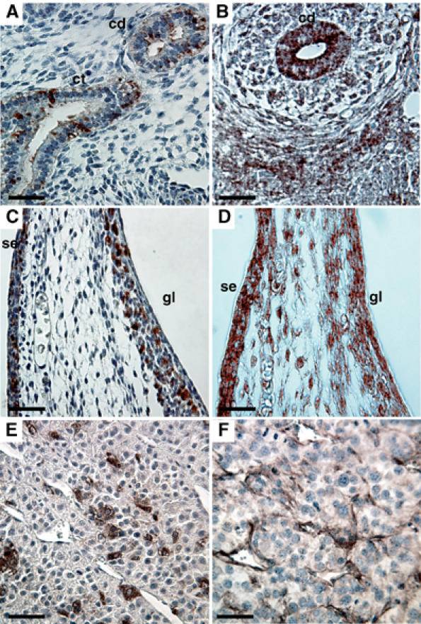
Tissue plasminogen activator (tPA) is a serine protease involved in the degradation of blood clots through the activation of plasminogen to plasmin. Here we report on the identification of tPA as a specific protease able to activate platelet-derived growth factor C (PDGF-C). The newly identified PDGF-C is secreted as a latent dimeric factor (PDGF-CC) that upon proteolytic removal of the N-terminal CUB domains becomes a PDGF receptor alpha agonist. The CUB domains in PDGF-CC directly interact with tPA, and fibroblasts from tPA-deficient mice fail to activate latent PDGF-CC. We further demonstrate that growth of primary fibroblasts in culture is dependent on a tPA-mediated cleavage of latent PDGF-CC, generating a growth stimulatory loop. Immunohistochemical analysis showed similar expression patterns of PDGF-C and tPA in developing mouse embryos and in tumors, indicating both autocrine and paracrine modes of activation of PDGF receptor-mediated signaling pathways. The identification of tPA as an activator of PDGF signaling establishes a novel role for the protease in normal and pathological tissue growth and maintenance, distinct from its well-known role in plasminogen activation and fibrinolysis.


Figures

Figure 1
Figure 1
Characterization of a PDGF-CC processing activity. (A) Endogenous expression of PDGF-CC from AG1523 fibroblasts detected by a PDGF-C-specific antibody. Reduced latent PDGF-C migrated as a 48 kDa species, while the released core domain migrated as a 22 kDa species. (B) Using an anti-His6 antibody, immunoreactivity was detected only in recombinant latent PDGF-C expressed in baculovirus-infected cells and not in conditioned medium from AG1523 cells. (C) Increasing concentrations of conditioned medium from AG1523 cells were incubated with fixed amounts of recombinant latent PDGF-CC. The reduced (R) and nonreduced (NR) recombinant proteins were analyzed by immunoblotting using an anti-His6 antibody. Under reducing conditions, the 48 kDa latent PDGF-C and the released 22 kDa core domain of PDGF-C were visualized. Under nonreducing conditions, the 90 kDa latent homodimer of PDGF-CC, the 60 kDa hemidimer, and the 35 kDa homodimeric core domain of PDGF-CC were visualized. (D) Quantification of the amounts of reduced full-length 48 kDa (▪) and cleaved 22 kDa (⧫) PDGF-C species. The results are mean±s.d. of five independent experiments. (E) Different protease inhibitors were preincubated with AG1523 medium, and then incubated with recombinant full-length PDGF-CC. Recombinant PDGF-CC incubated with serum-free medium (control) or AG1523 medium only (−) were used as controls. All lanes with incubations pretreated with serine protease inhibitors displayed reduced PDGF-C processing activity. An anti-His6 antibody was used. (F) List of the protease inhibitors used and the specificity of the inhibitors.
Figure 2
Figure 2
Cloning of candidate proteases from AG1523 fibroblastic cells. (A) Agarose gel electrophoresis of PCR products (arrowheads) amplified from AG1523 cDNA using degenerate oligonucleotide mixtures derived from trypsin-like serine protease domains. The amplified PCR fragments were cloned into the pCR2.1-TOPO vector and the nucleotide sequences of 18 clones were determined. (B) Histogram showing the identification of candidate proteases and distribution of the sequenced PCR-generated clones obtained from AG1523 cells.
Figure 3
Figure 3
tPA specifically cleaves latent PDGF-CC. Coexpression and functional analysis of tPA and NT on the proteolysis of PDGF-CC and PDGF-DD. (A, B, E) COS-1 cells were transfected with combinations of expression vectors encoding for PDGF-C or PDGF-D and different concentrations encoding for tPA and NT, respectively. Empty vector (mock) and the expression vectors alone were used as negative control. When coexpressed with PDGF-C, tPA released a 22 kDa fragment of PDGF-C (A, arrow), while tPA did not release the corresponding part of PDGF-D (B). In transfected cells, coexpressing NT and PDGF-C or PDGF-D, or mock transfection, did not release the core domains of PDGF-CC nor PDGF-DD. (C, D) In vitro cleavage of recombinant PDGF-CC (C) and PDGF-DD (D) using purified tPA in two different concentrations. PDGF-CC but not PDGF-DD is readily cleaved by tPA generating a 22 kDa band under reducing conditions, corresponding to the released core domain (lower arrowhead in C). Note the intermediate 32 kDa PDGF-C species (C, upper arrowhead), possibly due to cleavage by plasmin contamination in the tPA preparation (see Results). (E) Addition of the specific plasmin inhibitor α2-anti-plasmin (α2AP) into the cotransfection medium had no effect on the release of core PDGF-C by tPA nor had removal of Plg from the culture medium. N, normal FCS medium; D, Plg-depleted FCS medium.
Figure 4
Figure 4
tPA is the major PDGF-CC processing protease secreted from AG1523 cells and from primary mouse fibroblasts in culture. (A) Inhibition of cleavage of endogenous PDGF-CC produced by AG1523 cells using aprotinin and different concentrations of the specific tPA inhibitor tPA-STOP™. The inhibitors blocked processing of latent PDGF-CC showing that tPA accounts for the majority of the PDGF-C processing activity in conditioned media from AG1523 cells. (B) Serum-free media from wild-type and tPA-deficient fibroblasts were analyzed by immunoblotting. The results showed that both wild-type (+/+) and tPA-deficient (−/−) cells expressed latent PDGF-CC. However, tPA-deficient cells displayed a greatly reduced ability to process and activate the latent growth factor. tPA expression was analyzed by immunoblotting of conditioned media (middle panel). Agarose gel electrophoresis of PCR reactions from the genotyping of the animals used to establish the primary cultures of fibroblasts (lower panel). The immunoblot analyses were performed using protein-specific antibodies.
Figure 5
Figure 5
tPA-mediated proteolysis of latent PDGF-CC generates a PDGFR-α agonist. Conditioned serum-free media from transfected COS-1 cells were used to induce tyrosine phosphorylation of PDGFR-α expressed in PAE cells. (A) The 22 kDa fragment of PDGF-C, generated by tPA-mediated cleavage of latent PDGF-CC, induced efficient tyrosine phosphorylation of PDGFR-α as compared to mock, tPA, and PDGF-C controls as analyzed using antibodies against phosphotyrosine (PY99) (upper panel). The amount of precipitated PDGFR-α was monitored using antibodies to PDGFR-α (CED, middle panel). The amount of PDGF-C core domain in the media from the transfected cells was monitored by immunoblotting (lower panel). (B) Direct interaction of PDGF-CC with tPA. Ni-NTA beads coated with recombinant His6-tagged latent PDGF-CC, CUB domain, and core domains of PDGF-CC, or latent PDGF-DD, were incubated with purified tPA. Proteins eluted from the beads using a buffer containing 400 mM imidazole were analyzed by immunoblotting using specific antibodies. The results show that latent PDGF-CC interacts directly with tPA both via the CUB and the core domains. (C) Illustration of the cleavage site mutant. (D) Analysis of the cleavage site mutant of PDGF-CC using the cotransfection assay. Normal and mutant latent PDGF-CC forms were expressed in transfected COS-1 cells, without or with the coexpression of tPA. Analysis by immunoblotting showed that cleavage of latent PDGF-CC by tPA was abolished in the alanine cleavage site mutant (upper panel) suggesting that the tribasic site is the cleavage site for tPA. The expression of tPA was also monitored (lower panel).
Figure 6
Figure 6
An autocrine tPA-dependent growth stimulatory loop involving activation of latent PDGF-CC drives proliferation of fibroblasts in primary culture. Primary cultures of fibroblasts were established from wild-type and tPA-deficient animals. (A) Total cell numbers of wild-type (+/+) and tPA-deficient cells (−/−) after 36 h of culture in serum-free conditions (mean±s.d., n=4). Significantly less tPA-deficient cells were observed after the culture period (P<0.05). The tPA-deficient cells were stimulated to grow by the addition of activated PDGF-CC (mean±s.d., n=3). The seeding control was set to 100%. (B) Microphotographs showing wild-type and tPA-deficient fibroblasts following labeling with BrdU. Cell nuclei were visualized using DAPI (blue), while BrdU-labeled nuclei were identified by immunofluorescence using a specific antibody (red). (C) Quantification showed that significantly less tPA-deficient cells incorporated BrdU as compared to wild-type cells. Stimulation of the tPA-deficient cells with activated PDGF-CC or tPA enhanced BrdU incorporation, while wild-type cells were not markedly stimulated by this treatment (mean±s.d., n=3; n=2 for tPA treatment). *P<0.05, **P<0.01. (D) Activated PDGF-CC protein induced more efficient tyrosine phosphorylation of PDGFR-α in the tPA-deficient cells as compared to wild-type cells as analyzed using antibodies against phosphotyrosine (PY99) (upper panel). The amount of precipitated PDGFR-α was monitored using antibodies to PDGFR-α (CED, lower panel). These results show that growth of primary fibroblasts in culture is dependent on a growth stimulatory loop involving a tPA-dependent activation of latent PDGF-CC.
Figure 7
Figure 7
Colocalization of PDGF-CC and tPA. Immunohistochemical localization of PDGF-C (first column) and tPA (second column) in E14.5 mouse embryo and in T241 tumor xenografts. Tissue sections were stained using specific antibodies. (A, B) Developing kidney; overlapping staining for both PDGF-C and tPA was observed in the collecting ducts (cd). PDGF-C was also expressed in the collecting tubules (ct). (C, D) Skin of abdomen; colocalization of PDGF-C and tPA was seen in the germinal layer of the skin (gl) and in the surface ectoderm (se). (E, F) Expression of PDGF-C and tPA in T241 tumor xenografts. Scale bars, 50 μm.
Figure 8
Figure 8
Hypothetical mechanisms involved in the activation of PDGF-CC by tPA. (A) tPA binds to both the CUB domain and the growth factor domain of latent PDGF-CC. Released CUB domains might act as competitive inhibitors of the subsequent proteolytic activation of PDGF-CC. (B) A tPA-mediated activation of latent PDGF-CC drives proliferation of primary fibroblasts in culture.

Similar articles

Cited by

References

    1. Aase K, Abramsson A, Karlsson L, Betsholtz C, Eriksson U (2002) Expression analysis of PDGF-C in adult and developing mouse tissues. Mech Dev 110: 187–191 - PubMed
    1. Andrae J, Molander C, Smits A, Funa K, Nister M (2002) Platelet-derived growth factor-B and -C and active α-receptors in medulloblastoma cells. Biochem Biophys Res Commun 296: 604–611 - PubMed
    1. Bergsten E, Uutela M, Li X, Pietras K, Östman A, Heldin CH, Alitalo K, Eriksson U (2001) PDGF-D is a specific, protease-activated ligand for the PDGF β-receptor. Nat Cell Biol 3: 512–516 - PubMed
    1. Boucher P, Gotthardt M, Li WP, Anderson RG, Herz J (2003) LRP: role in vascular wall integrity and protection from atherosclerosis. Science 300: 329–332 - PubMed
    1. Bradford M (1976) A rapid and sensitive method for the quantification of microgram quantities of protein utilizing the principle of protein–dye binding. Anal Biochem 72: 248–254 - PubMed

Publication types

MeSH terms

Substances