. 2004 Oct;24(19):8576-85.
doi: 10.1128/MCB.24.19.8576-8585.2004.

FANCG is phosphorylated at serines 383 and 387 during mitosis

Affiliations

FANCG is phosphorylated at serines 383 and 387 during mitosis

Jun Mi et al. Mol Cell Biol. 2004 Oct.

Abstract

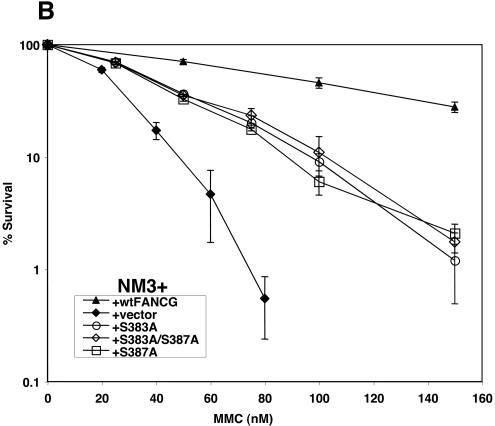
Fanconi anemia (FA) is an autosomal recessive disease marked by congenital defects, bone marrow failure, and high incidence of leukemia and solid tumors. Eight genes have been cloned, with the accompanying protein products participating in at least two complexes, which appear to be functionally dependent upon one another. Previous studies have described chromatin localization of the FA core complex, except at mitosis, which is associated with phosphorylation of the FANCG protein (F. Qiao, A. Moss, and G. M. Kupfer, J. Biol. Chem. 276:23391-23396, 2001). The phosphorylation of FANCG at serine 7 by using mass spectrometry was previously mapped. The purpose of this study was to map the phosphorylation sites of FANCG at mitosis and to assess their functional importance. Reasoning that a potential kinase might be cdc2, which was previously reported to bind to FANCC, we showed that cdc2 chiefly phosphorylated a 14-kDa fragment of the C-terminal half of FANCG. Mass spectrometry analysis demonstrated that this fragment contains amino acids 374 to 504. Kinase motif analysis demonstrated that three amino acids in this fragment were leading candidates for phosphorylation. By using PCR-directed in vitro mutagenesis we mutated S383, S387, and T487 to alanine. Mutation of S383 and S387 abolished the phosphorylation of FANCG at mitosis. These results were confirmed by use of phosphospecific antibodies directed against phosphoserine 383 and phosphoserine 387. Furthermore, the ability to correct FA-G mutant cells of human or hamster (where S383 and S387 are conserved) origin was also impaired by these mutations, demonstrating the functional importance of these amino acids. S387A mutant abolished FANCG fusion protein phosphorylation by cdc2. The FA pathway, of which FANCG is a part, is highly regulated by a series of phosphorylation steps that are important to its overall function.


Figures

FIG. 1.
FIG. 1.
FANCG is phosphorylated at mitosis. Whole-cell extracts from HeLa cells plus pMMP-vector, pMMP-FANCG, or pMMP-FANCG(S7A) cells asynchronous or synchronized into mitosis via nocodazole were prepared and run on SDS-PAGE. Immunoprecipitation (IP) was conducted with anti-Flag affinity gel. Immunoblotting with anti-FANCG antiserum showed the appearance of two more bands of higher mobility in mitotic cells (lanes 3 and 4) than for FANCG seen in asynchronous cells (lane 3). Mutation of FANCG at serine 7, a phosphorylation site that has been previously described (41a), does not account for mitotic phosphorylation (lane 4). Lane 1, vector-only control; lane 2, asynchronous plus wild-type FANCG; lane 3, mitotic plus wild-type FANCG; lane 4, mitotic plus S7A.
FIG. 2.
FIG. 2.
cdc2 phosphorylates a 14-kDa piece of the carboxyl terminus of FANCG. GST fusion proteins containing the amino half (N) and carboxyl half (C), respectively, of FANCG were thrombin cleaved. The resulting mixture was then subjected to in vitro kinase reaction, using beads containing cdc2 immunoprecipitated from mitotic HeLa cell extract. In vitro kinase reaction mixtures were subjected to SDS-PAGE. After transfer, the same filter was subjected both to autoradiography and to immunoblotting using anti-FANCG(C) antiserum. The immunoblot showed full-length FANCG(C) as well as a 14-kDa piece of FANCG(C). GST was also detected, because the original antibody was raised against a GST fusion protein. Autoradiography showed that not only full-length FANCG(C) but also a 14-kDa piece of FANCG(C) became phosphorylated by cdc2. A portion of the thrombin cleavage reaction mixture was run on SDS-PAGE and was Coomassie stained. The 14-kDa piece of FANCG(C) was excised and subjected to mass spectrometric analysis. The resulting spectrum of peptides corresponded to FANCG amino acids 374 to 504. ns, nonspecific.
FIG. 3.
FIG. 3.
S383 and S387 are phosphorylated at mitosis. pMMP-Flag-FANCG was mutated to alanine at S383, S387, or both by PCR-directed mutagenesis. The resulting constructs were then transduced into HeLa cells. Extracts from these cells synchronized to mitosis were prepared and immunoprecipitated (IP) with anti-Flag affinity gel. (A) Wild-type (WT) FANCG displayed two isoforms above the basal FANCG. Extra bands above the basal FANCG protein corresponding to phosphoproteins are marked by arrowheads. FANCG immunoblotting revealed that S383A and S387A mutations each eliminated one of the phospho-FANCG isoforms (lanes 4 and 5), while the double mutant eliminated both (lane 6). The T487A mutant did not alter the phospho-FANCG isoforms (lane 7). In all cases, the mutants coprecipitated FANCA. Lane 1, vector-only control; lane 2, wild-type FANCG, asynchronous; lane 3, wild-type mitotic; lane 4, S383A mitotic; lane 5, S387A mitotic; lane 6, S383A/S387A mitotic; lane 7, T487A mitotic. (B) Anti-phosphospecific antibodies to S383 and S387 were used to immunoblot a filter similar to that described for panel A. Anti-phosphoserine 383 specifically detected phosphorylated FANCG in all cell lines except FANCG(S383A) and FANCG (S383A/S387A). Similarly, anti-phosphoserine 387 antibody specifically detected phosphorylated FANCG in all cell lines except FANCG(S387A) and FANCG(S383A/S387A). Lane 1, preimmune control; lane 2, S383A; lane 3, S387A; lane 4, S383A/S387A; lane 5, T487A; lane 6, wild-type FANCG. (C) Asynchronous and mitotic HeLa whole-cell lysates were immunoprecipitated with FANCA antibody. Immunoblotting with phosphospecific antibodies revealed phosphoserine 383 and 387 on FANCG present only during mitosis. Lanes 1 and 5, mutant FA-G control; lanes 2 and 6, preimmune control; lanes 3 and 7, asynchronous; lanes 4 and 8, nocodazole-arrested mitotic cells. Asy, asynchronous; mit, mitotic.
FIG. 4.
FIG. 4.
S383 and S387 are functionally important in FANCG. (A) The mutant FANCG constructs used for experiments depicted in Fig. 3 were transduced into EUFA143 FA-G mutant cells. MMC cytotoxicity assays revealed that only wild-type FANCG (wtFANCG) fully corrected the mutant cells. (B) Whole-cell lysates of EUFA143 cells containing FANCG constructs were immunoblotted with FANCG antibody in order to demonstrate expression of FANCG. Lane 1, wild-type FANCG; lane 2, S383A/S387A; lane 3, S387A; lane 4, S383A; lane 5, vector-only control. Ku80 immunoblotting was performed on the same filter as a loading control. (C) The mutant FANCG constructs were transduced into 326SV FA-G mutant cells. MMC cytotoxicity assays revealed that only wild-type FANCG fully corrected the mutant cells. (D) Whole-cell lysates of 326SV cells containing FANCG constructs were immunoblotted with FANCG antibody in order to demonstrate equal expression of FANCG. Lane 1, vector-only control; lane 2, wild-type FANCG; lane 3, S383A/S387A; lane 4, S387A; lane 5, S383A. β-Tubulin immunoblotting was performed on the same filter as a loading control. (E) A total of 500,000 EUFA143 cells containing indicated FANCG constructs were treated with 1 μM MMC and were subsequently analyzed by FACS. All FA-G mutant constructs demonstrated marked increases in G2 accumulation similar to that of the mutant cell line compared to that of the cell line corrected with wild-type FANCG. The data presented are representative of three independent experiments.
FIG. 4.
FIG. 4.
S383 and S387 are functionally important in FANCG. (A) The mutant FANCG constructs used for experiments depicted in Fig. 3 were transduced into EUFA143 FA-G mutant cells. MMC cytotoxicity assays revealed that only wild-type FANCG (wtFANCG) fully corrected the mutant cells. (B) Whole-cell lysates of EUFA143 cells containing FANCG constructs were immunoblotted with FANCG antibody in order to demonstrate expression of FANCG. Lane 1, wild-type FANCG; lane 2, S383A/S387A; lane 3, S387A; lane 4, S383A; lane 5, vector-only control. Ku80 immunoblotting was performed on the same filter as a loading control. (C) The mutant FANCG constructs were transduced into 326SV FA-G mutant cells. MMC cytotoxicity assays revealed that only wild-type FANCG fully corrected the mutant cells. (D) Whole-cell lysates of 326SV cells containing FANCG constructs were immunoblotted with FANCG antibody in order to demonstrate equal expression of FANCG. Lane 1, vector-only control; lane 2, wild-type FANCG; lane 3, S383A/S387A; lane 4, S387A; lane 5, S383A. β-Tubulin immunoblotting was performed on the same filter as a loading control. (E) A total of 500,000 EUFA143 cells containing indicated FANCG constructs were treated with 1 μM MMC and were subsequently analyzed by FACS. All FA-G mutant constructs demonstrated marked increases in G2 accumulation similar to that of the mutant cell line compared to that of the cell line corrected with wild-type FANCG. The data presented are representative of three independent experiments.
FIG. 5.
FIG. 5.
Homologous hamster amino acids of FANCG are also functionally important. (A) FANCG amino acid sequences from human, hamster, and mouse were aligned. S383 and S387 have homologous amino acids in hamster and mouse, while T487 did not. (B) The mutant and wild-type FANCG (wtFANCG) constructs were transduced into mutant FA-G CHO cell lines. Cytotoxicity assays revealed that only wild-type FANCG was able to fully correct the mutant CHO cells. (C) Whole-cell lysates of CHO cells containing FANCG constructs were immunoblotted with FANCG antibody in order to demonstrate equal expression of FANCG. Ku80 immunoblotting was performed on the same filter as a loading control. Lane 1, CHO plus FANCG (wild type); lane 2, S383/S387A; lane 3, S387A; lane 4, S383A; lane 5, vector-only control.
FIG. 5.
FIG. 5.
Homologous hamster amino acids of FANCG are also functionally important. (A) FANCG amino acid sequences from human, hamster, and mouse were aligned. S383 and S387 have homologous amino acids in hamster and mouse, while T487 did not. (B) The mutant and wild-type FANCG (wtFANCG) constructs were transduced into mutant FA-G CHO cell lines. Cytotoxicity assays revealed that only wild-type FANCG was able to fully correct the mutant CHO cells. (C) Whole-cell lysates of CHO cells containing FANCG constructs were immunoblotted with FANCG antibody in order to demonstrate equal expression of FANCG. Ku80 immunoblotting was performed on the same filter as a loading control. Lane 1, CHO plus FANCG (wild type); lane 2, S383/S387A; lane 3, S387A; lane 4, S383A; lane 5, vector-only control.
FIG. 6.
FIG. 6.
cdc2 phosphorylates FANCG at S387. (A) FANCG(C) fusion protein was mutated by PCR-directed mutagenesis. The resulting proteins were then thrombin cleaved and subjected to in vitro kinase reaction with recombinant cdc2. Autoradiography revealed that GST-FANCG(S387A) and GST-FANCG(S387A/S387A) were unable to be phosphorylated. Lane 1, GST only; lane 2, wild-type FANCG(C); lane 3, S383A; lane 4, S387A; lane 5, S383A/S387A; lane 6, T487. (B) Wild-type and mutant peptides covering FANCG amino acids 378 to 392 were incubated in an in vitro kinase reaction mixture. A portion of each reaction mixture was spotted onto P81 paper at the indicated times and was subjected to scintillation counting. S387A and S383A/387A peptides completely eliminated cdc2 kinase activity.
FIG. 7.
FIG. 7.
cdc2 binds to FA core complex maximally at late G2. HeLa cells plus Flag-FANCA were synchronized to the G1-S border and then were released. Cells were collected at 0, 3, 6, and 9 h postrelease along with mitotic arrested cells. Lysates were prepared, and immunoprecipitations (IP) were performed with anti-Flag affinity gel. Immunoblotting revealed that cdc2 maximally was coprecipitated in late G2, 9 h after release. DNA histograms display synchronized cells at the indicated time points. Lane 1, vector-only control (Vec); lane 2, asynchronous cells (asy); lane 3, G1-S-arrested cells; lane 4, 3 h postrelease from G1-S; lane 5, 6 h; lane 6, 9 h; and lane 7, mitotic cells (mit).
FIG. 8.
FIG. 8.
FANCG mitotic mutants are aberrantly localized in mitosis. (A) An ECFP-FANCG construct was mutated by PCR-directed mutagenesis and was transfected into HeLa cells. Cells were then arrested in mitosis by using nocodazole. Cells were shaken from plates, collected, and cytospun onto slides. Fluorescent microscopy revealed that wild-type (wt) FANCG protein was perinuclear and in fine foci. The other mitotic mutants were in larger aggregates (S383A and double mutant) and/or dispersed in the nucleus (S387A and double mutant). (B) Chromatin extracts were prepared from mitotic HeLa cells containing the FANCG constructs shown in panel A. Lane 1, S383A; lane 2, S387A; lane 3, S383A/S387A; lane 4, wild-type FANCG; lane 5, vector only. Immunoblotting with FANCG antibody revealed that mutant FANCG was retained in chromatin. A Ku80 immunoblot on the same filter demonstrated equal loading. DAPI, 4,6-diamidino-2-phenylindole.
FIG. 8.
FIG. 8.
FANCG mitotic mutants are aberrantly localized in mitosis. (A) An ECFP-FANCG construct was mutated by PCR-directed mutagenesis and was transfected into HeLa cells. Cells were then arrested in mitosis by using nocodazole. Cells were shaken from plates, collected, and cytospun onto slides. Fluorescent microscopy revealed that wild-type (wt) FANCG protein was perinuclear and in fine foci. The other mitotic mutants were in larger aggregates (S383A and double mutant) and/or dispersed in the nucleus (S387A and double mutant). (B) Chromatin extracts were prepared from mitotic HeLa cells containing the FANCG constructs shown in panel A. Lane 1, S383A; lane 2, S387A; lane 3, S383A/S387A; lane 4, wild-type FANCG; lane 5, vector only. Immunoblotting with FANCG antibody revealed that mutant FANCG was retained in chromatin. A Ku80 immunoblot on the same filter demonstrated equal loading. DAPI, 4,6-diamidino-2-phenylindole.

Similar articles

Cited by

References

    1. Adachi, D., T. Oda, H. Yagasaki, K. Nakasato, T. Taniguchi, A. D. D'Andrea, S. Asano, and T. Yamashita. 2002. Heterogeneous activation of the Fanconi anemia pathway by patient-derived FANCA mutants. Hum. Mol. Genet. 11:3125-3134. - PubMed
    1. Akkari, Y. M., R. L. Bateman, C. A. Reifsteck, S. B. Olson, and M. Grompe. 2000. DNA replication is required to elicit cellular responses to psoralen-induced DNA interstrand cross-links. Mol. Cell. Biol. 20:8283-8289. - PMC - PubMed
    1. Alter, B. P., and N. S. Young. 1993. The bone marrow failure syndromes, p. 216-316. In D. G. Nathan and F. A. Oski (ed.), Hematology of infancy and childhood, vol. 1. W.B. Saunders, Philadelphia, Pa.
    1. Apostolou, S., S. A. Whitmore, J. Crawford, et al. 1996. Positional cloning of the Fanconi anaemia group A gene. Nat. Genet. 14:324-328. - PubMed
    1. Auerbach, A., M. Buchwald, and H. Joenje. 1997. Fanconi anemia, p. 317-332. In B. Vogelstein and K. Kinzler (ed.), Genetics of cancer. McGraw-Hill, New York, N.Y.

Publication types