. 2004 Oct;78(19):10238-48.
doi: 10.1128/JVI.78.19.10238-10248.2004.

Vaccinia virus mutants with alanine substitutions in the conserved G5R gene fail to initiate morphogenesis at the nonpermissive temperature

Affiliations

Vaccinia virus mutants with alanine substitutions in the conserved G5R gene fail to initiate morphogenesis at the nonpermissive temperature

Flavio G da Fonseca et al. J Virol. 2004 Oct.

Abstract

The initial characterization of the product of the vaccinia virus G5R gene, which is conserved in all poxviruses sequenced to date, is described. The G5 protein was detected in the core fraction of purified virions, and transcription and translation of the G5R open reading frame occurred early in infection, independently of DNA replication. Attempts to delete the G5R gene and isolate a replication-competent virus were unsuccessful, suggesting that G5R encodes an essential function. We engineered vaccinia virus mutants with clusters of charged amino acids changed to alanines and determined that several were unable to replicate at 40 degrees C but grew well at 37 degrees C. At the nonpermissive temperature, viral gene expression and DNA replication and processing were unperturbed. However, tyrosine phosphorylation and proteolytic cleavage of the A17 membrane protein and proteolytic cleavage of core proteins were inhibited at 40 degrees C, suggesting an assembly defect. The cytoplasm of cells that had been infected at the nonpermissive temperature contained large granular areas devoid of cellular organelles or virus structures except for occasional short crescent-shaped membranes and electron-dense lacy structures. The temperature-sensitive phenotype of the G5R mutants closely resembled the phenotypes of vaccinia virus mutants carrying conditionally lethal F10R protein kinase and H5R mutations. F10, although required for phosphorylation of A17 and viral membrane formation, was synthesized by the G5R mutants under nonpermissive conditions. An intriguing possibility is that G5 participates in the formation of viral membranes, a poorly understood event in poxvirus assembly.


Figures

FIG. 1.
FIG. 1.
Representation of portions of the VAC WR and vG5R-V5 genomes. (A) In the VAC WR genome, ORFs G4L, G5R, G5.5R, J1R, J2R (TK), and J3R are represented by boxes; the G4L, G5R, and G5.5 promoters (G4pr, G5pr, and G5.5pr, respectively) are indicated by arrowheads. In the VG5R-V5 genome, the G5R ORF was replaced by the neo gene (Neo) and the gus gene (Gus); a copy of the G5R gene containing a V5 epitope tag and under the control of its original promoter was inserted into the J2R (TK) gene site. (B) Plaque phenotypes of VAC WR and vG5R-V5. BS-C-1 cells were infected, incubated for 48 h, and then fixed and stained with crystal violet-ethanol solution.
FIG. 2.
FIG. 2.
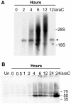
Temporal expression of the G5R gene. (A) Northern blot analysis. BS-C-1 cells were infected with vG5R-V5 in the absence or presence of 200 μM AraC and harvested at the indicated hours. Total RNA was extracted, resolved in a denaturing agarose gel, transferred to a nylon membrane, and hybridized to a 32P-labeled G5R gene probe. G5R transcripts are indicated by the arrowhead. The positions of 18S and 28S rRNAs are shown. (B) Western blot analysis. BS-C-1 cells that were uninfected (UN) or infected with vG5R-V5 in the presence or absence of 200 μM AraC were harvested at the indicated hours, and the lysates were analyzed by SDS-PAGE and Western blotting with a monoclonal antibody to the V5 epitope. The positions and masses (in kilodaltons) of marker proteins are indicated at the right.
FIG. 3.
FIG. 3.
Association of G5 with cores of purified virions. Virions purified from cells infected with vG5R-V5 by sedimentation through a sucrose cushion and two sucrose gradients (16) were incubated in Tris buffer alone (Ut) with 0.5% NP-40, or with 0.5% NP-40 plus 50 mM dithiothreitol (DTT). After centrifugation, the supernatant (S) and pellet (P) fractions were analyzed by Western blotting with a monoclonal antibody to V5. The masses (in kilodaltons) and positions of protein markers are indicated at the right.
FIG. 4.
FIG. 4.
Alanine-scanning mutagenesis. (A) The translated sequence of the G5R ORF is shown, with clusters of charged amino acids in light gray. The amino acid number of the first charged amino acid of each underlined cluster that was mutated is indicated. (B) Representation of portions of the genome of vG5R mutants (vG5R-Mut). Labeling is similar to that in Fig. 1A. The mutated G5R gene is flanked by the GFP and gpt genes. A hypothetical charge-to-alanine mutation is represented by the bar labeled mut within G5R.
FIG. 5.
FIG. 5.
Plaque formation at 31, 37, and 40°C. Confluent BS-C-40 cells were infected with VAC WR or G5R mutants generated through clustered charge-to-alanine mutagenesis. Cells were incubated for 48 h, fixed, and stained with crystal violet. The mutants are named according to the first charged amino acid of the mutated cluster, as shown in Fig. 4A. vG5Rwt contains an unmutated G5 ORF.
FIG. 6.
FIG. 6.
One-step virus growth. BS-C-40 monolayers in six-well plates were infected with 10 PFU of VAC WR, vG5R167, vG5R350, or vG5R167/325 per cell at 31, 37, or 40°C. After 1 h, the inocula were removed, the cells were washed twice with warm PBS, and fresh medium at the appropriate temperature was added. At various times after infection, cells were harvested, washed, lysed by freezing and thawing, and sonicated. Virus yields were then determined by plaque assay in BS-C-1 cells at 37°C.
FIG. 7.
FIG. 7.
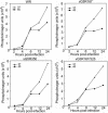
Viral DNA synthesis. BS-C-40 cells were infected at a multiplicity of 10 PFU per cell with VAC WR, vG5R167, vG5R167/325, or vG5R350 and incubated at 37 or 40°C. At the indicated times, total DNA was extracted, blotted onto a membrane, and hybridized to a 32P-labeled VAC DNA probe. Radioactivity was measured with a phosphorimager.
FIG. 8.
FIG. 8.
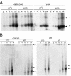
Processing of viral DNA into unit-length genomes. BS-C-40 cells were infected with 10 PFU of WR or vG5R350 per cell and incubated at 37 or 40°C for 2, 4, 6, 12, or 24 h. (A) Concatemer junction analysis. Cells were harvested, and total DNA was isolated, digested with BstEII, resolved in agarose gels, transferred to membranes, and hybridized to a 32P-labeled probe consisting of the linearized hairpin terminus of the VAC genome. Processed telomere fragments of 1.3 kbp (T) and concatemer junction fragments of 2.6 kbp (J) are indicated. (B) Pulsed-field analysis. Viral DNA was analyzed by pulsed-field gel electrophoresis, transferred to a membrane, and hybridized to a 32P-labeled VAC DNA probe. Gel wells (W) and the monomeric (M) and dimeric (D) forms of the genome are indicated.
FIG. 9.
FIG. 9.
Metabolic labeling of infected cells. BS-C-40 cells were infected with 10 PFU per cell of VAC WR or vG5R350 at 37 or 40°C and incubated for 2, 6, 12, or 24 h. At each time point, monolayers were washed with methionine-, cysteine-, and serum-deficient medium for 15 min. Cells were then pulse labeled for 30 min with 100 μCi of [35S]methionine and [35S]cysteine per ml, harvested, and analyzed by SDS-PAGE followed by autoradiography. The masses and positions of protein markers are indicated at the center of figure.
FIG. 10.
FIG. 10.
Localization of DNA and viral protein in viral factories. BS-C-40 cells were infected with 5 PFU per cell of VAC WR or vG5R350 and incubated at 37 or 40°C for 12 h. Cells were then fixed, permeabilized, and labeled with the DNA-specific dye DAPI and anti-H3 antibody (α-H3) followed by Texas Red-conjugated goat anti-rabbit immunoglobulin antibody. Arrowheads indicate representative cytoplasmic DNA factories adjacent to the larger nuclei. In some cases the factories overlap the nuclei and are not clearly resolved from them.
FIG. 11.
FIG. 11.
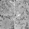
Electron microscopy of infected cells. BS-C-40 cells were infected with 5 PFU per cell of vG5R350 at 37°C (A) or 40°C (B and C) or with VAC WR at 40°C (D). At 12 h after infection, the cells were fixed and prepared for transmission electron microscopy (8). Electron micrographs are shown, with the scale indicated by the bars. Abbreviations; N, nucleus, M, mitochondrion; c, viral crescent; IV, immature virions; n, nucleoid within an immature virion; CEV, cell-associated enveloped virion; V, viroplasm. The arrow in panel C points to the lacy structure at the edge of the granular region.
FIG. 12.
FIG. 12.
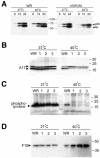
Cleavage and phosphorylation of viral proteins. (A) Cells were infected with 10 PFU per cell of WR or vG5R350 at 37 or 40°C for 8, 12, and 24 h. At each time point, cells were harvested, and lysates were prepared for SDS-PAGE and Western blotting with antiserum to the VAC p4b/4b protein. Arrowheads indicate the positions of p4b and 4b. The masses (in kilodaltons) and positions of protein markers are indicated at the center of figure. (B) Cells were infected with 10 PFU per cell of WR, vG5R167 (lane 1), vG5R167/325 (lane 2), or vG5R350 (lane 3) at 37 or 40°C. After 16 h, the cells were harvested,and lysates were analyzed by SDS-PAGE and Western blotting with antibody to A17. The positions and masses (in kilodaltons) of marker proteins are indicated at the right. The membrane used for panel B was stripped and reprobed with an antiphosphotyrosine antibody that recognizes phosphorylated A17 (C) and with F10 antibody (D).

Similar articles

Cited by

References

    1. Ansarah-Sobrinho, C., and B. Moss. 2004. Role of the I7 protein in proteolytic processing of vaccinia virus membrane and core components. J. Virol. 78:6335-6343. - PMC - PubMed
    1. Banham, A., and G. L. Smith. 1992. Vaccinia virus gene B1R encodes a 34-kDa serine/threonine protein kinase that localizes in cytoplasmic factories and is packaged into virions. Virology 191:803-812. - PubMed
    1. Baroudy, B. M., S. Venkatesan, and B. Moss. 1982. Structure and replication of vaccinia virus telomeres. Cold Spring Harbor Symp. Quant. Biol. 47:723-729. - PubMed
    1. Betakova, T., E. J. Wolffe, and B. Moss. 1999. Regulation of vaccinia virus morphogenesis: phosphorylation of the A14L and A17L membrane proteins and C-terminal truncation of the A17L protein are dependent on the F10L protein kinase. J. Virol. 73:3534-3543. - PMC - PubMed
    1. Black, E. P., N. Moussatche, and R. C. Condit. 1998. Characterization of the interactions among vaccinia virus transcription factors G2R, A18R, and H5R. Virology 245:313-322. - PubMed

MeSH terms

LinkOut - more resources