. 2005 Jan 15;385(Pt 2):613-23.
doi: 10.1042/BJ20040680.

Heterogeneous nuclear ribonucleoprotein K represses transcription from a cytosine/thymidine-rich element in the osteocalcin promoter

Affiliations

Heterogeneous nuclear ribonucleoprotein K represses transcription from a cytosine/thymidine-rich element in the osteocalcin promoter

Joseph P Stains et al. Biochem J. .

Abstract

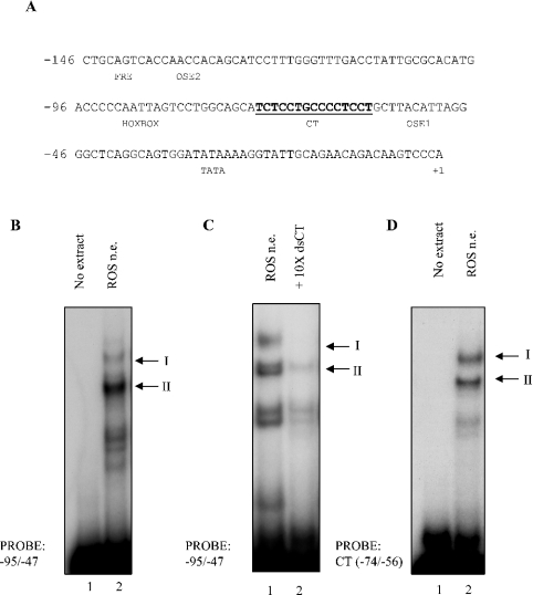
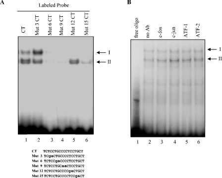
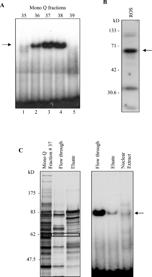
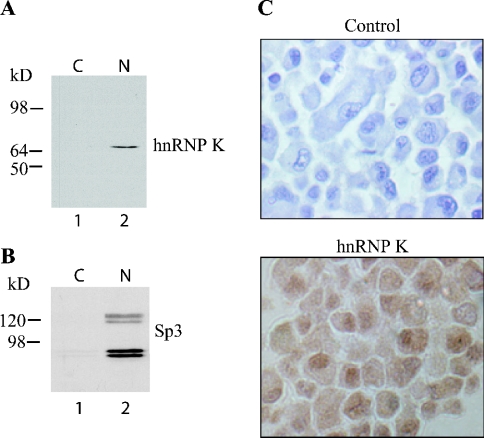
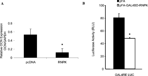
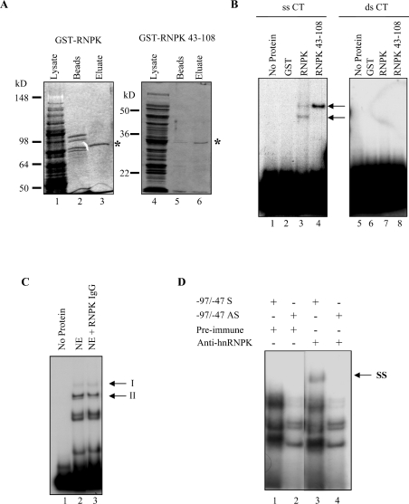
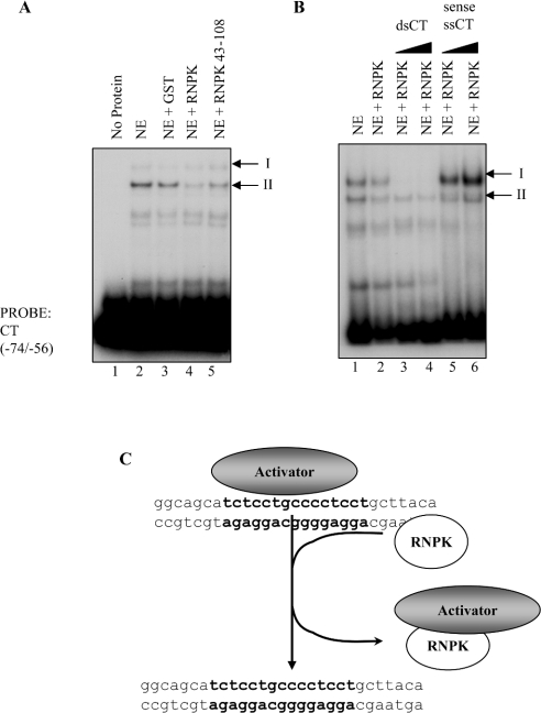
HnRNP K (heterogeneous nuclear ribonucleoprotein K) was biochemically purified from a screen of proteins co-purifying with binding activity to the osteocalcin promoter. We identify hnRNP K as a novel repressor of osteocalcin gene transcription. Overexpression of hnRNP K lowers the expression of osteocalcin mRNA by 5-fold. Furthermore, luciferase reporter assays demonstrate that overexpression of hnRNP K represses osteocalcin transcription from a CT (cytosine/thymidine)-rich element in the proximal promoter. Electrophoretic mobility-shift analysis reveals that recombinant hnRNP K binds to the CT-rich element, but binds ss (single-stranded), rather than ds (double-stranded) oligonucleotide probes. Accordingly, hnRNP K antibody can supershift a binding activity present in nuclear extracts using ss sense, but not antisense or ds oligonucleotides corresponding to the CT-rich -95 to -47 osteocalcin promoter. Importantly, addition of recombinant hnRNP K to ROS 17/2.8 nuclear extract disrupts formation of a DNA-protein complex on ds CT element oligonucleotides. This action is mutually exclusive with hnRNP K's ability to bind ss DNA. These results demonstrate that hnRNPK, although co-purified with a dsDNA-binding activity, does not itself bind dsDNA. Rather, hnRNP K represses osteocalcin gene transcription by inhibiting the formation of a transcriptional complex on the CT element of the osteocalcin promoter.


Figures

Figure 1
Figure 1. Identification of binding activities on a CT-rich stretch on the −95 to −47 osteocalcin promoter
(A) The −146 to +1 rat osteocalcin proximal promoter region is shown with the CT-rich DNA element shown in bold. Known transcription factor binding sites in the proximal promoter are indicated. Numbering is relative to the transcriptional start site. (B) EMSA was performed using ds −95 to −47 oligonucleotides incubated with (lane 2) or without (lane 1) nuclear extract (n.e.). Two binding activities were reproducibly observed and are denoted complex I and complex II, as indicated. (C) EMSA was performed with ds −95 to −47 osteocalcin probes in the presence (lane 2) or absence (lane 1) of a 10-fold molar excess of unlabelled, ds CT (−74 to −56) oligonucleotides. (D) EMSA was performed using the CT-rich −74 to −56 region of the osteocalcin promoter, incubated with (lane 2) or without (lane 1) nuclear extract.
Figure 2
Figure 2. Identification of the core binding cognate, but not the factor in the fast migrating complex
(A) EMSA was performed using radiolabelled CT and mutated CT oligonucleotides (lanes 1–6). The identity of the various mutated CT oligonucleotides used as probes is listed below. Mutated bases are in lower case letters. (B) EMSA was performed using radiolabelled ds −95 to −47 oligonucleotides incubated with (lanes 2–6) or without (lane 1) nuclear extract. Extracts were incubated with anti-c-fos (lane 3), c-jun (lane 4), ATF-1 (lane 5) and ATF-2 (lane 6) antibodies.
Figure 3
Figure 3. Elution profile of − 95 to − 47 binding activities and protein fractionation at various purification steps
(A) EMSA of binding activities recovered from a NaCl gradient Mono Q FPLC fractionation, using the −95 to−47 region of the osteocalcin promoter as a probe. (B) Southwestern hybridizations were performed using concatamerized −95 to −47 oligonucleotide probes. (C) Left panel: silver stain of eluate from fraction 37 MonoQ column, flow through and eluate after further purification through a CM-Sepharose column. Black rectangle indicates the ∼65 kDa band submitted for microsequencing. Right panel: EMSA, using the flow-through and eluate fractions from the CM-Sepharose column and the starting nuclear extract of ROS 17/2.8 cells, were performed using the −95 to −47 probe.
Figure 4
Figure 4. HnRNP K is expressed in osteoblastic cells
(A) Cytosolic and nuclear extracts from ROS 17/2.8 cells were immunoblotted and probed with an anti-hnRNP K antibody, revealing expression primarily in the nucleus. (B) Immunoblotting with Sp3 reveals the specificity of the nuclear fraction. (C) Immunocytochemistry of ROS 17/2.8. Note that ROS17/2.8 cells show immunoreactivity (brown stain) for hnRNP K, which is more abundant in the nucleus than in the cytosol. In control cells, the primary antibody was omitted (×200 magnification).
Figure 5
Figure 5. HnRNP K inhibits transcription of the osteocalcin gene
(A) ROS17/2.8 cells were transiently transfected with RNPK-pcDNA3 or empty vector (pcDNA3). Total RNA was isolated from the cells 72 h post-transfection, and reverse transcribed. The cDNA was subjected to real-time PCR using osteocalcin-specific primers. Relative expression levels were determined by normalizing to GAPDH. Results (means±S.D.) are from a representative experiment performed in triplicate. Each experiment was repeated at least three times. *P<0.05 compared with control. (B) The function of hnRNP K was examined using a one-hybrid assay. Briefly, MC3T3-E1 cells were transiently co-transfected with GAL4RE-LUC (a GAL4 response element containing luciferase reporter) plasmid and GAL4BD–hnRNP K fusion protein expression plasmid (GAL4BD–RNPK). As a control, empty plasmid was used in a parallel experiment. Cells were analysed for luciferase activity 72 h post-transfection. Representative data (means±S.D.) are from an experiment performed in triplicate. Each experiment was repeated at least three times. *P<0.05 compared with control.
Figure 6
Figure 6. Overexpression of hnRNP K decreases the transcriptional activity of the osteocalcin promoter through a CT-rich element
(A) ROS17/2.8 cells were transiently co-transfected with CT-RSVLUC, mtCT-RSVLUC or ‘empty’ RSVLUC heterologous promoter reporter and pcDNA3 or mycRNPK-pcDNA3. Cells were analysed for luciferase activity 72 h post-transfection. Inset, overexpression of hnRNP K in the transfected cells was confirmed by immunoblotting using anti-hnRNP K and anti-myc antibodies. GAPDH antibodies were used as a control for loading. (B) ROS17/2.8 cells were transiently co-transfected with −92 OCLUC, −92ΔCT OCLUC homologous promoter reporter or ‘promoterless’ pGL2 basic and pcDNA3 or pcDNA3-mycRNPK. Expression of hnRNP K reduces reporter expression from the −92 osteocalcin promoter (−92 OCLUC). All data (means±S.D.) are from a representative experiment performed in triplicate. Each experiment was repeated at least three times. *P<0.05 compared with control.
Figure 7
Figure 7. HnRNP K binds ss sense but not ds osteocalcin CT element
(A) Coomassie Blue-stained SDS/PAGE of bacterially expressed GST-RNPK (left panel) and GST-RNPK 43–108 (right panel). A predominant approx. 90 kDa band corresponding to the predicted molecular mass of a GST-hnRNPK fusion protein is observed in the eluted fraction (lane 3, *). An approx. 33 kDa band corresponding to the predicted molecular mass of a GST-truncated hnRNPK fusion protein is observed in the eluted fraction (lane 6, *). Lanes 1 and 4, induced bacterial cell extract; lanes 2 and 5, glutathione–Sepharose bead fraction. Lanes 3 and 6, eluate fractions. (B) EMSA was performed using ss sense (lanes 1–4) or ds (lanes 5–8) CT element oligonucleotides. Bacterially expressed GST fusion proteins were analysed for DNA-binding activity. Lanes 1 and 5, no protein; lanes 2 and 6, 200 ng GST; lanes 3 and 7, 200 ng GST-RNPK; lanes 4 and 8, 200 ng GST-RNPK amino acids 43–108. Arrow denotes band of interest. (C) EMSA was performed using ds CT oligonucleotides (lanes 2 and 3). Anti-hnRNP K antibodies were co-incubated with nuclear extract (lane 3) to detect hnRNP K binding by supershift. Lane 1, no extract; NE, nuclear extract. (D) EMSA was performed using ss sense (lanes 1 and 3) or antisense (lanes 2 and 4) −95 to −47 oligonucleotides. Incubation of nuclear extract with an anti-hnRNP K antibody produced a supershift (SS) of binding using sense (lane 3), but not antisense (lane 4) oligonucleotides. Band migration pattern is compared with nuclear extract incubated with preimmune serum (lanes 1 and 2).
Figure 8
Figure 8. HnRNP K disrupts DNA-binding activity on the CT element without directly binding DNA
(A) EMSA was performed using ds CT element oligonucleotides. Crude nuclear extract was analysed for DNA-binding activity in the presence or absence of 200 ng recombinant GST, RNPK or RNPK amino acids 43–108. The binding activity of complex II is markedly diminished by the addition of recombinant RNPK (lane 4) or RNPK amino acids 43–108 (lane 5) to the nuclear extract, but not by GST (lane 3). Lane 1, contains no extract; lane 2, contains nuclear extract alone. (B) EMSAs were performed using radiolabelled ds CT element oligonucleotides. Lane 1, contains ROS17/2.8 nuclear extract. Incubation of nuclear extract with recombinant GST-RNPK disrupts formation of the faster migrating complex II (lane 2 versus lane 1). Preincubation of hnRNP K with 50- or 100-fold unlabelled ds CT oligonucleotides had greatly reduced the DNA-binding activities of complexes I and II, as expected (lanes 3 and 4). However, when GST-RNPK was preincubated with 50- or 100-fold unlabelled ss sense CT oligonucleotides (lanes 5 and 6 respectively), the ability of hnRNP K to disrupt complex II was attenuated. (C) HnRNP K can disrupt the formation of a protein–DNA complex on the osteocalcin CT element without directly binding to the DNA. In fact, the interaction between hnRNP K and ss DNA appears to be mutually exclusive to its ability to compete off a protein–DNA complex from the osteocalcin promoter. We hypothesize that the resulting repression of osteocalcin transcription caused by this interaction is probably due to the subtraction of an activator from the osteocalcin promoter.

Similar articles

Cited by

References

    1. Romberg R. W., Werness P. G., Riggs B. L., Mann K. G. Inhibition of hydroxyapatite crystal growth by bone-specific and other calcium-binding proteins. Biochemistry. 1986;25:1176–1180. - PubMed
    1. Hoang Q. Q., Sicheri F., Howard A. J., Yang D. S. Bone recognition mechanism of porcine osteocalcin from crystal structure. Nature (London) 2003;425:977–980. - PubMed
    1. Ducy P., Karsenty G. Two distinct osteoblast-specific cis-acting elements control expression of a mouse osteocalcin gene. Mol. Cell. Biol. 1995;15:1858–1869. - PMC - PubMed
    1. Ducy P., Zhang R., Geoffroy V., Ridall A. L., Karsenty G. Osf2/Cbfa1: a transcriptional activator of osteoblast differentiation. Cell (Cambridge, Mass.) 1997;89:747–754. - PubMed
    1. Towler D. A., Rutledge S.-J. C., Rodan G. A. Msx-2/Hox 8.1: a transcriptional regulator of the rat osteocalcin promoter. Mol. Endocrinol. 1994;8:1484–1493. - PubMed

Publication types

MeSH terms