

The neuropeptide pigment-dispersing factor coordinates pacemaker interactions in the Drosophila circadian system
- PMID: 15356209
- PMCID: PMC6729918
- DOI: 10.1523/JNEUROSCI.2370-04.2004
The neuropeptide pigment-dispersing factor coordinates pacemaker interactions in the Drosophila circadian system
Abstract
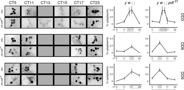
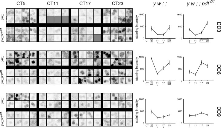
In Drosophila, the neuropeptide pigment-dispersing factor (PDF) is required to maintain behavioral rhythms under constant conditions. To understand how PDF exerts its influence, we performed time-series immunostainings for the PERIOD protein in normal and pdf mutant flies over 9 d of constant conditions. Without pdf, pacemaker neurons that normally express PDF maintained two markers of rhythms: that of PERIOD nuclear translocation and its protein staining intensity. As a group, however, they displayed a gradual dispersion in their phasing of nuclear translocation. A separate group of non-PDF circadian pacemakers also maintained PERIOD nuclear translocation rhythms without pdf but exhibited altered phase and amplitude of PERIOD staining intensity. Therefore, pdf is not required to maintain circadian protein oscillations under constant conditions; however, it is required to coordinate the phase and amplitude of such rhythms among the diverse pacemakers. These observations begin to outline the hierarchy of circadian pacemaker circuitry in the Drosophila brain.
Figures


Similar articles
-
The neuropeptide PDF acts directly on evening pacemaker neurons to regulate multiple features of circadian behavior.PLoS Biol. 2009 Jul;7(7):e1000154. doi: 10.1371/journal.pbio.1000154. Epub 2009 Jul 21. PLoS Biol. 2009. PMID: 19621061 Free PMC article.
-
A subset of dorsal neurons modulates circadian behavior and light responses in Drosophila.Neuron. 2007 Mar 1;53(5):689-701. doi: 10.1016/j.neuron.2007.01.034. Neuron. 2007. PMID: 17329209 Free PMC article.
-
Electrical silencing of PDF neurons advances the phase of non-PDF clock neurons in Drosophila.J Biol Rhythms. 2008 Apr;23(2):117-28. doi: 10.1177/0748730407312984. J Biol Rhythms. 2008. PMID: 18375861
-
Circadian clock genes in Drosophila: recent developments.Indian J Exp Biol. 2003 Aug;41(8):797-804. Indian J Exp Biol. 2003. PMID: 15248475 Review.
-
Circadian pathway: the other shoe drops.Curr Biol. 2005 Dec 20;15(24):R987-9. doi: 10.1016/j.cub.2005.11.053. Curr Biol. 2005. PMID: 16360675 Review.
Cited by
-
Daily rhythms in locomotor circuits in Drosophila involve PDF.J Neurophysiol. 2013 Aug;110(3):700-8. doi: 10.1152/jn.00126.2013. Epub 2013 May 15. J Neurophysiol. 2013. PMID: 23678016 Free PMC article.
-
UBR4/POE facilitates secretory trafficking to maintain circadian clock synchrony.Nat Commun. 2022 Mar 24;13(1):1594. doi: 10.1038/s41467-022-29244-1. Nat Commun. 2022. PMID: 35332162 Free PMC article.
-
DN1(p) circadian neurons coordinate acute light and PDF inputs to produce robust daily behavior in Drosophila.Curr Biol. 2010 Apr 13;20(7):591-9. doi: 10.1016/j.cub.2010.02.056. Epub 2010 Apr 1. Curr Biol. 2010. PMID: 20362452 Free PMC article.
-
Pigment Dispersing Factor Is a Circadian Clock Output and Regulates Photoperiodic Response in the Linden Bug, Pyrrhocoris apterus.Front Physiol. 2022 Apr 29;13:884909. doi: 10.3389/fphys.2022.884909. eCollection 2022. Front Physiol. 2022. PMID: 35574487 Free PMC article.
-
Drosophila pacemaker neurons require g protein signaling and GABAergic inputs to generate twenty-four hour behavioral rhythms.Neuron. 2010 Dec 9;68(5):964-77. doi: 10.1016/j.neuron.2010.11.017. Neuron. 2010. PMID: 21145008 Free PMC article.
References
-
- Akten B, Jauch E, Genova GK, Kim EY, Edery I, Raabe T, Jackson FR (2003) A role for CK2 in the Drosophila circadian oscillator. Nat Neurosci 6: 251-257. - PubMed
-
- Blanchardon E, Grima B, Klarsfeld A, Chelot E, Hardin PE, Preat T, Rouyer F (2001) Defining the role of Drosophila lateral neurons in the control of circadian rhythms in motor activity and eclosion by targeted genetic ablation and PERIOD protein overexpression. Eur J Neurosci 13: 871-888. - PubMed
-
- Colwell CS, Michel S, Itri J, Rodriguez W, Tam J, Lelievre V, Hu Z, Liu X, Waschek JA (2003) Disrupted circadian rhythms in VIP- and PHI-deficient mice. Am J Physiol Regul Integr Comp Physiol 285: 939-949. - PubMed
Publication types
MeSH terms
Substances
Grants and funding
LinkOut - more resources
Full Text Sources
Molecular Biology Databases
Research Materials