. 2004;3(4):15.
doi: 10.1186/jbiol10. Epub 2004 Aug 23.

The phosphatidylserine receptor has essential functions during embryogenesis but not in apoptotic cell removal

Affiliations

The phosphatidylserine receptor has essential functions during embryogenesis but not in apoptotic cell removal

Jens Böse et al. J Biol. 2004.

Abstract

Background: Phagocytosis of apoptotic cells is fundamental to animal development, immune function and cellular homeostasis. The phosphatidylserine receptor (Ptdsr) on phagocytes has been implicated in the recognition and engulfment of apoptotic cells and in anti-inflammatory signaling. To determine the biological function of the phosphatidylserine receptor in vivo, we inactivated the Ptdsr gene in the mouse.

Results: Ablation of Ptdsr function in mice causes perinatal lethality, growth retardation and a delay in terminal differentiation of the kidney, intestine, liver and lungs during embryogenesis. Moreover, eye development can be severely disturbed, ranging from defects in retinal differentiation to complete unilateral or bilateral absence of eyes. Ptdsr -/- mice with anophthalmia develop novel lesions, with induction of ectopic retinal-pigmented epithelium in nasal cavities. A comprehensive investigation of apoptotic cell clearance in vivo and in vitro demonstrated that engulfment of apoptotic cells was normal in Ptdsr knockout mice, but Ptdsr-deficient macrophages were impaired in pro- and anti-inflammatory cytokine signaling after stimulation with apoptotic cells or with lipopolysaccharide.

Conclusion: Ptdsr is essential for the development and differentiation of multiple organs during embryogenesis but not for apoptotic cell removal. Ptdsr may thus have a novel, unexpected developmental function as an important differentiation-promoting gene. Moreover, Ptdsr is not required for apoptotic cell clearance by macrophages but seems to be necessary for the regulation of macrophage cytokine responses. These results clearly contradict the current view that the phosphatidylserine receptor primarily functions in apoptotic cell clearance.


Figures

Figure 1
Figure 1
Targeted inactivation of the phosphatidylserine receptor gene. (a) Ptdsr gene-targeting strategy. Homologous recombination in ES cells results in the deletion of exons I and II of the murine Ptdsr gene through replacement of a loxP-flanked neomycin phosphotransferase gene (neo), thereby ablating the reading frame of the encoded protein. Coding exons I-VI are shown as filled boxes, and deleted exons are colored green. Restriction sites are: A, AatII; B, BamHI; EI, EcoRI; EV, EcoRV; K, KpnI; R, RsrII; S, SacII; Sc, ScaI, X, XhoI. The probe sites are red boxes labeled: C, 5' outside probe; D, 3' outside probe. (b) Southern blot analysis of genomic DNA extracted from wild-type (+/+) and Ptdsr+/- (+/-) animals, digested with BamHI and hybridized with the 5' outside probe to confirm germ-line transmission of the mutant Ptdsr allele. 'Wild-type' indicates the BamHI fragment of 17.2 kb from the wild-type Ptdsr allele; 'mutant' indicates the BamHI fragment of 11.6 kb from the targeted Ptdsr allele. (c) PCR genotyping of embryos and animals from intercrosses of heterozygous Ptdsr+/- using a wild-type and a mutant allele-specific primer combination, respectively. (d) Northern blot analysis of total RNA isolated from E13.5 wild-type, Ptdsr+/- and Ptdsr -/- embryos. (e) Western blot analysis of protein from homogenates of E13.5 wild-type, Ptdsr+/- and Ptdsr -/- embryos using a Ptdsr-specific antibody. Developmental abnormalities at (f,g) E15.5 and (h) birth; in this and all subsequent figures wild-type littermates are located on the left and homozygous mutant mice on the right. The Ptdsr -/- embryos show exencephaly (f) or prosencephalic hernia in the forebrain region (arrowhead, neonate 2; h), uni-or bilateral absence of the eyes (f,g and neonate 2 in h, and arrow, neonate 3 in h), an abnormal head shape with proboscis (g), edema (arrowheads in f and g), and general anemia (asterisk, neonate 3 in h).
Figure 2
Figure 2
Expression analysis of Ptdsr during embryonic development. (a) Schematic representation of the construction of the Ptdsr gene-trap mouse line used for expression analysis at different embryonic stages. Gray and bright blue boxes represent regulatory elements of the gene-trap, and β-geo, the β-galactosidase/neomycin phosphotransferase fusion protein-expression cassette [48,51]. Restriction enzyme nomenclature is as in Figure 1 (b) Whole-mount β-galactosidase staining of heterozygous Ptdsr gene-trap embryos at mid-gestation. Expression of Ptdsr is highest in neural tissues and somites, in the branchial arches, the developing limbs, the heart, the primitive gut and the developing eye. (c-e) Sectioning of E12.5 β-galactosidase-stained embryos confirms expression of Ptdsr in (c) the neural tube; (inset in c) neural epithelium; (d) somites; and (e) eyes. Expression in the eye is restricted to developing neural retinal and lens cells. (f) Expression analysis of adult tissues by northern blot. Expression of Ptdsr in the muscle (asterisk) was detected only on long-term exposures of the filter (> 48 h). A β-actin hybridization was used to confirm equal loading of RNA samples. Scale bar, 100 μm.
Figure 3
Figure 3
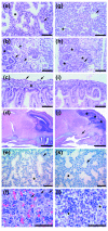
Histological analysis of wild-type and Ptdsr -/-organs during embryogenesis. (a-f) Wild-type embryos and (g-l) Ptdsr -/- littermates were isolated at various embryonic stages, serially sectioned sagittally and analyzed for developmental abnormalities in detail after H&E staining. At E16.5, the lungs of (g) Ptdsr -/- embryos had sacculation just starting, and well-formed alveoli (asterisks) or epithelium-lined bronchioles (arrows) were scarce compared to (a) wild-type lungs. At E16.5, the glomeruli (arrows) in the kidney of (h) Ptdsr -/- embryos were underdeveloped compared to (b) wild-type, collecting tubules (arrowheads) were missing and undifferentiated blastemas (asterisks) were more abundant. The jejunum had no intramural ganglia in Ptdsr -/-embryos (i; and arrows in c); and a well-developed submucosa (asterisk in c) was missing. Brain sections at E18.5 show that (j) Ptdsr -/-embryos may have herniation (arrow) of the hypothalamus through the ventral skull (secondary palate), most likely through Rathke's pouch, and a severe malformation of the cortex (asterisks) compared to (d) wild-type embryos. At E18.5, (e) wild-type and (k) Ptdsr -/- lungs showed normal sacculation and formation of alveoli (asterisks) and bronchioles (arrow). (f) Wild-type neonatal liver had significant numbers of megakaryocytes (arrows), compared to (l) homozygous mutant littermates, and higher numbers of erythropoietic islands and of mature erythrocytes. Hepatocellular vacuoles are due to glycogen stores (asterisks) that were not metabolized in perinatally dying Ptdsr -/- animals, in contrast to wild-type newborns. Scale bar, 100 μm, except for (d) and (j), 1 mm.
Figure 4
Figure 4
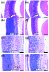
Morphology of wild-type and Ptdsr -/- retinas. Serial sagittal sections of (a-d) wild-type and (e-h) Ptdsr -/- retina were analyzed for developmental abnormalities at (a,e) E12.5, (b,f) E16.5, (c,g) E18.5, and (d,h) P0. Normal patterning of the retina was observed in Ptdsr -/-embryos, with an outer granular layer (OGL), outer plexiform layer (OPL), inner granular layer (IGL) and inner plexiform layer (IPL). Note that the IGL in Ptdsr -/- retinas is less thick than that in wild-type littermates in comparing (c,g) and (d,h). Morphometric analysis (numbered lines) of wild-type and Ptdsr -/- retinas confirmed the initial finding of a thinner retina in Ptdsr -/- animals than in wild-type (all values in μm). Scale bar, 50 μm.
Figure 5
Figure 5
Histological analysis of eye development in severely affected eyeless Ptdsr -/- embryos. (a) In anophthalmic Ptdsr -/- embryos, unilateral or bilateral absence of the eyes could be detected. (b-d) Serial H&E-stained sagittal sections of homozygous mutant embryos at (b) E17.5 and (c,d) E18.5 show complex malformation of the optic cup and lack of any lens structure. Careful examination of adjacent sections (b-d) reveals an ectopic misplacement of retinal-pigmented epithelium in the maxillary sinus. Not only is the deposition of pigment clearly visible (higher magnification insets) but also the induction of proliferation of underlying tissues and the change in morphology of the maxillary sinus (d). Scale bar, 100 μm in (b-d).
Figure 6
Figure 6
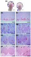
Analysis of programmed cell death and involvement of macrophages in the removal of apoptotic cells in wild-type and Ptdsr -/-embryos. (a) Whole-mount TUNEL staining (blue) of limb buds from wild-type and Ptdsr -/- embryos at E13.5 show no differences in the amount or localization of apoptotic cells during the beginning regression of the interdigital web. Serial sagittal sections stained for activated caspase 3 (aCasp3; red) in (b-d) wild-type and (f-h) Ptdsr -/- embryos at E12.5 show apoptotic cells in the neural tube (b,f), the mesonephros (c,g) and the developing paravertebral ganglia (d,h). Tissue distribution and total number of apoptotic cells was indistinguishable between genotypes and was confirmed by the comparison of consecutive sections of wild-type and Ptdsr -/-embryos from different developmental stages. Analysis of macrophage numbers and location by F4/80 staining (brown) of consecutive sections in paravertebral ganglia of (e) wild-type and (i) homozygous mutant embryos revealed that macrophages (arrows) are not located close to apoptotic cells during embryonic development. (For comparison, see also Additional data file 1, Figure S1, with the online version of this article). Scale bar, 100 μm.
Figure 7
Figure 7
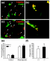
Phagocytosis of apoptotic cells by fetal liver-derived macrophages (FLDMs). FLDMs from (a,b) wild-type and (c,d) Ptdsr -/- embryos were cultured for 60 min with TAMRA-stained (red) apoptotic thymocytes (treated with staurosporine) from C57BL/6J mice and then stained with F4/80 (green). Macrophages of both genotypes have phagocytosed apoptotic cells (arrowheads). (e) Quantification of phagocytosis of apoptotic cells by wild-type or Ptdsr -/-macrophages revealed no differences in the percentage of macrophages that had engulfed apoptotic cells, whether or not apoptosis had been induced by staurosporine. Microscopic analysis (b,d) and quantification of the number of apoptotic cells phagocytosed by single macrophages and (f) calculation of the average number of cells phagocytosed per macrophage failed to reveal differences in the efficacy of removal of apoptotic cells between wild-type and Ptdsr -/- FLDMs.
Figure 8
Figure 8
Cytokine production by FLDMs upon stimulation with lipopolysaccharide (LPS) and apoptotic cells. FLDMs from wild-type and Ptdsr -/- embryos were incubated (a,b,d) with medium (0), LPS (10 ng/ml), apoptotic cells (ratio 1:10) or in combination with LPS and apoptotic cells or (c) with LPS (100 ng/ml) alone. Culture supernatants were harvested after 22 h (a,b,d) or at the indicated time points (c). TNF-α and TGF-β1 were quantified by ELISA and IL-10 by cytometric bead array (CBA) assay. Data are presented as mean ± SEM from at least three independent experiments, each carried out in triplicate. *, significant difference between genotypes, p < 0.05; **, significant difference between genotypes, p < 0.01; Wilcoxon-signed rank test.

Similar articles

Cited by

References

    1. Jacobson MD, Weil M, Raff MC. Programmed cell death in animal development. Cell. 1997;88:347–354. doi: 10.1016/S0092-8674(00)81873-5. - DOI - PubMed
    1. Baehrecke EH. How death shapes life during development. Nat Rev Mol Cell Biol. 2002;3:779–787. doi: 10.1038/nrm931. - DOI - PubMed
    1. Vaux DL, Korsmeyer SJ. Cell death in development. Cell. 1999;96:245–254. doi: 10.1016/S0092-8674(00)80564-4. - DOI - PubMed
    1. Savill J, Fadok V. Corpse clearance defines the meaning of cell death. Nature. 2000;407:784–788. doi: 10.1038/35037722. - DOI - PubMed
    1. Lauber K, Blumenthal SG, Waibel M, Wesselborg S. Clearance of apoptotic cells: getting rid of the corpses. Mol Cell. 2004;14:277–287. doi: 10.1016/S1097-2765(04)00237-0. - DOI - PubMed

Publication types