. 2004 Sep 1;24(35):7632-9.
doi: 10.1523/JNEUROSCI.2123-04.2004.

Severe defects in dorsal thalamic development in low-density lipoprotein receptor-related protein-6 mutants

Affiliations

Severe defects in dorsal thalamic development in low-density lipoprotein receptor-related protein-6 mutants

Cheng-Ji Zhou et al. J Neurosci. .

Abstract

Mice with mutations in the Wnt coreceptor low-density lipoprotein receptor-related protein-6 (LRP6) have a smaller and severely disorganized dorsal thalamus and lack thalamocortical projections. Using molecular markers, we showed that most dorsal thalamic and epithalamic neurons were missing, and most of the major dorsal thalamic nuclei were not identifiable. However, the ventral thalamus was essentially unaffected, although the dorsal thalamic defect leads to rostral displacement of portions of the ventral thalamus. Analysis of younger embryos showed that epithalamic and dorsal thalamic neurons were not produced at early stages of development, whereas ventral thalamic neurons were still produced. These defects were accompanied by improper formation of the boundary between dorsal and ventral thalamus, the zona limitans interthalamica (ZLI). Furthermore, the expression of an early marker of posterior forebrain development that marks the compartment from the midbrain-hindbrain junction to the ZLI (including the future dorsal thalamus, pretectum, and midbrain) was disrupted, supporting the idea that diencephalic development is abnormal from very early in embryogenesis. This study provides compelling in vivo evidence that thalamic development requires normal activity of the LRP6-mediated canonical Wnt signaling pathway.


Figures
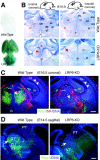
Figure 1.
Figure 1.
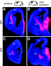
Disruption of the thalamus in LRP6 knock-out (KO) mice. A, X-gal staining (blue) in heterozygous (LRP6+/-) shows that LRP6 is ubiquitously expressed in entire developing brain at E12.5. B, Nissl staining in E18.5 coronal brain sections shows anatomic defects of thalamus in mutants. These defects are most severe at rostral levels where all lateral thalamic structures are missing, and only the medial thalamic regions remained with a disorganized ventricular zone (red arrowhead). Note the extra choroid plexus-like structure in the third ventricle (red star). At caudal levels, the major posterior dorsal thalamic nuclei are not identifiable (red arrow). C, Double immunofluorescence for Prox1 (green; a dorsal thalamic neuronal marker) and neurofilament (red) in coronal sections at E16.5 shows that most dorsal thalamic neurons (large white arrows) are missing and that there are few axonal projections to the cortical subplate (where thalamocortical axons project prominently at this age) (white arrowheads) in the mutants. In addition, the habenular recess is larger in the mutant (asterisk), and the fibers normally coursing adjacent to the habenular ventricular zone (small white arrows) are missing or misplaced in the mutant. D, Immunofluorescence of Prox1 (green) in sagittal brain sections of E14.5 mice shows a similar phenotype in the dorsal thalamus (white arrows). 3V, Third ventricle; Cx, cerebral cortex; CM, central medial thalamic nuclei; dLG, dorsal lateral geniculate nucleus; DT, dorsal thalamus; Hb, habenular nuclei; Hi, hippocampal formation; hit, habenulo-interpenduncular tract; p1, 2, 3, prosomeres 1, 2, 3; PT, pretectum; PV, paraventricular thalamic nuclei; Rt, reticular thalamic nucleus; VL, ventral lateral thalamic nuclei; vLG, ventral lateral geniculate nucleus; VM,ventral medial thalamic nuclei; VP, ventral posterior thalamic nuclei; VT, ventral thalamus; ZI, zona incerta. Scale bars, 50 μm.
Figure 2.
Figure 2.
LRP6 mutants fail to produce thalamocortical projections. A-D, E18.5 wild-type (A, B) and mutant (C, D) brains after placement of DiI crystal in the thalamus. Wild-type mice, shown at two coronal levels, have fibers from the dorsal thalamus projecting through the internal capsule to the cortex dorsally. In mutants, the fibers that leave the thalamus get stalled and terminate in the striatum. Str, Striatum; CX, cortex; DT, dorsal thalamus.
Figure 3.
Figure 3.
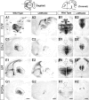
Molecular anatomy of the dorsal thalamus in LRP6 mutants. A-H, In situ hybridization of dorsal thalamic markers Tcf4 (A, B), Gbx2 (C, D), Id4 (E, F), and RORα (G, H) are used to examine the details of the thalamic defects in sagittal and coronal sections at middle-late gestation. The black dashed line area (A-H) indicates the entire dorsal thalamic region as defined by the expression of Tcf4 in adjacent sections. The arrows (A2, C2, E2, G2, H2) are used to show the location of residual staining for each marker in the small remaining dorsal thalamic structure. In LRP6 knock-out (KO) mice, the anterior, ventral, and lateral regions of the dorsal thalamus are most affected. RORα is a marker for thalamic sensory relay neurons (25) (G, H), and these neurons are especially severely affected in the mutant brains. Cx, Cerebral cortex; DT, dorsal thalamus; Hi, hippocampus; hit, habenulo-interpenduncular tract; Hy, hypothalamus; imt, intermediate thalamic neuroepithelium; ldn, laterodorsal nuclei; lhn, lateral habenular nucleus; p1, 2, 3, prosomere 1, 2, 3; Pa, pallidal ganglion; PT, pretectum; SE, septal ganglion; Ta, anterior thalamic region; Te, epithalamus; Ti, internal thalamic region; vln, ventral lateral nuclei; VT, ventral thalamus. Scale bars, 100 μm.
Figure 4.
Figure 4.
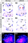
Analysis of ventral thalamic markers in middle-late gestation. A1-B2, Reelin expression in coronal brain sections (E15.5) by in situ hybridization shows that the caudal ventral thalamus appears quite normal (B1, B2), whereas the rostral ventral thalamus may have been partially displaced by the absence of the dorsal thalamus rostrally (A2, red stars). Note that the reelin expression pattern in other regions, including pretectum and hypothalamus of the wild type and mutants, is also comparable. C1, C2, Immunofluorescence of GAD65 (red) in E17.5 sagittal brain sections (with a DNA staining in blue) shows that the GAD65-immunopositive ventral thalamusis largely intact, where as the rostral portion of the ventral thalamusis shifted dorsally in the mutants. This is consistent with the reelin expression in the mutant rostral ventral thalamus (A2). Note that the anatomical structure of the tectum (dorsal midbrain) appears normal in the mutants. The white dashed line area indicates the normal and defective dorsal thalamus (C1, C2). 3V, Third ventricle; Cb, cerebellum; Cx, cerebral cortex; DT, dorsal thalamus; Hi, hippocampus; Hy, hypothalamus; Pa, pallidal ganglion; PT, pretectum; SE, septal ganglion; Te, epithalamus; VT, ventral thalamus. Scale bars, 100 μm.
Figure 5.
Figure 5.
Analysis of regional thalamic neuronal cell fate early in thalamic development. The top panels are coronal sections of normal thalamus at E12 stained withβ4, Gbx2, Netrin, and Dlx2 that mark differentiated neurons of the epithalamus (Te), dorsal thalamus (DT), ZLI, and ventral thalamus (VT) from dorsal to ventral, respectively. In the bottom panels, no β4 (in Te), Gbx2 (in DT), or Netrin (in ZLI) are expressed in the LRP6-knock-out (KO) thalamus. However, the expression of ventral neuronal marker Dlx (Dlx2 by in situ hybridization and Dll by immunofluorescence) is slightly displaced into what should be the dorsal thalamic region (see schematic panel at bottom right). Scale bars, 50 μm.
Figure 6.
Figure 6.
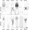
Progenitor zones and compartmentalization of the thalamus. Tcf4 and Wnt3 mark the ventricular zone of prosomere 2 (p2), including DT and Te, Shh and Wnt5a mark the p2/p3 boundary, and SFRP2 and Wnt7b mark the ventricular zone of p3 (VT) and eminentia thalami (EMT). In LRP6 knock-out (KO) mutant brains, only weak expression of Tcf4 and Wnt3 is seen and no expression of Shh or Wnt5a. However, the ventral thalamic ventricular zone markers are intact. It is difficult to be sure whether the ventral thalamic ventricular zone is truly expanded or somewhat displaced rostrally because of the smaller size of the rostral dorsal thalamus. Scale bars, 50 μm.
Figure 7.
Figure 7.
Early diencephalic defects in LRP6 mutants. A1-C2, Whole-mount in situ hybridization for Shh and Gbx2 in E10.5 and E11.5 brains. In E11.5 wild-type brains, Shh extends from the ventral floorplate in two spurs that project to the roofplate, whereas in mutant brains, this domain of expression stops less than midway (dashed arrow line). Gbx2 expression is visible weakly at E10.5 and much more strongly by E11.5 in wild-type mice but is essentially absent in mutant brains at these time points. Note that the telencephalon was dissected free to allow ease of visualization of the diencephalon. D1, D2, Whole-mount in situ hybridization of Irx3 in E9.5 embryos shows a defect in the caudal dorsal diencephalic domain in the mutants even before thalamic neuronal differentiation (see E10.5 in C1 for the earliest on set of Gbx2 expression). The arrow indicates the future ZLI, the boundary between dorsal and ventral thalamus. The arrowhead indicates the isthmus, the midbrain-hindbrain junction (which appears intact). The Irx3 expression territory between the ZLI and isthmus in the mutants is much smaller than in wild type. Dien, Diencephalon; DT, dorsal thalamus; is, isthmus; Mesen, mesencephalon; Meten, metencephalon; os, optic stalk; p1, 2, 3, prosomere 1, 2, 3; PT, pretectum; Telen, telencephalon; VT, ventral thalamus.

Similar articles

Cited by

References

    1. Bagri A, Gurney T, He X, Zou YR, Littman DR, Tessier-Lavigne M, Pleasure SJ (2002) The chemokine SDF1 regulates migration of dentate granule cells. Development 129: 4249-4260. - PubMed
    1. Bloch-Gallego E, Ezan F, Tessier-Lavigne M, Sotelo C (1999) Floor plate and netrin-1 are involved in the migration and survival of inferior olivary neurons. J Neurosci 19: 4407-4420. - PMC - PubMed
    1. Bramblett DE, Copeland NG, Jenkins NA, Tsai MJ (2002) BHLHB4 is a bHLH transcriptional regulator in pancreas and brain that marks the dimesencephalic boundary. Genomics 79: 402-412. - PubMed
    1. Braun MM, Etheridge A, Bernard A, Robertson CP, Roelink H (2003) Wnt signaling is required at distinct stages of development for the induction of the posterior forebrain. Development 130: 5579-5587. - PubMed
    1. Caspary T, Anderson KV (2003) Patterning cell types in the dorsal spinal cord: what the mouse mutants say. Nat Rev Neurosci 4: 289-297. - PubMed

Publication types

MeSH terms