Comparative Study
. 2004 Sep;165(3):795-805.
doi: 10.1016/S0002-9440(10)63342-1.

Calpain mediates calcium-induced activation of the erk1,2 MAPK pathway and cytoskeletal phosphorylation in neurons: relevance to Alzheimer's disease

Affiliations
Comparative Study

Calpain mediates calcium-induced activation of the erk1,2 MAPK pathway and cytoskeletal phosphorylation in neurons: relevance to Alzheimer's disease

Veeranna et al. Am J Pathol. 2004 Sep.

Abstract

Aberrant phosphorylation of the neuronal cytoskeleton is an early pathological event in Alzheimer's disease (AD), but the underlying mechanisms are unclear. Here, we demonstrate in the brains of AD patients that neurofilament hyperphosphorylation in neocortical pyramidal neurons is accompanied by activation of both Erk1,2 and calpain. Using immunochemistry, Western blot analysis, and kinase activity measurements, we show in primary hippocampal and cerebellar granule (CG) neurons that calcium influx activates calpain and Erk1,2 and increases neurofilament phosphorylation on carboxy terminal polypeptide sites known to be modulated by Erk1,2 and to be altered in AD. Blocking Erk1,2 activity either with antisense oligonucleotides to Erk1,2 mRNA sequences or by specifically inhibiting its upstream activating kinase MEK1,2 markedly reduced neurofilament phosphorylation. Calpeptin, a cell-permeable calpain inhibitor, blocked both Erk1,2 activation and neurofilament hyperphosphorylation at concentrations that inhibit calpain-mediated cleavage of brain spectrin. By contrast, inhibiting Erk1,2 with U-0126, a specific inhibitor of Mek1,2, had no appreciable effect on ionomycin-induced calpain activation. These findings demonstrate that, under conditions of calcium injury in neurons, calpains are upstream activators of Erk1,2 signaling and are likely to mediate in part the hyperphosphorylation of neurofilaments and tau seen at early stages of AD as well as the neuron survival-related functions of the MAP kinase pathway.


Figures
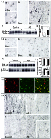
Figure 1
Figure 1
1-1: Calpains are activated in AD brains. Vibratome sections (30 μm) of control and AD prefrontal neocortices (Lamina III) were immunohistochemically stained as described in Materials and Methods using a polyclonal antibody that recognizes total m-calpain (A), and a polyclonal antibody (C-18) that recognizes the active form of m-calpain (B) . Western blot analysis was carried out on cytosolic fractions of AD and control prefrontal neocortices using a monoclonal antibody to μ-calpain. The blots were developed by alkaline phosphatase-based chemiluminescence (C). The relative densities of the bands were measured by scanning the films using O-foto software followed by quantitation using NIH image and presented in bar graph. (D; n = 10; P < 0.05. The L/H ratio reflects the relative degree of activation of calpain in AD and control brain samples and represents the ratio between the active calpain (low molecular weight band at 76 kd = L) and the inactive precursor at high molecular mass (high molecular weight band at 80 kd = H). 1-2: Erk1,2 Map kinases are activated in AD brains. Vibratome sections (30 μm) of control and AD prefrontal neocortices (Lamina III) were immunohistochemically stained as described in Materials and Methods using (A) a monoclonal antibody that recognizes total Erk1,2 and (B) a polyclonal antibody that recognizes the phosphorylated forms of Erk1,2. Western blot analysis was carried out on whole homogenates of AD and control prefrontal neocortices using polyclonal Erk1,2, and phospho-Erk1,2 antibodies (C). The blots were developed by alkaline phosphatase-based chemiluminescence. The relative densities of the bands were measured by scanning the films using O-foto software followed by quantitation using Scan Analysis program and shown in the bar graph (D; n = 5). The measurement of P44 Erk1 and P42 Erk2 bands was done separately using the same total area for different samples. 1-3: Active calpain and active Erk1,2 are co-localized in AD brain. Paraffin sections (6 μm) from prefrontal cortex were double-immunofluorescence-labeled as described in Materials and Methods. Using a polyclonal antibody (C-18) that recognizes the active form of m-calpain and a monoclonal antibody that recognizes total Erk1as primary antibodies. Alexa 564 (red) and Alexa 468 (green) conjugated secondary antibodies were used to probe active calpain (A; red) and active Erk1,2 (B; green). The sections were imaged using a confocal microscope. The overlay displaying yellow (C) illustrates co-localization of active Erk1,2 and active calpain. 1−4. Neurofilament phosphorylation is enhanced in neocortical pyramidal neurons in AD brain. Vibratome sections (30 μm) of control and AD prefrontal neocortex (Lamina III) were immunohistochemically stained as described in Materials and Methods using (A) SMI 32 monoclonal antibody that recognizes dephospho-epitopes on the neurofilament-H and NF-M tail domains; (B) SMI-31 monoclonal antibody that specifically recognizes the phosphorylated tail domains of these neurofilament subunits and strongly decorates neurites and some perikarya (inset) in AD brains.
Figure 2
Figure 2
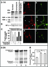
2-1: Calpeptin inhibits ionomycin-induced calpain activation and NF-M phosphorylation in primary hippocampal neurons in culture. A: Cytosol of hippocampal neurons, untreated (lane 1) or treated with ionomycin (0.5 μmol/L; lane 2) or ionomycin with calpeptin (20 μmol/L; lane 3), were subjected to 7% SDS-PAGE followed by Western blot analysis. A α-spectrin monoclonal antibody that specifically reacts with brain spectrin (fodrin) was used at 1:1000 dilution. This antibody also reacts with a 150-kd calpain-specific cleavage product of fodrin and a caspase-3-specific cleavage product of fodrin migrating at 120 kd; fodrin breakdown products (FBP 150 and FBP 120) both indicated by arrows. B: Western blot analysis, showing unaltered NF-L in hippocampal neurons under the three experimental conditions shown in Figure 2–1A. C: The bar graph presents densitometric analyses of fodrin and the 150-kd fodrin cleavage product from multiple immunoblot analyses. The values represent the mean ± SEM from three separate experiments.*, P < 0.05. D–I: Hippocampal neurons (8 DIV), untreated (D and E) or treated with 0.5 μmol/L ionomycin (F and G) or ionomycin with calpeptin (20 μmol/L; H and I), were then immunostained with a polyclonal antibody to NF-L (D, F, H) or a monoclonal antibody to phospho-NF-M (E, G, I). The arrows in F and G point to the processes that show hyperphosphorylated NF-M. 2-2: Inhibition of Erk1,2 has no effect on ionomycin-induced calpain activation in primary hippocampal neurons in culture. A: Cytosol from hippocampal neurons, untreated (lane 1) or treated with ionomycin (0.5 μmol/L; lane 2) or ionomycin with Mek1,2 inhibitor, U-0126 (10 μmol/L; lane 3), were subjected to 7% SDS-PAGE followed by Western blot analysis, using an α-spectrin monoclonal antibody that specifically reacts with brain spectrin (fodrin) and a calpain-specific cleavage product of fodrin migrating at 150 kd. B: Densitometric analysis of the 150-kd calpain cleavage product of fodrin from immunoblot analyses as represented in A. The level of the 150-kd product is expressed as a percentage of total fodrin-related immunoreactivity to correct for unavoidable minor differences in total protein loading from different hippocampal cultures. The values represent the mean ± SEM from three separate experiments.*, P < 0.05.
Figure 3
Figure 3
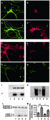
The Mek1,2 inhibitor, U-0126, calpeptin, and Erk1,2 antisense oligonucleotides attenuate Erk1,2 activation and NF-phosphorylation in hippocampal neurons. A–H: Hippocampal neurons (14 DIV) were either untreated (A and B) or treated with 0.5 μmol/L ionomycin (C and D) were immunostained with a monoclonal antibody to β tubulin (A and C) and a polyclonal antibody to phospho-Erk 1,2 (B and D). These neurons were also treated with sense (E and G) and antisense (F and H) oligonucleotides against Erk1,2 mRNA as described in Materials and Methods and were immunostained with a goat polyclonal antibody (Santa Cruz) against Erk1,2 (E and F) and with a monoclonal antibody to phospho-NF-M (G and H). I: Recombinant Erk2 (1 μg) was incubated with the immunprecipitate of Mek1,2, from lysates of ionomycin-treated hippocampal neurons, in the absence (lane 2) and presence (lane 3) of U-0126 (10 μmol/L) or calpeptin (lane 4; 20 μΜ) in a kinase reaction mixture detailed in the text, followed by SDS-PAGE and silver stain. Lane 1 represents the immunoprecipitate of Mek1,2. J: Autoradiogram of Erk2 shown in I. K: Representive image taken of a Western blot for a KSPXXXK fusion protein using the monoclonal antibody RT-97, specific for phosphorylated NF-H and NF-M. The KSPXXXK fusion protein was incubated in a kinase reaction mixture containing the immunoprecipitate of Mek1,2 and recombinant Erk2, in the absence (lane 2) and presence (lane 3) of U-0126 (10 μmol/L) or calpeptin (lane 4; 20 μΜ). Lane 1 represents the mixture of immunoprecipitate of Mek1,2 and Erk2, and lane 5 represents KSPXXXK fusion protein alone. L: Representive image taken of a Western blot for hippocampal neuron cell lysates (15 μg) in vehicle-treated (lane 1), ionomycin (0.5 μmol/L; lane 2), ionomycin and U-0126- (0.5 μmol/L and 10 μmol/L; lane 3) and calpeptin-treated (20 μmol/L; lane 4) neurons for 24 hours, that were probed for Erk1,2 and phospho-Erk1,2 expression. M: Bar graph shows the relative band intensities of p-Erk1,2. Values are expressed as mean ± SEM for three separate experiments. The ionomycin-treated neurons (lane 2) showed a significantly higher activation compared to vehicle-treated controls (lane 1; P < 0.001, n = 3) and the neurons treated with ionomycin together with calpeptin (lane 4; P < 0.05, n = 3).
Figure 4
Figure 4
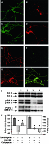
The Mek1,2 inhibitor, U-0126, and calpeptin attenuate ionomycin-induced Erk1,2 activation and NF-phosphorylation in cerebellar granule neurons. A–D: CG neurons (7 DIV) either vehicle-treated (A, B, E, F) or treated with 0.5 μmol/L ionomycin (C, D, G, H), were immunostained with antibodies against β-tubulin (A and C), p-Erk1,2 (B and D), NF-L (E and G), and phospho-NF-M (F and H). E: The CG neurons (7 DIV), either vehicle-treated (lane 1) or treated with ionomycin (0.5 μmol/L; lane 2) or ionomycin together with either U-0126 (10 μmol/L; lane 3) or calpeptin (20 μmol/L; lane 4) for 24 hours, were subjected to SDS-PAGE (equal protein loading, 15 μg) and Western blot analysis with either anti-Erk1,2, anti p-Erk1,2, or SMI 31 against phospho-NF-H and neurofilament M. F: Bar graph shows the relative band intensities of p-Erk1,2 (left) and phospho-NF-M (right). The values are the mean ± SEM of three to five separate experiments. *, P < 0.05.
Figure 5
Figure 5
Calpeptin reverses ionomycin-induced Erk1,2 activation in CG neurons but has no direct effect on Erk1,2 activity in vitro. A: CG neurons (7 DIV), were either vehicle-treated (bar 1), or treated with ionomycin (0.5 μmol/L; bar 2), or ionomycin together with calpeptin (20 μmol/L; bar 3) for 24 hours were then lysed. Erk1,2 were immuno-precipitated using anti-Erk1,2 polyclonal antibody, and kinase activity was measured using myelin basic protein as a substrate. The relative kinase activity is expressed as a percentage of the vehicle-treated control, represented as 100%. Values are means ± SEM for five separate experiments, P < 0.05. B: A time course of Erk2 kinase activity in the presence and absence of calpeptin (20 μmol/L). In vitro kinase activity was monitored using a synthetic peptide as described in Materials and Methods. Values are means ± SEM for five determinations at each time point.

Similar articles

Cited by

References

    1. Sternberger NH, Sternberger LA, Ulrich J. Aberrant neurofilament phosphorylation in Alzheimer disease. Proc Natl Acad Sci USA. 1985;82:4274–4276. - PMC - PubMed
    1. Trojanowski JQ, Walkenstein N, Lee VM. Expression of neurofilament subunits in neurons of the central and peripheral nervous system: an immunohistochemical study with monoclonal antibodies. J Neurosci. 1986;6:650–660. - PMC - PubMed
    1. Julien J. Neurofilament functions in health and disease. Curr Opin Neurobiol. 1999;9:554–560. - PubMed
    1. Lavedan C, Buchholtz S, Nussbaum R, Albin R, Polymeropoulos M. A mutation in the human neurofilament M gene in Parkinson’s disease that suggests a role for the cytoskeleton in neuronal degeneration. Neurosci Lett. 2002;322:57–61. - PubMed
    1. Al-Chalabi A, Miller C. Neurofilaments and neurological disease. Bioessays. 2003;25:346–355. - PubMed

Publication types

MeSH terms