. 2004 Aug 18;24(33):7366-77.
doi: 10.1523/JNEUROSCI.1739-04.2004.

BDNF regulates the translation of a select group of mRNAs by a mammalian target of rapamycin-phosphatidylinositol 3-kinase-dependent pathway during neuronal development

Affiliations

BDNF regulates the translation of a select group of mRNAs by a mammalian target of rapamycin-phosphatidylinositol 3-kinase-dependent pathway during neuronal development

Gerhard M Schratt et al. J Neurosci. .

Abstract

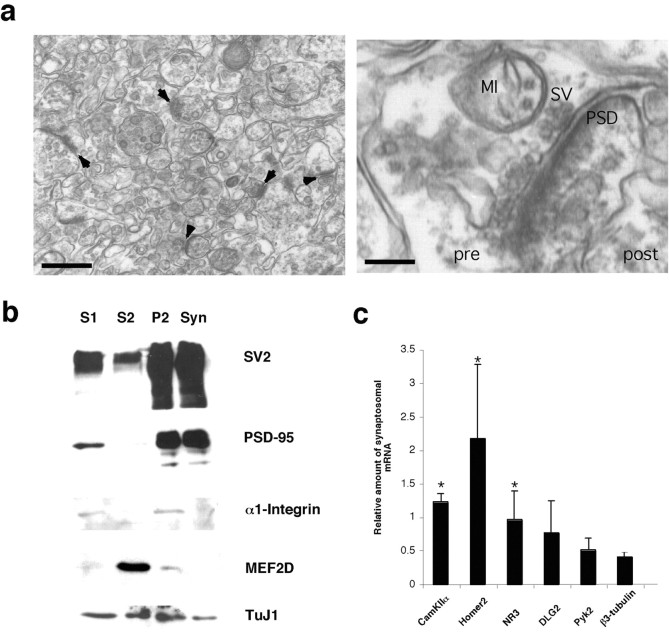
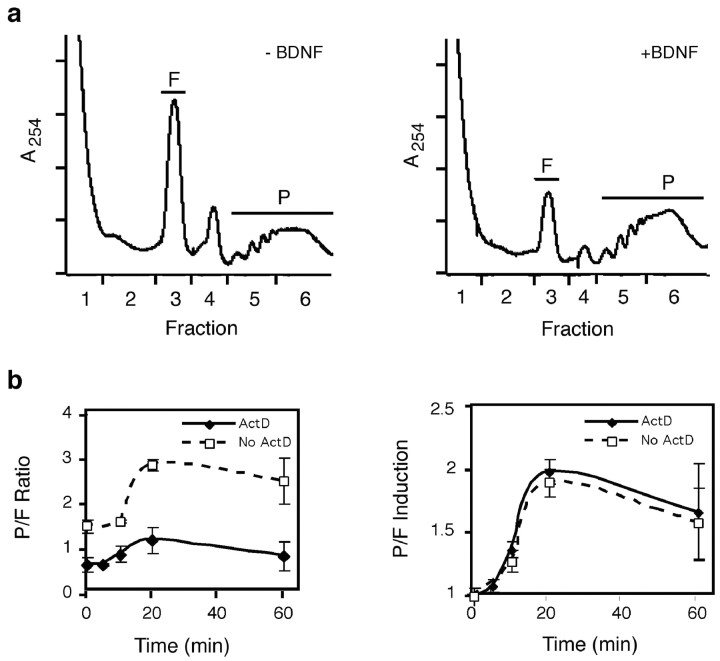
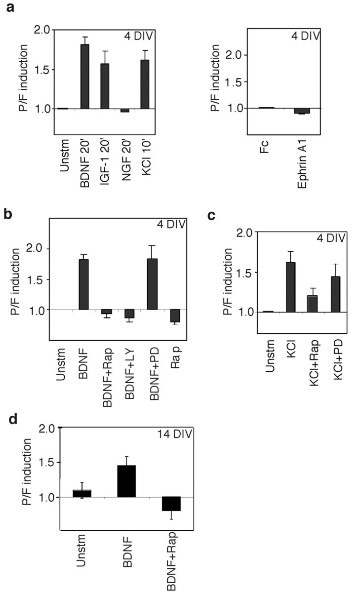
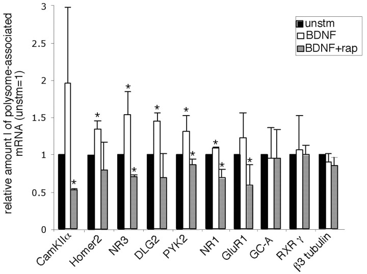
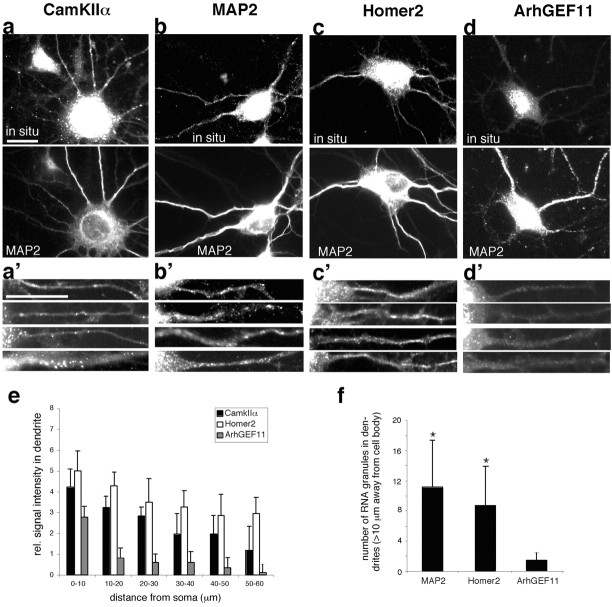
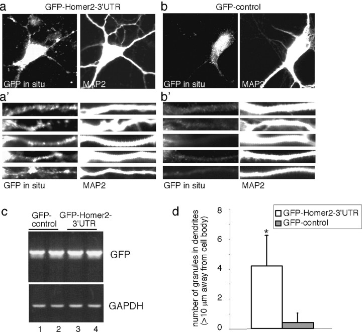
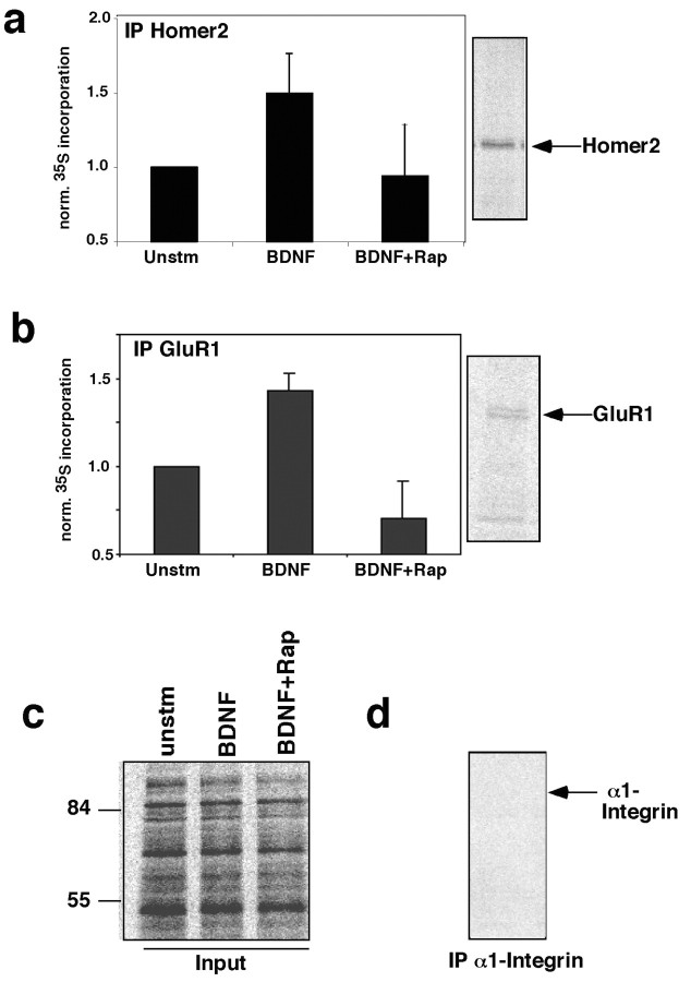
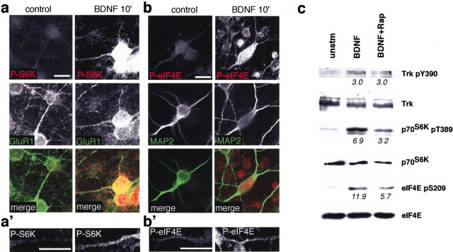
Local regulation of mRNA translation plays an important role in axon guidance, synaptic development, and neuronal plasticity. Little is known, however, regarding the mechanisms that control translation in neurons, and only a few mRNAs have been identified that are locally translated within axon and dendrites. Using Affymetrix gene arrays to identify mRNAs that are newly associated with polysomes after exposure to BDNF, we identified subsets of mRNAs for which translation is enhanced in neurons at different developmental stages. In mature neurons, many of these mRNAs encode proteins that are known to function at synapses, including CamKIIalpha, NMDA receptor subunits, and the postsynaptic density (PSD) scaffolding protein Homer2. BDNF regulates the translation of Homer2 locally in the synaptodendritic compartment by activating translational initiation via a mammalian target of rapamycin-phosphatidylinositol 3-kinase-dependent pathway. These findings suggest that BDNF likely regulates synaptic function by inducing the local synthesis of numerous synaptic proteins. The local translation of the cytoskeleton-associated protein Homer2 in particular might have important implications for growth cone dynamics and dendritic spine development.


Figures

Figure 4.
Figure 4.
Homer2 mRNA is enriched in synaptoneurosomes. a, Electron micrographs of a representative synaptoneurosome preparation at low (bar = 500 nm) and high (bar = 200 nm) magnification. Arrowheads in the left panel point to juxtaposed presynaptic and postsynaptic membranes. The right panel shows an individual synaptoneurosome, with a presynaptic mitochondrion (MI), synaptic vesicles (SV), and the postsynaptic density (PSD). b, Enrichment of synaptic proteins in synaptoneurosomes. The synaptoneurosome preparation was performed as described in Materials and Methods, and samples were taken at each stage of the procedure. Aliquots (20 μg) of each fraction were resolved by SDS-PAGE, and Western blotting was performed with antibodies to SV2, PSD95, α1-integrin, MEF2D, and TuJ1 as a loading control. The lanes are labeled as follows: S1, whole lysate before centrifugation; S2, supernatant from P2 spin; P2, pellet from P2 spin (14,000 × g); Syn, synaptosomal fraction. c, Relative abundance of the indicated messages in synaptoneurosomes was determined by quantitative RT-PCR analysis. Values represent the ratio of the relative mRNA amount in synaptoneurosomes to the relative mRNA amount in total brain at P15. Error bars represent the SE of three independent experiments. Asterisk indicates that the respective value is significantly different from the β3-tubulin control (p < 0.05).
Figure 1.
Figure 1.
BDNF treatment increases protein translation in neurons. a, Effect of BDNF treatment on polysome profile. E18 rat cortical neurons at 4 DIV were treated with or without BDNF for 20 min and harvested. Lysates were applied to a 15-45% linear sucrose gradient, centrifuged, and fractionated with continuous optical density measurement. F, Free ribosomal subunit fractions; P, polysome fractions. b, Time course of the polysome shift after BDNF treatment. After 2 hr serum starvation, neurons were treated with or without actinomycin D (ActD) for 40 min before BDNF treatment. The normalized ratio of the integral of the F and P curves was plotted as a function of time of BDNF stimulation. P/F induction represents the relative change in P/Fratio in stimulated cells to that of untreated neurons. Error bars represent the SE of at least five independent experiments.
Figure 2.
Figure 2.
Multiple stimuli can regulate mRNA translation in neurons. a, Effect of growth factor treatment on the polysome profile. Neurons were treated with growth factors or stimulated with 60 mm KCl for the indicated times and harvested as in Figure 1. b, PI3K-mTOR signaling is required for the BDNF-induced polysome shift. Neurons were starved, pretreated with actinomycin D, and incubated with the indicated inhibitors for 20 min before BDNF stimulation. Cells were harvested 20 min after BDNF addition. Rap, Rapamycin (20 ng/ml); LY, 25 μm LY294002; PD, 60 μm PD98059. c, Depolarization stimulates mRNA translation through distinct mechanisms. Neurons were starved, pretreated with actinomycin D, and incubated with inhibitors for 20 min before 60 mm KCl stimulation. Inhibitor treatment was performed as described above. Error bars represent the SE of at least three independent experiments. d, BDNF induces a polysome shift in cortical neurons at 14 DIV. Neurons were treated as in b, polysome fractionation was performed, and the P/F ratios were calculated. Error bars represent the SE of three independent experiments.
Figure 3.
Figure 3.
Association of specific neuronal mRNAs with polysomes after BDNF stimulation. Cultured E18 rat cortical neurons at 14 DIV were serum starved, preblocked with actinomycin D, and treated with or without BDNF for 20 min. When indicated, 20 ng/ml rapamycin was added 20 min before BDNF stimulation (Rap+BDNF). After sucrose gradient separation and fractionation, 1 μg of RNA from combined polysome fractions was purified and reverse transcribed to generate cDNA. Relative mRNA levels were determined using quantitative real-time PCR (unstimulated = 1). Error bars represent the SE of three independent experiments. Asterisk indicates that the respective value is significantly different from unstimulated control (p < 0.05).
Figure 5.
Figure 5.
Homer2 mRNA is present in dendrites of hippocampal neurons in culture. Hippocampal neurons at 7 DIV were processed for in situ hybridization with specific probes against CamKIIα, MAP2, Homer2, and ArhGEF11 (a-d, top panel). Costaining for MAP2 protein was used to visualize dendrites (a-d, bottom panel). Higher-magnification images illustrate the presence of RNA granules in multiple dendrites of CamKIIα, MAP2, and Homer2 samples (a′-c′) and their almost complete absence in the ArhGEF11 control sample (d′). Representative examples from one of three independent experiments are shown. Scale bar, 20 μm. e, Mean intensity values for 10 μm dendritic segments were calculated for each mRNA as described in Materials and Methods. Error bars represent the SE of intensity values derived from at least 12 randomly chosen dendrites per individual mRNA. f, The number of granules for each mRNA was determined in the same population of dendrites as in e. Dendrites from MAP2 and Homer2 samples contain significantly more RNA granules than dendrites from the ArhGEF11 control (p < 0.05).
Figure 6.
Figure 6.
The 3′UTR of Homer2 is sufficient for dendritic targeting. Hippocampal neurons at 7 DIV were transfected with GFP-Homer2-3′UTR and GFP-control RNA and processed for in situ hybridization with a probe specific for GFP (a, b, left panel). Costaining for MAP2 protein was used to visualize dendrites (a, b, right panel). Higher-magnification images illustrate the presence of RNA granules in multiple dendrites of GFP-Homer2-3′UTR-transfected neurons (a′, leftpanel) and their almost complete absence in GFP-control-transfected neurons (b′, right panel). Representative examples from one of three independent experiments are shown. Scale bar, 20 μm. c, Total RNA was prepared from GFP-control (lanes 1, 2) and GFP-Homer2-3′UTR (lanes 3, 4)-transfected neurons and used for semiquantitative RT-PCR analysis with primers specific for GFP (top panel) or GAPDH (bottom panel) as a loading control. d, The number of RNA granules in distal dendritic segments was determined for neurons transfected with the indicated RNAs. Values represent the mean of at least 12 dendrites for each construct. Asterisks indicate that the respective values are significantly higher than the value for the GFP-control construct (p < 0.05).
Figure 7.
Figure 7.
BDNF treatment increases the synthesis of Homer2 and GluR1 protein in synaptoneurosomes in a rapamycin-sensitive manner. Synaptoneurosomes were prepared as above, incubated at 37°C, and stimulated with BDNF for 0 or 60 min in the presence of 100 μCi/ml [35S]methionine. The stimulation was performed in the presence or absence of rapamycin. Lysates were immunoprecipitated with antibodies to Homer2 (a), GluR1 (b), and α1-integrin (d), resolved on SDS-PAGE gel, and visualized by autoradiography. Autoradiographs of lysates before immunoprecipitation are shown in c. The quantification of autoradiographs from three independent experiments is shown (unstimulated = 1). 35S incorporation into Homer2 and GluR1 protein is significantly higher in BDNF-treated samples compared with unstimulated control samples (p < 0.05).
Figure 8.
Figure 8.
BDNF stimulation induces the local phosphorylation of multiple translation factors in neuronal dendrites in a rapamycin-sensitive manner. a, b, BDNF stimulation induces the local phosphorylation of translation factors in dendrites. E18 cortical neurons at 7 DIV were serum starved for 4 hr before stimulating with BDNF (50 ng/ml) for 10 min. Samples were fixed and immunocytochemistry was performed with anti-phospho-p70S6K (Thr 389) (a) and anti-phospho-eIF4E (Ser209) (b) together with anti GluR1 (a) or anti-MAP2 (b) to stain dendrites. Higher-magnification images (a′, b′) illustrate the presence of phosphorylated translation factors in representative dendrites. Scale bar, 20 μm. c, BDNF-induced phosphorylation of translation factors in synaptoneurosomes is rapamycin sensitive. Synaptoneurosomes were treated with BDNF (100 ng/ml) for 10 min, in the presence of rapamycin (20 ng/ml) when indicated. Synaptosomal lysates were prepared and analyzed for the phosphorylation of translation factors by Western blotting as described above. Induction of phosphorylated protein relative to the unstimulated control is given below each blot. Results are representative of three independent experiments.
Figure 9.
Figure 9.
Local phosphorylation of translation factors near synapses. Hippocampal neurons (14 DIV) were stimulated as in Figure 8 before processing for immunocytochemistry with anti-phospho-Trk (a, c), anti-phospho-eIF4E (d, f), and anti-phosphop-70S6K (g, i) antibodies. Synaptic regions were visualized by costaining with anti-PSD-95 (b, c, e, f) and anti-synapsin (h, i) antibodies. Yellow signal in merged images indicates colocalization of phosphorylated proteins with synaptic marker proteins. Scale bar, 20 μm.

Similar articles

Cited by

References

    1. Aakalu G, Smith WB, Nguyen N, Jiang C, Schuman EM (2001) Dynamic visualization of local protein synthesis in hippocampal neurons. Neuron 30: 489-502. - PubMed
    1. Aizawa H, Wakatsuki S, Ishii A, Moriyama K, Sasaki Y, Ohashi K, Sekine-Aizawa Y, Sehara-Fujisawa A, Mizuno K, Goshima Y, Yahara I (2001) Phosphorylation of cofilin by LIM-kinase is necessary for semaphorin 3A-induced growth cone collapse. Nat Neurosci 4: 367-373. - PubMed
    1. Bonni A, Greenberg ME (1997) Neurotrophin regulation of gene expression. Can J Neurol Sci 24: 272-283. - PubMed
    1. Brittis PA, Lu Q, Flanagan JG (2002) Axonal protein synthesis provides a mechanism for localized regulation at an intermediate target. Cell 110: 223-235. - PubMed
    1. Campbell DS, Holt CE (2001) Chemotropic responses of retinal growth cones mediated by rapid local protein synthesis and degradation. Neuron 32: 1013-1026. - PubMed

Publication types

MeSH terms