. 2004 Nov 1;383(Pt. 3):475-82.
doi: 10.1042/BJ20040914.

Vif is an auxiliary factor of the HIV-1 reverse transcriptase and facilitates abasic site bypass

Affiliations

Vif is an auxiliary factor of the HIV-1 reverse transcriptase and facilitates abasic site bypass

Reynel Cancio et al. Biochem J. .

Abstract

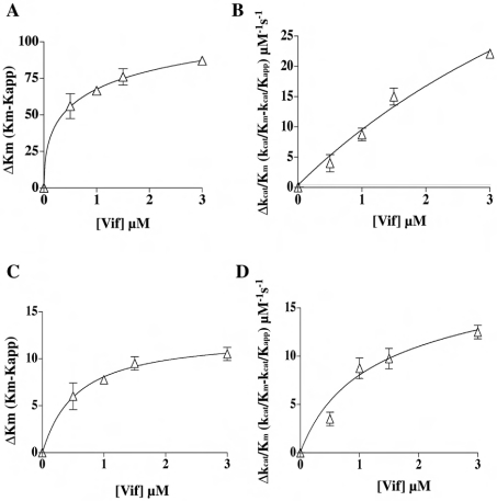
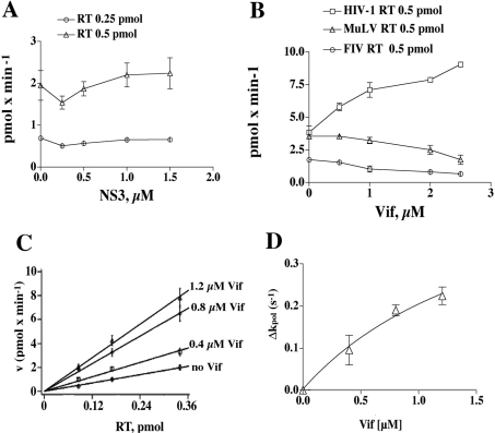
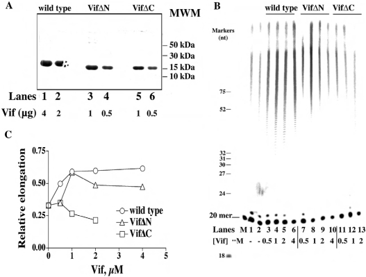
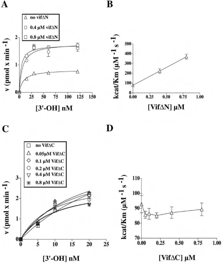
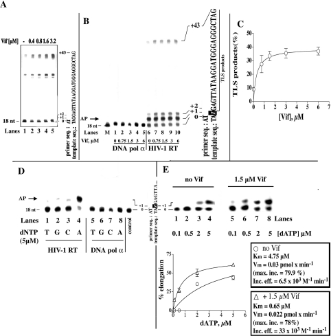
The HIV-1 accessory protein Vif was found to modulate the RNA- and DNA-dependent DNA synthesis activity of the viral RT (reverse transcriptase) in two ways: (i) it stimulated the binding of the viral RT to the primer by increasing the association rate kcat/K(m) and by decreasing the thermodynamic barrier DeltaH([ES]) for complex formation, and (ii) it increased the polymerization rate of HIV-1 RT. A Vif mutant lacking the final 56 amino acids at the C-terminus failed to stimulate the viral RT. On the other hand, another Vif mutant lacking the first 43 amino acids at the N-terminus, which are involved in RNA binding and interaction with the viral protease, was able to stimulate RT activity. In addition, Vif was found to promote the bypass of an abasic site by HIV-1 RT.


Figures

Figure 1
Figure 1. HIV-1 Vif increases the affinity of the viral RT for a 3′-hydroxy primer during both RDS and DDS
(A) The variation of the Km value for the 3′-hydroxy primer (ΔKm) under RDS conditions is expressed as the difference between the Km value determined in the absence of Vif and in its presence (Kapp). The points were fitted to the hyperbolic equation ΔKmmax/(1+Kd,vif/[Vif]), where Δmax denotes the maximum difference in the kinetic parameters extrapolated at infinite Vif concentration. (B) The variation of the kcat/Km value (Δkcat/Km) under RDS conditions is expressed as the difference between the kcat/Km value determined in the absence of Vif and that determined in its presence (kcat/Kapp). The points were fitted to the hyperbolic equation Δkcat/Kmmax/(1+Kd,vif/[Vif]). The fitting parameters were r2=0.9735 and SSE (sum of squares of errors)=15.76. (C, D) As in (A) and (B) respectively, but under DDS conditions. The fitting parameters were r2=0.9559 and SSE=9.
Figure 2
Figure 2. HIV-1 Vif specifically stimulates the reaction rate (kpol) of HIV-1 RT
(A) Increasing amounts of recombinant HCV NS3 were titrated in the presence of 0.25 (○) or 0.5 (▵) pmol of HIV-1 RT, under RDS conditions. The NS3 concentrations used were 0.25, 0.5, 1 and 1.5 μM. (B) Increasing amounts of recombinant Vif were titrated in the presence of HIV-1 RT (□), FIV RT (○) or MuLV RT (▵). Vif concentrations used were 0.5, 1, 2 and 2.5 μM. (C) The dependence of the reaction velocity (pmol·min−1) under RDS conditions from the RT concentration was tested in the absence or in the presence of 0.4, 0.8 or 1.2 μM Vif, as indicated. (D) The slopes of the curves shown in (A), representing the reaction rate kpol. The variation of kpol under RDS conditions (Δkpol) is expressed as the difference between the values determined in the absence and in the presence of Vif. The points were fitted to the hyperbolic equation Δkpolmax/(1+Kd,vif/[Vif]), where Δmax denotes the maximum difference in the kinetic parameters extrapolated at infinite Vif concentration. Fitting parameters were r2=0.9605, SSE (sum of squares of errors)=0.0025.
Figure 3
Figure 3. Purification and characterization of the N- and C-terminal domain deletion mutants of HIV-1 Vif
(A) SDS/PAGE of samples taken after the final step of purification (Mono-S) of recombinant wild-type Vif and the mutants VifΔN and VifΔC. Increasing amounts of Vif were subjected to denaturing PAGE (10% gel) and stained with Coomassie Blue. MWM denotes molecular-mass markers (B) 7 M Urea/17% polyacrylamide sequencing gel analysis of the products synthesized by HIV-1 RT on poly(rA)·[5′-32P]oligo(dT)20 in the absence or in the presence of wild-type Vif and the VifΔN and VifΔC mutants. The concentrations of recombinant Vif proteins used are indicated. The positions corresponding to the 20-mer primer, as well as of various oligonucleotide size markers, are indicated on the left. (C) Quantification of the products synthesized in the experiment shown in (B) was performed after scanning densitometry with the program ImageQuant. The intensities of the signals corresponding to all the products above the 20-mer primer were normalized to the total intensities of the corresponding lane, and the relative incorporation (r.i..; expressed as % of total input) values were plotted against Vif concentration. Data were fitted to the hyperbolic equation r.i. (%)=%incmax/(1+Kd,vif/[Vif]), where %incmax denotes maximal percentage incorporation extrapolated at infinite Vif concentration.
Figure 4
Figure 4. The C-terminal domain of Vif is required for stimulation of HIV-1 RT activity
(A) The variation in the velocity of the reaction catalysed by HIV-1 RT under RDS conditions, in the absence or in the presence of increasing amounts of the VifΔN protein, was plotted as a function of the nucleic acid substrate concentration (expressed as available 3′-hydroxy ends). Data were fitted to the Michelis–Menten equation of the form v=Vmax/(1+Km/[3′-hydroxy]). Poly(rA)·oligo(dT) concentrations used were 15, 30, 60 and 120 nM. (B) The maximal reaction rate (kcat) was calculated from the Vmax values derived from the experiments shown in (A) according to the relationship kcat=Vmax/[E0], where [E0] is the total input enzyme concentration. These kcat values were used to calculate the kcat/Km values in the absence and in the presence of VifΔN, which were then plotted against the VifΔN concentration. (C) Experiments were performed as in (A), but in the absence or in the presence of increasing amounts of VifΔC protein. Poly(rA)·oligo(dT) concentrations used were 5, 10 and 20 nM. (D) kcat/Km values in the absence or in the presence of VifΔC were calculated as in (B) and plotted against VifΔ concentration.
Figure 5
Figure 5. Vif stimulates TLS by HIV-1 RT in the presence of an abasic site
(A) 7 M Urea/20% (w/v) polyacrylamide sequencing gel analysis of the products synthesized by HIV-1 RT on the undamaged 18/73-mer DNA template in the presence of Vif. (B) 7 M Urea/20% (w/v) polyacrylamide sequencing gel analysis of the products synthesized by HIV-1 RT (lanes 6–10) or human DNA polymerase α (lanes 1–5) in the absence or in the presence of Vif and in the presence of the 18/73-mer DNA template containing an abasic site at position +1. The concentrations of recombinant Vif protein used are indicated. The positions corresponding to the 18-mer primer and to the full-length products, as well as the sequence of the template strand, are indicated on the right. The arrow on the left (AP) indicates the position of the products corresponding to incorporation opposite the lesion. (C) Quantification of the TLS products synthesized in the experiment shown in (B) was performed after scanning densitometry with the program ImageQuant. The intensity of the signal corresponding to the TLS products were normalized to the total intensity of the corresponding lane, and the relative incorporation (TLS products; expressed as % of total input) was plotted against Vif concentration. Data were fitted to the hyperbolic equation TLS (%)=%TLSmax/(1+Kd,vif/[Vif]), where %TLSmax denotes maximal percentage TLS extrapolated at infinite Vif concentration. (D) 7 M Urea/20% polyacrylamide sequencing gel analysis of the products synthesized by HIV-1 RT (lanes 1–4) or human DNA polymerase α (lanes 5–8) under single nucleotide incorporation conditions. Reactions were performed in the absence of Vif and in the presence of the 18/73-mer DNA template containing an abasic site at position +1. The single nucleotide added in each reaction is indicated. The position corresponding to the 18-mer primer is indicated on the left, and the sequence of the template strand is shown on the right. The arrow on the left indicates the position of the products corresponding to incorporation opposite the lesion. (E) Upper panel: 7 M Urea/20% polyacrylamide sequencing gel analysis of the products synthesized by HIV-1 RT in the absence or in the presence of Vif and in the presence of the 18/73-mer DNA template containing an abasic site at position +1, under single nucleotide incorporation conditions. The concentrations of the single-nucleotide dATP used in each reaction are indicated below the gel. The positions corresponding to the 18-mer primer (0) and to the first incorporated nucleotide (+1), as well as the sequence of the template strand, are indicated on the left. Lower panel: quantification of products synthesized (see upper panel) was performed after scanning densitometry with the program ImageQuant. The intensity of the signal corresponding to the +1 products was normalized to the total intensity of the corresponding lane, and the relative elongation values (expressed as % of elongated primer ends compared with total input) were plotted against dATP concentration. Data were fitted to the Michelis–Menten equation in the form: v=Vmax/(1+Km/[dATP]). The calculated Km, Vmax (Vm) and Vmax/Km (incorporation efficiency) values are indicated on the right.

Similar articles

Cited by

References

    1. Frankel A. D., Young J. A. HIV-1: fifteen proteins and an RNA. Annu. Rev. Biochem. 1998;67:1–25. - PubMed
    1. Gabuzda D. H., Li H., Lawrence K., Vasir B. S., Crawford K., Langhoff E. Essential role of vif in establishing productive HIV-1 infection in peripheral blood T lymphocytes and monocyte/macrophages. J. Acquired Immune Defic. Syndr. 1994;7:908–915. - PubMed
    1. Inubushi R., Adachi A. Cell-dependent function of HIV-1 Vif for virus replication. Int. J. Mol. Med. 1999;3:473–476. - PubMed
    1. Simon J. H., Miller D. L., Fouchier R. A., Soares M. A., Peden K. W., Malim M. H. The regulation of primate immunodeficiency virus infectivity by Vif is cell species restricted: a role for Vif in determining virus host range and cross-species transmission. EMBO J. 1998;17:1259–1267. - PMC - PubMed
    1. Simon J. H., Carpenter E. A., Fouchier R. A., Malim M. H. Vif and the p55(Gag) polyprotein of human immunodeficiency virus type 1 are present in colocalizing membrane-free cytoplasmic complexes. J. Virol. 1999;73:2667–2674. - PMC - PubMed

Publication types

MeSH terms