Oxidative damage to methyl-CpG sequences inhibits the binding of the methyl-CpG binding domain (MBD) of methyl-CpG binding protein 2 (MeCP2)
- PMID: 15302911
- PMCID: PMC514367
- DOI: 10.1093/nar/gkh739
Oxidative damage to methyl-CpG sequences inhibits the binding of the methyl-CpG binding domain (MBD) of methyl-CpG binding protein 2 (MeCP2)
Abstract
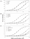
Cytosine methylation in CpG dinucleotides is believed to be important in gene regulation, and is generally associated with reduced levels of transcription. Methylation-mediated gene silencing involves a series of DNA-protein and protein-protein interactions that begins with the binding of methyl-CpG binding proteins (MBPs) followed by the recruitment of histone-modifying enzymes that together promote chromatin condensation and inactivation. It is widely known that alterations in methylation patterns, and associated gene activities, are often found in human tumors. However, the mechanisms by which methylation patterns are altered are not currently understood. In this paper, we investigate the impact of oxidative damage to a methyl-CpG site on MBP binding by the selective placement of 8-oxoguanine (8-oxoG) and 5-hydroxymethylcytosine (HmC) in a MBP recognition sequence. Duplexes containing these specific modifications were assayed for binding to the methyl-CpG binding domain (MBD) of one member of the MBP family, methyl-CpG binding protein 2 (MeCP2). Our results reveal that oxidation of either a single guanine to 8-oxoG or of a single 5mC to HmC, significantly inhibits binding of the MBD to the oligonucleotide duplex, reducing the binding affinity by at least an order of magnitude. Oxidative damage to DNA could therefore result in heritable, epigenetic changes in chromatin organization.
Figures


Similar articles
-
5-halogenated pyrimidine lesions within a CpG sequence context mimic 5-methylcytosine by enhancing the binding of the methyl-CpG-binding domain of methyl-CpG-binding protein 2 (MeCP2).Nucleic Acids Res. 2005 May 25;33(9):3057-64. doi: 10.1093/nar/gki612. Print 2005. Nucleic Acids Res. 2005. PMID: 15917437 Free PMC article.
-
The solution structure of the domain from MeCP2 that binds to methylated DNA.J Mol Biol. 1999 Sep 3;291(5):1055-65. doi: 10.1006/jmbi.1999.3023. J Mol Biol. 1999. PMID: 10518942
-
Altered chromatin structure associated with methylation-induced gene silencing in cancer cells: correlation of accessibility, methylation, MeCP2 binding and acetylation.Nucleic Acids Res. 2001 Nov 15;29(22):4598-606. doi: 10.1093/nar/29.22.4598. Nucleic Acids Res. 2001. PMID: 11713309 Free PMC article.
-
Methyl-CpG binding proteins in the nervous system.Cell Res. 2005 Apr;15(4):255-61. doi: 10.1038/sj.cr.7290294. Cell Res. 2005. PMID: 15857580 Review.
-
Methyl-CpG-binding proteins. Targeting specific gene repression.Eur J Biochem. 2001 Jan;268(1):1-6. doi: 10.1046/j.1432-1327.2001.01869.x. Eur J Biochem. 2001. PMID: 11121095 Review.
Cited by
-
Tissue type is a major modifier of the 5-hydroxymethylcytosine content of human genes.Genome Res. 2012 Mar;22(3):467-77. doi: 10.1101/gr.126417.111. Epub 2011 Nov 21. Genome Res. 2012. PMID: 22106369 Free PMC article.
-
Epigenetic Regulation of Oxidative Stress in Ischemic Stroke.Aging Dis. 2016 May 27;7(3):295-306. doi: 10.14336/AD.2015.1009. eCollection 2016 May. Aging Dis. 2016. PMID: 27330844 Free PMC article. Review.
-
Placental DNA hypomethylation in association with particulate air pollution in early life.Part Fibre Toxicol. 2013 Jun 7;10:22. doi: 10.1186/1743-8977-10-22. Part Fibre Toxicol. 2013. PMID: 23742113 Free PMC article.
-
Frequent expression of MAGE1 tumor antigens in bronchial epithelium of smokers without lung cancer.Exp Ther Med. 2011 Jan;2(1):137-142. doi: 10.3892/etm.2010.176. Epub 2010 Dec 2. Exp Ther Med. 2011. PMID: 22977481 Free PMC article.
-
Recognition and cleavage of 5-methylcytosine DNA by bacterial SRA-HNH proteins.Nucleic Acids Res. 2015 Jan;43(2):1147-59. doi: 10.1093/nar/gku1376. Epub 2015 Jan 6. Nucleic Acids Res. 2015. PMID: 25564526 Free PMC article.
References
-
- Ehrlich M. and Wang,F.Y.-H. (1981) 5-Methylcytosine in eukaryotic DNA. Science, 212, 1350–1357. - PubMed
-
- Riggs A.D. and Jones,P.A. (1983) 5-Methylcytosine, gene regulation and cancer. Adv. Cancer Res., 40, 1–30. - PubMed
-
- Doerfler W. (1983) DNA methylation and gene activity. Ann. Rev. Biochem., 52, 93–124. - PubMed
-
- Antequera F., Boyes,J. and Bird,A. (1990) High levels of de novo methylation and altered chromatin structure at CpG islands in cell lines. Cell, 62, 503–514. - PubMed
-
- Jaenisch R. and Bird,A. (2003) Epigenetic regulation of gene expression: how the genome integrates intrinsic and environmental signals. Nature Genet., 33, 245–254. - PubMed
Publication types
MeSH terms
Substances
Grants and funding
LinkOut - more resources
Full Text Sources
Other Literature Sources
Miscellaneous

