. 2004 Jul 28;24(30):6776-84.
doi: 10.1523/JNEUROSCI.1826-04.2004.

Neuronal nicotinic synapse assembly requires the adenomatous polyposis coli tumor suppressor protein

Affiliations

Neuronal nicotinic synapse assembly requires the adenomatous polyposis coli tumor suppressor protein

Murali Krishna Temburni et al. J Neurosci. .

Abstract

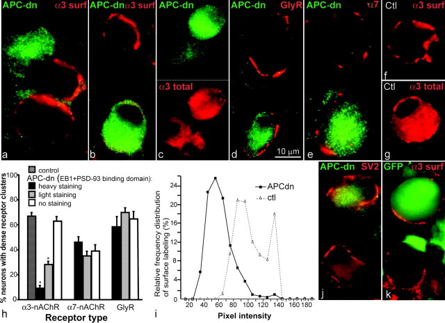
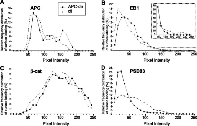
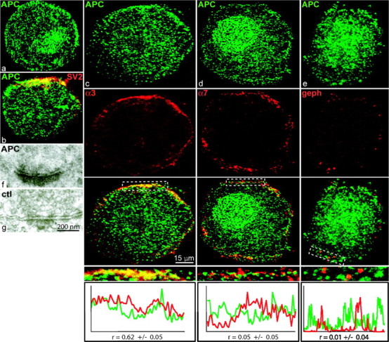
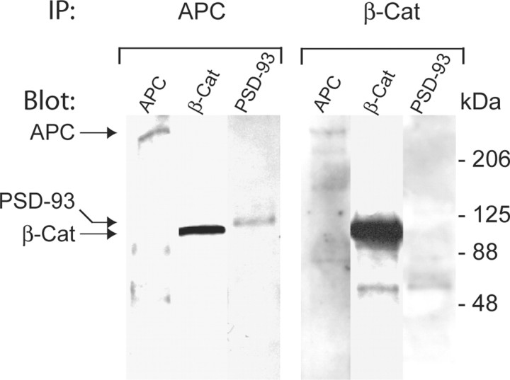
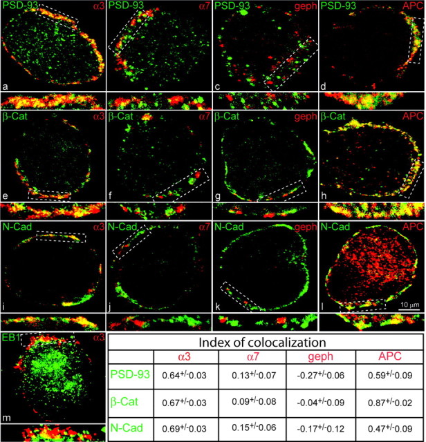
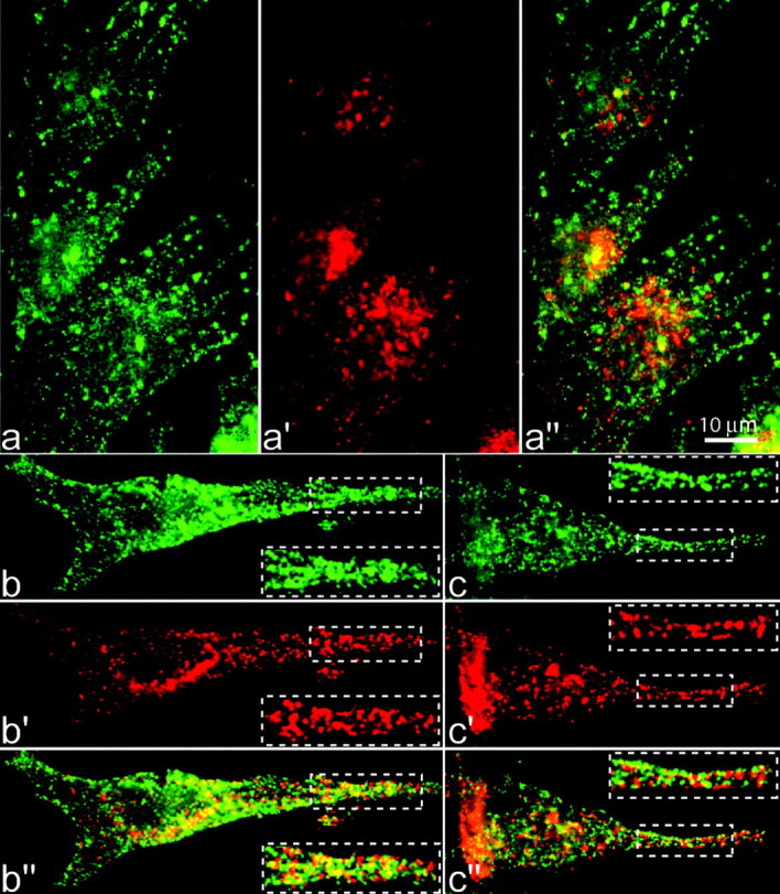
Normal cognitive and autonomic functions require nicotinic synaptic signaling. Despite the physiological importance of these synapses, little is known about molecular mechanisms that direct their assembly during development. We show here that the tumor-suppressor protein adenomatous polyposis coli (APC) functions in localizing alpha3-nicotinic acetylcholine receptors (nAChRs) to neuronal postsynaptic sites. Our quantitative confocal microscopy studies indicate that APC is selectively enriched at cholinergic synapses; APC surface clusters are juxtaposed to synaptic vesicle clusters and colocalize with alpha3-nAChRs but not with the neighboring synaptic glycine receptors or perisynaptic alpha7-nAChRs on chick ciliary ganglion (CG) neurons. We identify PSD (postsynaptic density)-93, beta-catenin, and microtubule end binding protein EB1 as APC binding partners. PSD-93 and beta-catenin are also enriched at alpha3-nAChR postsynaptic sites. EB1 shows close proximity to and partial overlap with alpha3-nAChR and APC surface clusters. We tested the role of APC in neuronal nicotinic synapse assembly by using retroviral-mediated in vivo overexpression of an APC dominant-negative (APC-dn) peptide to block the interaction of endogenous APC with both EB1 and PSD-93 during synapse formation in CG neurons. The overexpressed APC-dn led to dramatic decreases in alpha3-nAChR surface levels and clusters. Effects were specific to alpha3-nAChR postsynaptic sites; synaptic glycine receptor and perisynaptic alpha7-nAChR clusters were not altered. In addition, APC-dn also reduced surface membrane-associated clusters of PSD-93 and EB1. The results show that APC plays a key role in organizing excitatory cholinergic postsynaptic specializations in CG neurons. We identify APC as the first nonreceptor protein to function in localizing nAChRs to neuronal synapses in vivo.


Figures

Figure 5.
Figure 5.
In vivo overexpression of an APC dominant-negative peptide reduced surface clusters (surf) of α3-nAChRs, but not of α7-nAChRs or GlyRs, on CG neurons. Epifluorescence micrographs show double-labeled E11-E13 CG frozen sections. a-c, α3-nAChR (a, b; red) surface clusters were dramatically reduced in neurons overexpressing the HA-tagged APC-dn (green) compared with those on nearby uninfected neurons [internal control (Ctl)], whereas intracellular α3-nAChR labeling (c; representing the biosynthetic pool) was not detectably altered. d, e, In sharp contrast to α3-nAChRs, GlyR (d) and α7-nAChR (e) surface clusters were not apparently altered in neurons overexpressing the APC-dn compared with those on nearby uninfected control neurons. Uninfected control CG age-matched and processed in parallel showed typical α3-nAChR surface clusters (f) and internal staining (g). As additional indications of specificity, there were no detectable changes in presynaptic terminal morphology (j) [synaptic vesicle (SV2) labeling; red] or α3-nAChR surface clusters on retroviral GFP-infected neurons (k; as a negative control). HA staining (green) shows that retroviral infection was restricted to CG neurons and occasionally a few glial cells (small HA-positive cells surrounding the neuronal somata). h, Histogram showing the proportion of the neuron population with large densely stained neurotransmitter receptor surface clusters for APC-dn overexpressing CG neurons and uninfected control neurons. For infected CGs, individual neurons were classified into three groups on the basis of APC-dn expression levels as judged by the pixel intensity of HA immunolabeling:heavy, light, or not detectable. Cells considered to be heavily labeled had pixel intensities ranging from 105 to 200, lightly labeled ranging from 50 to 105, and not detectable labeling had pixel intensity levels below 30 after subtracting background levels. Bars represent the mean (±SEM) from the analysis of >427 randomly selected neurons (1125 APC-dn infected neurons for α3-nAChR clusters) from three or more embryos. The asterisk indicates significant difference from controls (p < 0.001; ANOVA). i, Graph showing substantial decreases in the pixel intensity of α3-nAChR surface labeling on heavily APC-dn infected neurons versus uninfected control neurons at matched ages. Pixel intensities were measured along randomly chosen∼3 μm segments of labeled neuronal surface membrane (n = 3 or more segments per neuron; 4-6 neurons). The values were binned into incremental groups of 10 pixel intensity steps (from 0 to 9, 10 to 19..., up to saturation). We then calculated the percentage of pixels that belonged to each pixel intensity category and plotted the data as relative frequency polygons.
Figure 6.
Figure 6.
In vivo overexpression of the APC dominant-negative peptide led to decreases in EB1 and PSD-93 surface membrane-associated labeling in CG neurons. A-D, The graphs show decreases in the pixel intensity of EB1 (B) and PSD-93 (D) surface membrane-associated labeling in APC-dn infected neurons versus uninfected control CG neurons. Inset, To enhance visualization of the decrease in EB1 surface membrane-associated labeling, we separately calculated and graphed the relative frequency distribution of pixel intensities in the higher range only (pixel intensities from 140 to 255). In contrast, APC (A) and β-catenin (β-cat) (C) surface labeling was not detectably different in infected versus uninfected control CG neurons (n = 3 or more labeled surface membrane segments per neuron; 4-6 neurons for each immunolabel).
Figure 1.
Figure 1.
APC localization relative to synapses and the different neurotransmitter receptor types on the CG neuron surface. Confocal micrographs of immunolabeled acutely dissociated E18 CG neurons. a, APC localized in patches along a portion of the neuronal surface membrane and small clusters throughout the cytoplasm and nucleus. The nuclear labeling was confirmed by 4′, 6′-diamidino-2-phenylindole staining (data not shown). b, Double-labeled neuron showing that APC surface clusters are juxtaposed to synaptic vesicle clusters (SV2 labeling in red; presynaptic terminal). c, Double-labeled neuron showing strong colocalization of APC and α3-nAChR surface clusters (predominance of yellow fluorescent patches). d, e, In contrast, APC and α7-nAChRs (d) and gephyrin (geph; e) (the GlyR directly associated protein) showed little overlap at the neuron surface (predominance of distinct red or green patches). Insets, Threefold magnification views of the boxed regions. Bottom panels, Graphs show red and green fluorescence intensity profiles for the boxed regions. Staining intensities of surface clusters covaried and correlated with each other for α3-nAChRs with APC but not for α7-nAChRs and GlyRs/gephyrin with APC (n = 2-3 surface areas per cell; 4-6 neurons). f, g, Electron micrographs show that APC is localized at the postsynaptic density (f) (APC detected by horseradish peroxidase); unlabeled control (ctl) synapse is included for comparison (g).
Figure 2.
Figure 2.
APC, β-catenin (β-Cat), and PSD-93 form a complex in the postsynaptic density in vivo. The psds were isolated from E18 chick brain (without the cerebellum and brainstem). The psd preparation was immunoprecipitated (IP) with anti-APC antibody, and the immunoprecipitate was probed with antibodies that recognize β-catenin and PSD-93. In addition, the psds were immunoprecipitated with anti-β-catenin antibody and probed with antibodies that recognize APC and PSD-93. APC coprecipitated both β-catenin and PSD-93, whereas β-catenin coprecipitated APC, but not PSD-93, from the psds. In addition, β-catenin is more abundant than APC in the coprecipitates, likely because of multiple β-catenin molecules that bind to one APC molecule via the 15 amino acid and 20 amino acid repeat domains (Rubinfeld et al., 1995; Fearnhead et al., 2001).
Figure 3.
Figure 3.
Localization of APC binding partners at nicotinic cholinergic postsynaptic sites. a, d, e, h, i, l, Confocal micrographs of double-labeled acutely dissociated E18 CG neurons showing the APC binding partners PSD-93 and β-catenin (β-Cat) and the cellular adhesion molecule N-cadherin (N-Cad) predominantly colocalized with α3-nAChR (a, e, i) and APC (d, h, l) surface clusters (overlap, yellow). b, c, f, g, j, k, In contrast, little colocalization was seen with α7-nAChRs (b, f, j) and gephyrin (geph) (c, g, k). m, In addition, the APC binding partner EB1 showed close proximity to and partial overlap with α3-nAChR surface clusters. However, EB1 did not accumulate much at the surface membrane. Instead, EB1 labeling was predominantly cytoplasmic. Insets, Twofold magnification views of boxed regions. The table shows that the fluorescence staining intensities of surface clusters covaried and correlated with each other for PSD-93, β-catenin, and N-cadherin with α3-nAChRs and APC but not with α7-nAChRs or GlyRs/gephyrin (n = 2-3 surface areas per cell; 4-6 neurons).
Figure 4.
Figure 4.
Efficacy of the overexpressed APC dominant-negative construct tested in chick fibroblasts in vitro. a-c, Epifluorescence micrographs of double-labeled chick DF1 fibroblasts showing that the overexpressed APC-dn (a) led to drastic decreases in endogenous APC (green) and EB1 (red) surface membrane-associated clusters compared with uninfected control fibroblasts (b, c). The APC clusters were distributed in radial arrays throughout the cytoplasm of the APC-dn infected cells (a). To distinguish endogenous APC from the exogenous APC-dn (APC C-terminal peptide fragment), we used the anti-APC antibody that recognizes the N-terminal epitope. Insets, Twofold magnification views of boxed regions in control uninfected fibroblasts (b, c).

Similar articles

Cited by

References

    1. Barth AI, Siemers KA, Nelson WJ (2002) Dissecting interactions between EB1, microtubules and APC in cortical clusters at the plasma membrane. J Cell Sci 115: 1583-1590. - PMC - PubMed
    1. Berrueta L, Tirnauer JS, Schuyler SC, Pellman D, Bierer BE (1999) The APC-associated protein EB1 associates with components of the dynactin complex and cytoplasmic dynein intermediate chain. Curr Biol 9: 425-428. - PubMed
    1. Bienz M (2002) The subcellular destinations of APC proteins. Nat Rev Mol Cell Biol 3: 328-338. - PubMed
    1. Brakeman JS, Gu SH, Wang XB, Dolin G, Baraban JM (1999) Neuronal localization of the Adenomatous polyposis coli tumor suppressor protein. Neuroscience 91: 661-672. - PubMed
    1. Brenman JE, Christopherson KS, Craven SE, Mc-Gee AW, Bredt DS (1996) Cloning and characterization of postsynaptic density 93, a nitric oxide synthase interacting protein. J Neurosci 16: 7407-7415. - PMC - PubMed

Publication types

MeSH terms

LinkOut - more resources