. 2004 Aug 4;23(15):2942-51.
doi: 10.1038/sj.emboj.7600285. Epub 2004 Jul 22.

Activation of a vinculin-binding site in the talin rod involves rearrangement of a five-helix bundle

Affiliations

Activation of a vinculin-binding site in the talin rod involves rearrangement of a five-helix bundle

Evangelos Papagrigoriou et al. EMBO J. .

Abstract

The interaction between the cytoskeletal proteins talin and vinculin plays a key role in integrin-mediated cell adhesion and migration. We have determined the crystal structures of two domains from the talin rod spanning residues 482-789. Talin 482-655, which contains a vinculin-binding site (VBS), folds into a five-helix bundle whereas talin 656-789 is a four-helix bundle. We show that the VBS is composed of a hydrophobic surface spanning five turns of helix 4. All the key side chains from the VBS are buried and contribute to the hydrophobic core of the talin 482-655 fold. We demonstrate that the talin 482-655 five-helix bundle represents an inactive conformation, and mutations that disrupt the hydrophobic core or deletion of helix 5 are required to induce an active conformation in which the VBS is exposed. We also report the crystal structure of the N-terminal vinculin head domain in complex with an activated form of talin. Activation of the VBS in talin and the recruitment of vinculin may support the maturation of small integrin/talin complexes into more stable adhesions.


Figures

Figure 1
Figure 1
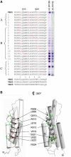
Topology and molecular surfaces of talin 482–789. (A) Ribbon diagram of the talin 482–789 topology (centre) with helices labelled H1–H9 from the N-terminus. Charged molecular surface representations of the same orientation as the ribbon diagram are shown at the far right and the opposing view 180° rotated is shown at the far left. (B) The talin 482–789 sequence annotated with three-dimensional structural features using the program JOY (Mizuguchi et al, 1998). Boxed regions span residues within α-helices and are coloured to match the equivalent helix in the topology diagram above. Key to residue annotation is as follows: solvent inaccessible, upper case; solvent accessible, lower case; positive Φ, italic; hydrogen bond to other side chain, tilde; hydrogen bond to main-chain amide, bold; hydrogen bond to main-chain carbonyl, underline.
Figure 2
Figure 2
Vinculin head binding to talin peptide spot arrays. (A) Analysis of Vh′ binding to talin VBS1 and a series of 24 VBS1 synthetic peptide 25-mers with alanine and glutamic acid residues substituted into various positions. For each peptide, the substituted positions are highlighted in red. Peptides A1–A5 represent triple and quadruple alanine substitutions. Peptides B1–B13 represent single and double alanine substitutions. Peptides C1–C6 represent single glutamic acid substitutions. The VBS-like peptides were spot-synthesized on a nitrocellulose membrane (Frank and Overwin, 1996) with 0.5 nmol of peptide per spot and bound Vh′ was detected using a polyclonal GST antibody. The blot was assayed with 50 and 150 nM Vh′ domain with each spot shown to the right of the respective peptide sequence. For comparison, the talin rod VBS2 and VBS3 sequences are shown aligned with VBS1 (Bass et al, 1999). (B) Two views of the talin 482–655 structure are illustrated with residues implicated in Vh′ binding from the peptide substitution experiments highlighted. Residues are colour coded red for a reduction in binding and green for those that were equivalent to the wild type or result in an apparent increase in binding. Helices H1, H2, H3 and H5 are shown as cylinders and the VBS1 helix H4 is shown as a helical ribbon. On the left-hand view, helices H1, H2 and H5 are transparent to illustrate the VBS side chains on the buried face of H4.
Figure 3
Figure 3
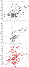
NMR analysis of the talin/vinculin interaction. Two-dimensional [15N/1H]HSQC spectra of uniformly 15N-labelled talin polypeptides. (A) Free talin (482–655). (B) Free talin (482–636). (C) Superposition of the spectra of talin (482–636) free in solution (black) and in the presence of 1.5 molar excess of Vh′ (red).
Figure 4
Figure 4
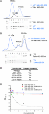
Binding of Vh′ to activated talin mutants. Superdex-75 (10/30) gel filtration chromatography of wild-type and mutant talin polypeptides in the presence/absence of the Vh′ domain. In each case, 0.5 ml fractions were collected and analysed using a Coomassie-stained 4–12% gradient SDS–PAGE gel, which is shown beneath each elution profile. The molecular weight markers of 28 and 17 kDa are indicated. (A) The profile for talin 482–636 is shown as a black line and the mixture with Vh′ as a blue line. Molecular weights as estimated from standards are indicated. An excess of Vh′ is revealed as a 41 kDa peak in the blue profile. (B) Talin 482–655 and a double mutant L608A–L615A complex with Vh′ are shown as black and blue lines, respectively. (C) Tabulated summary of the gel filtration experiments shown in panels A and B and equivalent experiments performed on double alanine mutants V619A–L623A, Q610A–K613A and E621A–R624A where elution profiles are not shown. (D) Binding of GST-Vh′ to microtitre wells coated with the activated talin 482–636 polypeptide. The effects of preincubating GST-Vh′ with increasing concentrations of various talin polypeptides are shown.
Figure 5
Figure 5
Structure and comparison of Vh′/VBS1 and talin 482–655. (A) Complex crystal structure of Vh′ residues (green) and talin VBS1 (blue). (B) Topological equivalence of the VBS1 helix (blue) within the talin 482–655 structure (top) and the Vh′/VBS1 complex structure (bottom). (C) Cα backbone traces of the superposed talin 482–655 (blue) and the structure of the Vh′/VBS1 complex (orange). The structures were superposed based on 15 Cα atoms spanning the five turns of the VBS1 helix. The helices from talin 482–655 are numbered.
Figure 6
Figure 6
Limited proteolysis of talin and vinculin domains. (A) SDS–PAGE. Lane A1: talin 482–655 wild type untreated; A2: trypsin treated; B1: talin 482–655 L608A/L615A double mutant; B2: trypsin treated; C1: talin 482–636; C2: trypsin treated. Talin 482–655 is resistant to proteolysis, whereas the activated mutant talin 482–655 L608A/L615A and talin 482–636 are increasingly susceptible and most likely exist in an unfolded or a partially unfolded state. The reduction in molecular weight observed in lane A2 results from the cleavage of the talin 482–655 histidine tag as confirmed by N-terminal sequencing. (B) SDS–PAGE. Lane A1: vinculin 1–258 (Vh′) untreated; A2: Vh′ trypsin treated; B1; Vh′, talin 482–636 1:1 mixture; B2: trypsin treated. Vh′ is sensitive to proteolysis and is degraded by trypsin. When Vh′ is incubated with talin 482–636, Vh′ becomes partially protease resistant. The remaining structure of the talin helices is degraded. All proteolysis experiments were carried out using a 1:10 wt/wt trypsin incubated for 1 h at 20°C.
Figure 7
Figure 7
Schematic diagram showing the binding/activation of talin and vinculin. Talin is shown bound to integrins via the talin head domain and to F-actin via the C-terminal region of the talin rod. The structure of residues 482–655 (which contains the vinculin-binding site VBS1) is indicated using the same colour scheme as in Figure 1. In this conformation, talin is unable to bind vinculin because the residues involved in vinculin binding to helix 4 (blue) are buried in the hydrophobic core of the five-helix bundle. In order to bind vinculin, talin must undergo a major conformational change in which VBS1 is released from the core of the talin 482–655 structure. The talin VBS1 helix is then available to activate vinculin by inducing a conformational change within Vh′, which displaces Vt, allowing Vt to bind F-actin. Upon binding Vh′, the talin VBS inserts itself into the Vh′ N-terminal domain converting this four-helix bundle (orange) into a stable five-helix bundle.

Similar articles

Cited by

References

    1. Albiges-Rizo C, Frachet P, Block MR (1995) Down regulation of talin alters cell adhesion and the processing of the α5β1 integrin. J Cell Sci 108: 3317–3329 - PubMed
    1. Bakolitsa C, dePereda JM, Bagshaw CR, Critchley DR, Liddington RC (1999) Crystal structure of the vinculin tail suggests a pathway for activation. Cell 99: 603–613 - PubMed
    1. Barsukov IL, Prescot A, Bate N, Patel B, Floyd DN, Bhanji N, Bagshaw CR, Letinic K, Di Paolo G, De Camilli P, Roberts GC, Critchley DR (2003) Phosphatidylinositol phosphate kinase type 1gamma and beta1-integrin cytoplasmic domain bind to the same region in the talin FERM domain. J Biol Chem 278: 31202–31209 - PubMed
    1. Bass MD, Patel B, Barsukov IG, Fillingham IJ, Mason R, Smith BJ, Bagshaw CR, Critchley DR (2002) Further characterization of the interaction between the cytoskeletal proteins talin and vinculin. Biochem J 362: 761–768 - PMC - PubMed
    1. Bass MD, Smith BJ, Prigent SA, Critchley DR (1999) Talin contains three similar vinculin-binding sites predicted to form an amphipathic helix. Biochem J 341: 257–263 - PMC - PubMed

Publication types