. 2004 Aug;24(15):6635-44.
doi: 10.1128/MCB.24.15.6635-6644.2004.

Gene targeting reveals a widespread role for the high-mobility-group transcription factor Sox11 in tissue remodeling

Affiliations

Gene targeting reveals a widespread role for the high-mobility-group transcription factor Sox11 in tissue remodeling

Elisabeth Sock et al. Mol Cell Biol. 2004 Aug.

Abstract

The high-mobility-group domain-containing transcription factor Sox11 is expressed transiently during embryonic development in many tissues that undergo inductive remodeling. Here we have analyzed the function of Sox11 by gene deletion in the mouse. Sox11-deficient mice died at birth from congenital cyanosis, likely resulting from heart defects. These included ventricular septation defects and outflow tract malformations that ranged from arterial common trunk to a condition known as double outlet right ventricle. Many other organs that normally express Sox11 also exhibited severe developmental defects. We observed various craniofacial and skeletal malformations, asplenia, and hypoplasia of the lung, stomach, and pancreas. Eyelids and the abdominal wall did not close properly in some Sox11-deficient mice. This phenotype suggests a prime function for Sox11 in tissue remodeling and identifies SOX11 as a potentially mutated gene in corresponding human malformation syndromes.


Figures

FIG. 1.
FIG. 1.
Targeted disruption of Sox11 in mice. (A) Schematic representation of the targeting construct (top), the Sox11 wild-type locus (middle), and the mutant locus (bottom). The Sox11 open reading frame is shown as a box, with flanking regions shown as bars. Regions of homology between the wild-type locus and the targeting vector are depicted as black bars, and surrounding genomic regions that are not contained in the targeting construct are depicted as white bars. Plasmid backbone sequences of the targeting construct are indicated by thick lines. Restriction sites for BamHI (B) and EcoRI (E) are shown, as are the locations of the 5′ and 3′ probes and the start codon of the Sox11 gene (ATG). LacZ, gene for β-galactosidase; Neo, neomycin resistance selection cassette; Tk, herpes simplex virus thymidine kinase gene cassette. (B) Southern blot analysis of DNAs from wild-type (wt) and heterozygous (+/−) ES cells digested with EcoRI for use with the 5′ probe or with BamHI for use with the 3′ probe. The sizes of bands corresponding to the wild-type and targeted alleles are indicated. (C) PCR-based genotyping of wild-type (wt), heterozygous (+/−), and Sox11-deficient (−/−) embryos at 18.5 dpc. The sizes of DNA fragments (in base pairs) in the DNA size marker (m) are indicated to the left of the agarose gel. c, control without added genomic DNA.
FIG. 2.
FIG. 2.
Developmental expression of β-galactosidase marker gene integrated into Sox11 gene locus. β-Galactosidase activity was detected colorimetrically with the X-Gal substrate in whole-mount (A to E) and cryotome-sectioned (F to K) heterozygous embryos at 8.5 dpc (A), 9.5 dpc (B), 10.5 dpc (C and D), 12.5 dpc (E to G), and 14.5 dpc (H to K). No X-Gal staining was detected in wild-type littermates under identical conditions. Abbreviations: ba, branchial arches; drg, dorsal root ganglia; ef, earflap; el, eye lid; ge, ganglionic eminence; gt, gut; hb, hindbrain; ht, heart; lb, limb bud; ma, mammary anlage; mz, mantle zone of the spinal cord; ov, otic vesicle; pa, pancreas; sc, spinal cord; so, somite; sp, spleen; st, stomach; sym, sympathetic chain ganglia; vb, vibrissae; vz, ventricular zone of the spinal cord.
FIG. 3.
FIG. 3.
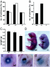
Distribution of genotypes and birth weights in Sox11-deficient embryos and control littermates. (A) Embryos isolated between 8.5 and 15.5 dpc (filled bars [n = 262]) and between 16.5 and 18.5 dpc (open bars [n = 222]) were grouped according to genotype as wild-type (wt), heterozygous (+/−), or Sox11-deficient (−/−). The relative contribution of each genotype is indicated. (B) Relative genotype distribution of 3-week-old mice (n = 98). (C) Body weights were determined (in grams) for embryos at 18.5 dpc after delivery by caesarean section (17 wild-type embryos, 31 heterozygous embryos, and 25 Sox11-deficient embryos). The mean body weight (± standard error of the mean [SEM]) is presented for each genotype. (D) Gross morphology of embryos at 18.5 dpc after delivery by caesarean section. Note the smaller size, facial malformations, and cyanotic appearance of the Sox11-deficient embryo. (E to G) Eyelid closure defects in Sox11-deficient mice (F and G) compared to a control littermate (E) at 18.5 dpc.
FIG. 4.
FIG. 4.
Lung hypoplasia in Sox11-deficient mice. (A and B) Appearance of the lung and the five separate lung lobes at 18.5 dpc. In counterclockwise orientation starting at the top, the lobes are as follows: cranial right lobe, middle right lobe, caudal right lobe, accessory right lobe, and left lobe. (C to F) Hematoxylin-eosin staining of paraffin-embedded sections of the left lobe at low (C and D) and high (E and F) magnifications. (A, C, and E) Wild-type embryo; (B, D, and F) severely affected Sox11-deficient littermate. Note that neither embryo had breathed and that the lungs had not been inflated with air.
FIG. 5.
FIG. 5.
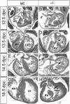
Ventricular septation defects in Sox11-deficient mice. Hematoxylin-eosin-stained paraffin-embedded sections (horizontal plane) of the heart at atrioventricular valve levels in wild-type embryos (wt) and age-matched Sox11-deficient littermates (−/−) at 12.5, 13.5, 14.5, and 18.5 dpc. From 13.5 dpc onwards, the ventricular septum should completely separate the left ventricle (LV) from the right ventricle (RV). Arrows mark the ventricular septation defect.
FIG. 6.
FIG. 6.
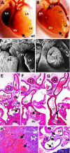
Developmental defects of cardiac outflow tract in Sox11-deficient mice. (A to D) Outer appearance of the heart in wild-type (wt) embryos (A and C) and Sox11-deficient (−/−) littermates (B and D) at 18.5 dpc, as recorded by light (A and B) and scanning electron (C and D) microscopy. In wild-type embryos, the pulmonary trunk (PT) and aorta (A) were clearly separated and originated from their typical positions. In Sox11-deficient embryos, a single arterial common trunk (CT) that originated from the right ventricle (RV) and was continuous with the descending aorta (A), which was situated on the left side, was observed. Arrows in panels A and B mark the interventricular sulcus. RA, right atrium; LA, left atrium; LV, left ventricle. (E and F) Histology of consecutive serial sections (horizontal plane) of the cardiac outflow tract in Sox11-deficient embryos at 18.5 dpc. A common arterial trunk (CT) continues from the right ventricle (RV). Pulmonary arteries (PA) and coronary arteries (CA) originate from the common trunk (E and F), which is continuous with the descending aorta (G). A, aorta; Es, esophagus; Tr, trachea; LA, left atrium. Arrows mark the positions of the semilunar valves. (H) Hemorrhagic infarction and necrosis in the lung of a Sox11-deficient embryo (18.5 dpc), with a complete absence of pulmonary arteries. Br, bronchiole. (I) Stenosis of the pulmonary trunk (PT) in a Sox11-deficient embryo at 18.5 dpc. A, aorta; CA, coronary artery; RA, right atrium; Thy, thymus. Arrows mark the positions of the semilunar valves.
FIG. 7.
FIG. 7.
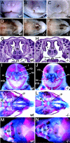
Cleft palate and cranial malformations in Sox11-deficient embryos. (A to F) Outer appearance of snout (A to C [frontal view]) and palate (D to F [ventral view after removal of mandibles]) in wild-type (A and D) and Sox11-deficient (B, C, E, and F) embryos at 18.5 dpc. The cleft lip in Sox11-deficient embryos can be one-sided (B) or two-sided (C) and is associated with a cleft palate (F). Arrows in panel E mark the mild palatal cleft observed in a Sox11-deficient embryo without an obvious cleft lip. (G and H) Histology of coronal sections of wild-type (wt) and Sox11-deficient (−/−) heads at 18.5 dpc. (I to N) Alizarin red and alcian blue staining of cranial bones and cartilage in wild-type (wt) embryos (I, K, and M) and Sox11-deficient (−/−) littermates (J, L, and N) at 18.5 dpc. (I and J) Frontal view of skull; (K and L) ventral view of skull; (M and N) dorsal view of skull. Abbreviations: f, frontal bone; ip, interparietal bone; m, maxilla; mb, mandible; nb, nasal bone; nc, nasal capsule; ns, nasal septum; p, palatine bone; ps, palatal shelves; pt, pterygoid bone; so, supraoccipital bone; t, tongue; v, vomeronasal organ.
FIG. 8.
FIG. 8.
Skeletal malformations in Sox11-deficient embryos. (A to C) Alizarin red (bone) and alcian blue (cartilage) staining of rib cages (ventral view) of wild-type (wt) embryo (A) and Sox11-deficient (−/−) littermates (B and C) at 18.5 dpc. Insets show magnifications of the caudal sternum. xp, xiphoid process of sternum. (D) Alizarin red (bone) and alcian blue (cartilage) staining of spinal column (dorsal view) of Sox11-deficient embryos at 18.5 dpc at thoracolumbar level. The asterisk marks the missing 13th rib. Arrows point to additional rib studs on the first lumbar vertebra. Two spinal columns are shown, with each being half-sided. (E and F) Lumbar spinal columns of wild-type (wt) and Sox11-deficient embryos at 18.5 dpc from the ventral aspect showing malformations of vertebral bodies. (G and H) Kinked tail of Sox11-deficient mouse at 18.5 dpc shown by alizarin red and alcian blue staining (G) and by outer appearance (H). The arrow in panel G points to the fused caudal vertebrae. (I and J) Hindlimbs of wild-type (wt) and Sox11-deficient (−/−) mice at 18.5 dpc showing reduced bone formation of talus (white arrow in panel I) and phalanges (black arrowheads in panel J). (K) Clavicles from Sox11-deficient (−/−) (top) and wild-type (wt) (bottom) siblings at 18.5 dpc showing abnormal ventral curvature of the Sox11-deficient clavicle.
FIG. 9.
FIG. 9.
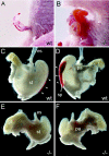
Abdominal wall closure defects, asplenia, and stomach hypoplasia in Sox11-deficient embryos. (A and B) Abdominal body wall malformations in Sox11-deficient mice (B) compared to their control littermates (A) at 18.5 dpc. (C to F) Appearance of stomach (st) with spleen (sp; white arrowheads) and pancreas (pa) of wild-type (C and D) and Sox11-deficient (E and F) mice at 18.5 dpc from ventral (C and E) and dorsal (D and F) aspects. es, esophagus; gt, gut.

Similar articles

Cited by

References

    1. Abu-Issa, R., G. Smyth, I. Smoak, K.-I. Yamamura, and E. N. Meyera. 2002. Fgf8 is required for pharyngeal arch and cardiovascular development in the mouse. Development 129:4613-4625. - PubMed
    1. Bernstein, E. 2004. The cardiovascular system, p. 413-437. In R. E. Behrman, R. M. Kliegman, and A. M. Arvin (ed.), Nelson textbook of pediatrics, 17th ed. Saunders, Philadelphia, Pa.
    1. Bowles, J., G. Schepers, and P. Koopman. 2000. Phylogeny of the SOX family of developmental transcription factors based on sequence and structural indicators. Dev. Biol. 227:239-255. - PubMed
    1. Brewer, S., W. Feng, J. Huang, S. Sullivan, and T. Williams. 2004. Wnt1-Cre-mediated deletion of AP-2alpha causes multiple neural crest-related defects. Dev. Biol. 267:132-152. - PubMed
    1. Brewer, S., X. Jiang, S. Donaldson, T. Williams, and H. M. Sucov. 2002. Requirement for AP-2α in cardiac outflow tract morphogenesis. Mech. Dev. 110:139-149. - PubMed

Publication types

MeSH terms