. 2004 Jul 5;166(1):73-83.
doi: 10.1083/jcb.200310098.

Monoubiquitination and endocytosis direct gamma-secretase cleavage of activated Notch receptor

Affiliations

Monoubiquitination and endocytosis direct gamma-secretase cleavage of activated Notch receptor

Neetu Gupta-Rossi et al. J Cell Biol. .

Erratum in

  • J Cell Biol. 2004 Nov 8;167(3):following 562

Abstract

Activation of mammalian Notch receptor by its ligands induces TNFalpha-converting enzyme-dependent ectodomain shedding, followed by intramembrane proteolysis due to presenilin (PS)-dependent gamma-secretase activity. Here, we demonstrate that a new modification, a monoubiquitination, as well as clathrin-dependent endocytosis, is required for gamma-secretase processing of a constitutively active Notch derivative, DeltaE, which mimics the TNFalpha-converting enzyme-processing product. PS interacts with this modified form of DeltaE, DeltaEu. We identified the lysine residue targeted by the monoubiquitination event and confirmed its importance for activation of Notch receptor by its ligand, Delta-like 1. We propose a new model where monoubiquitination and endocytosis of Notch are a prerequisite for its PS-dependent cleavage, and discuss its relevance for other gamma-secretase substrates.


Figures

Figure 1.
Figure 1.
Endocytosis is required for γ-secretase cleavage of Notch ΔE. (A) Endocytosis is required for nuclear localization of ΔE derivatives. HeLa cells were transiently transfected with ΔE (A and C) and/or a dominant-negative form of GFP-fused Eps15 (B and C, EPS15DN). In A and B, living cells were first incubated with CY3-Tf for 1 h at 37°C. Eps15DN was visualized directly by the fluorescence emitted by GFP (B1, B3, C1, and C3), whereas ΔE was detected using an anti-myc antibody (Alexa 488® labeled in green: A1 and A3; CY3 labeled in red: C2 and C3). CY3-Tf reveals the internalization of transferrin receptor (in red: A2, A3, B2, and B3). Panels numbered 3 are merges of the corresponding panels 1 and 2. (B) γ-Secretase processing of ΔE is inhibited by overexpression of dominant-negative forms of dynamin2 (dynK44A) and Eps15 (Eps15DN). 293T cells were transfected with plasmids encoding ΔE (lanes 1–4), ΔE along with dynK44A (lanes 5 and 6) or Eps15DN (lanes 7 and 8), or ICv (lane 9). Cells were pulse labeled with [35S]methionine and chased for 1 h. 50 μM MW167 (lanes 3 and 4) was added as indicated in the Materials and methods. Anti-myc immunoprecipitated cell extracts were analyzed by SDS-PAGE followed by autoradiography. ΔETM indicates the ΔE membrane-anchored form, asterisks mark 95- and 150-kD migrating products corresponding respectively to dynK44A (lanes 5 and 6) and Eps15DN (lanes 7 and 8). The ratio between ICv and ΔETM was quantified in this representative experiment using phosphorimager analysis to 38, 9, and 20%, corresponding respectively to lanes 2, 6, and 8. The open arrowhead shows a slower migrating band stabilized in the presence of MW167 (lanes 3 and 4).
Figure 1.
Figure 1.
Endocytosis is required for γ-secretase cleavage of Notch ΔE. (A) Endocytosis is required for nuclear localization of ΔE derivatives. HeLa cells were transiently transfected with ΔE (A and C) and/or a dominant-negative form of GFP-fused Eps15 (B and C, EPS15DN). In A and B, living cells were first incubated with CY3-Tf for 1 h at 37°C. Eps15DN was visualized directly by the fluorescence emitted by GFP (B1, B3, C1, and C3), whereas ΔE was detected using an anti-myc antibody (Alexa 488® labeled in green: A1 and A3; CY3 labeled in red: C2 and C3). CY3-Tf reveals the internalization of transferrin receptor (in red: A2, A3, B2, and B3). Panels numbered 3 are merges of the corresponding panels 1 and 2. (B) γ-Secretase processing of ΔE is inhibited by overexpression of dominant-negative forms of dynamin2 (dynK44A) and Eps15 (Eps15DN). 293T cells were transfected with plasmids encoding ΔE (lanes 1–4), ΔE along with dynK44A (lanes 5 and 6) or Eps15DN (lanes 7 and 8), or ICv (lane 9). Cells were pulse labeled with [35S]methionine and chased for 1 h. 50 μM MW167 (lanes 3 and 4) was added as indicated in the Materials and methods. Anti-myc immunoprecipitated cell extracts were analyzed by SDS-PAGE followed by autoradiography. ΔETM indicates the ΔE membrane-anchored form, asterisks mark 95- and 150-kD migrating products corresponding respectively to dynK44A (lanes 5 and 6) and Eps15DN (lanes 7 and 8). The ratio between ICv and ΔETM was quantified in this representative experiment using phosphorimager analysis to 38, 9, and 20%, corresponding respectively to lanes 2, 6, and 8. The open arrowhead shows a slower migrating band stabilized in the presence of MW167 (lanes 3 and 4).
Figure 2.
Figure 2.
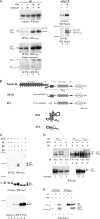
PSs interact with a monoubiquitinated form of ΔE, ΔE u . (A) Analysis of PS1–Notch interaction. 293T cells were transfected with ΔE (lanes 1–4), ΔE+CT (lanes 5 and 6), and PS1 (lanes 3, 4, and 6) expression vectors for 24 h, then treated with MW167 for 3 h when indicated. Extracts or PS1 immunoprecipitates (IP) were analyzed by SDS-PAGE (4–12%, lanes 1–4; 5%, lanes 5 and 6) and immunoblotted with antibodies against myc, Notch, or the loop domain of PS1 (recognizing PS1FL and PS1CTF). ΔETM and ΔE+CTTM represent the membrane-anchored forms encoded by the expression vectors; ΔEu and ΔE+CTU are the novel modified forms revealed by this assay. (B) Constructs used in this paper. Top, Notch derivatives: FL, ΔE, and ICv forms, and the epitopes recognized by the antibodies used are represented. Note that pepex and V1744 respectively recognize the membrane-anchored ΔE and the γ-secretase cleavage product ICv, whereas anti-myc and anti-Notch (not depicted) antibodies detect all the derivatives. FLTM represents the TM subunit of Notch heterodimer resulting from furin cleavage. Bottom, PS2 and its derivative, ΔC4. ΔC4 encodes the NH2-terminal 168 aa of PS2, containing its two first TM domains. (C) ΔC4 specifically interacts with ΔE-derived forms. 293T cells were transfected with either Notch FL (lanes 1 and 2), ΔE (lanes 3 and 4), or ICv (lanes 5 and 6), along with ΔC4 (even-numbered lanes). Total cell extracts (middle and bottom) and their immunoprecipitates obtained with anti-PS2 antibody (top) were analyzed by Western blotting using anti-myc (top and middle) or V1744 (bottom). (D) ΔEu results from monoubiquitination of ΔE. 293T cells were transiently transfected with ΔE, along with ΔC4 and either WT ubiquitin (HA-tagged, lanes 1 and 2; VSV-tagged, lanes 4 and 4′) or VSV-tagged UbKO (lanes 5 and 5′). In the top panel, cell extracts were immunoprecipitated with anti-PS2 (lanes 1, 4′, and 5′), anti-HA (lanes 2 and 3), or anti-VSV (lanes 4 and 5) before SDS-PAGE analysis. In the bottom panel, 2% of total protein lysates used for IP were loaded. Extracts from WTVSV or KOVSV Ub-transfected cells (bottom, lanes 4 and 5, respectively) were divided into two halves for IP with either VSV (lanes 4 and 5) or PS2 (lanes 4′ and 5′). Immunoblots were performed using anti-myc antibody. Asterisks indicate the ΔEu form in whole-cell extracts. (E) Dimerization of ΔE molecules. 293T cells were transfected with myc-tagged ΔE and/or an untagged, COOH-terminally truncated ΔE, ΔER5, as indicated. The corresponding cell extracts were immunoblotted with anti-Notch antibody directly (lanes 1–3) or after anti-myc immunoprecipitation (lanes 4–6). The products derived from each construct are indicated: ΔEu, ΔETM, and ICv; and ΔER5u, ΔER5TM, and ICvR5. White lines indicate that intervening lanes have been spliced out.
Figure 3.
Figure 3.
Lysine 1749 monoubiquitination precedes γ-secretase cleavage of ΔE. (A) Sequence alignment of the juxtamembrane region of various Notch receptors, beginning at the first residue of ΔE (not including the signal peptide). aa 1704–1753 of murine Notch l (first line) were compared with homologous and orthologous sequences (m: mouse, h: human, c: chicken, x: Xenopus, d: Drosophila). The TM region is boxed in gray and arrows mark the TACE and γ-secretase cleavage sites. The double-headed arrow spans the immunopeptide sequence used to generate the pepex antibody (see Materials and methods). Sequence changes in K1749R and LLFF are underlined and italicized. Note that except Notch 4, all Notch sequences contain a conserved juxtamembrane lysine residue (aa 1749 of mNotch1, arrow in bold). (B) Subcellular localization of mutant forms of ΔE. HeLa cells were transiently transfected by ΔE (panel 1), LLFF (panel 2), or K1749R (panel 3) expression vectors. Cells were fixed, Triton-permeabilized, and stained for fluorescence microscopy using the anti-myc antibody revealed by Alexa 488®–coupled secondary antibody. (C) Monoubiquitination of K1749 is required for γ-secretase cleavage. 293T cells were transfected with ΔC4 (lanes 1–3), various ΔE forms (WT: lanes 1, 4, and 5; LLFF: lanes 2 and 7; K1749R: lanes 3 and 6), or ICv (lane 8). In lanes 1–3, cell extracts were subjected to immunoprecipitation with anti-PS2 antibody, analyzed by immunoblotting with anti-ubiquitin antibody (top), and reprobed with anti-Notch antibody (bottom). In lanes 4–8, cell extracts were directly blotted with V1744 (top) or anti-myc (bottom) antibodies. White lines indicate that intervening lanes have been spliced out.
Figure 3.
Figure 3.
Lysine 1749 monoubiquitination precedes γ-secretase cleavage of ΔE. (A) Sequence alignment of the juxtamembrane region of various Notch receptors, beginning at the first residue of ΔE (not including the signal peptide). aa 1704–1753 of murine Notch l (first line) were compared with homologous and orthologous sequences (m: mouse, h: human, c: chicken, x: Xenopus, d: Drosophila). The TM region is boxed in gray and arrows mark the TACE and γ-secretase cleavage sites. The double-headed arrow spans the immunopeptide sequence used to generate the pepex antibody (see Materials and methods). Sequence changes in K1749R and LLFF are underlined and italicized. Note that except Notch 4, all Notch sequences contain a conserved juxtamembrane lysine residue (aa 1749 of mNotch1, arrow in bold). (B) Subcellular localization of mutant forms of ΔE. HeLa cells were transiently transfected by ΔE (panel 1), LLFF (panel 2), or K1749R (panel 3) expression vectors. Cells were fixed, Triton-permeabilized, and stained for fluorescence microscopy using the anti-myc antibody revealed by Alexa 488®–coupled secondary antibody. (C) Monoubiquitination of K1749 is required for γ-secretase cleavage. 293T cells were transfected with ΔC4 (lanes 1–3), various ΔE forms (WT: lanes 1, 4, and 5; LLFF: lanes 2 and 7; K1749R: lanes 3 and 6), or ICv (lane 8). In lanes 1–3, cell extracts were subjected to immunoprecipitation with anti-PS2 antibody, analyzed by immunoblotting with anti-ubiquitin antibody (top), and reprobed with anti-Notch antibody (bottom). In lanes 4–8, cell extracts were directly blotted with V1744 (top) or anti-myc (bottom) antibodies. White lines indicate that intervening lanes have been spliced out.
Figure 4.
Figure 4.
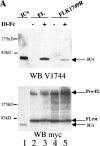
Lysine 1749 is critical for Notch signaling. (A) 293T cells were transfected with either ICv (lane 1), Notch FL (lanes 2 and 3), or FLK1749R (lanes 4 and 5). Notch FL activation was performed by overnight incubation with either control conditioned media (indicated as “−” in lanes 2 and 4) or clustered Dl-Fc– containing media (+, lanes 3 and 5). Total cell extracts were analyzed by V1744 Western blotting (top) and were reprobed with anti-myc antibody (bottom). Full-length Notch precursor is designated Pro-FL. (B) HeLa cells were transiently transfected with Notch FL (panels 1–4) or FLK1749R (panels 5–8). Ligand-binding assay was performed for 1 h with either control conditioned media (CM) or clustered Dl-Fc containing media (Dl-Fc). Notch was detected using an anti-myc antibody revealed by an Alexa 488®–coupled secondary antibody. Even-numbered panels show phase-contrast sections (differential interference contrast), odd-numbered panels an overlay of differential interference contrast and fluorescence. The bottom diagram represents the quantitative analysis of nuclear staining of Dl-Fc–activated cells (n = 28 and 23 for Notch FL and FLK1749R, respectively); 68% of WT Notch-expressing cells exhibited at least 40% nuclear staining when activated by the ligand, whereas none of the FLK1749R cells did.
Figure 4.
Figure 4.
Lysine 1749 is critical for Notch signaling. (A) 293T cells were transfected with either ICv (lane 1), Notch FL (lanes 2 and 3), or FLK1749R (lanes 4 and 5). Notch FL activation was performed by overnight incubation with either control conditioned media (indicated as “−” in lanes 2 and 4) or clustered Dl-Fc– containing media (+, lanes 3 and 5). Total cell extracts were analyzed by V1744 Western blotting (top) and were reprobed with anti-myc antibody (bottom). Full-length Notch precursor is designated Pro-FL. (B) HeLa cells were transiently transfected with Notch FL (panels 1–4) or FLK1749R (panels 5–8). Ligand-binding assay was performed for 1 h with either control conditioned media (CM) or clustered Dl-Fc containing media (Dl-Fc). Notch was detected using an anti-myc antibody revealed by an Alexa 488®–coupled secondary antibody. Even-numbered panels show phase-contrast sections (differential interference contrast), odd-numbered panels an overlay of differential interference contrast and fluorescence. The bottom diagram represents the quantitative analysis of nuclear staining of Dl-Fc–activated cells (n = 28 and 23 for Notch FL and FLK1749R, respectively); 68% of WT Notch-expressing cells exhibited at least 40% nuclear staining when activated by the ligand, whereas none of the FLK1749R cells did.
Figure 5.
Figure 5.
Kinetics of ΔE endocytosis compared with LLFF and K1749R. HeLa cells were transiently transfected with ΔE (A panels), LLFF (B panels), or K1749R (C panels). Living cells were incubated with an antibody directed against the extracellular part of Notch ΔE (pepex, in green: A1–A4, B1–B3, and C1–C3) and CY3-Tf (in red: A1–A3) at 4°C (T0), or were incubated at 37°C for either 20 min (T20; A2, A4, B2, and C2) or 45 min (T45). Cells were fixed, saponin permeabilized, and incubated with anti-myc (in blue: A3, B4, and C4) or anti-clathrin antibodies (in red: A4) and the appropriate dye-labeled secondary antibodies. Same fields are presented in B3 and B4, and in C3 and C4. The inset in A4 shows a higher magnification of the boxed region and highlights ΔE and clathrin colocalization (in yellow). Preparations were analyzed by confocal microscopy (0.3-μm sections).

Similar articles

Cited by

References

    1. Annaert, W.G., L. Levesque, K. Craessaerts, I. Dierinck, G. Snellings, D. Westaway, P.S. George-Hyslop, B. Cordell, P. Fraser, and B. De Strooper. 1999. Presenilin 1 controls γ-secretase processing of amyloid precursor protein in pre-Golgi compartments of hippocampal neurons. J. Cell Biol. 147:277–294. - PMC - PubMed
    1. Benmerah, A., M. Bayrou, N. Cerf-Bensussan, and A. Dautry-Varsat. 1999. Inhibition of clathrin-coated pit assembly by an Eps15 mutant. J. Cell Sci. 112:1303–1311. - PubMed
    1. Bland, C.E., P. Kimberly, and M.D. Rand. 2003. Notch-induced proteolysis and nuclear localization of the Delta ligand. J. Biol. Chem. 278:13607–13610. - PubMed
    1. Brou, C., F. Logeat, N. Gupta, C. Bessia, O. LeBail, J.R. Doedens, A. Cumano, P. Roux, R.A. Black, and A. Israël. 2000. A novel proteolytic cleavage involved in Notch signaling: The role of the disintegrin-metalloprotease TACE. Mol. Cell. 5:207–216. - PubMed
    1. Buttner, C., S. Sadtler, A. Leyendecker, B. Laube, N. Griffon, H. Betz, and G. Schmalzing. 2001. Ubiquitination precedes internalization and proteolytic cleavage of plasma membrane-bound glycine receptors. J. Biol. Chem. 276:42978–42985. - PubMed

Publication types

MeSH terms