. 2004 Sep;15(9):4003-10.
doi: 10.1091/mbc.e04-04-0293. Epub 2004 Jun 23.

The cochaperone HspBP1 inhibits the CHIP ubiquitin ligase and stimulates the maturation of the cystic fibrosis transmembrane conductance regulator

Affiliations

The cochaperone HspBP1 inhibits the CHIP ubiquitin ligase and stimulates the maturation of the cystic fibrosis transmembrane conductance regulator

Simon Alberti et al. Mol Biol Cell. 2004 Sep.

Abstract

The CHIP ubiquitin ligase turns molecular chaperones into protein degradation factors. CHIP associates with the chaperones Hsc70 and Hsp90 during the regulation of signaling pathways and during protein quality control, and directs chaperone-bound clients to the proteasome for degradation. Obviously, this destructive activity should be carefully controlled. Here, we identify the cochaperone HspBP1 as an inhibitor of CHIP. HspBP1 attenuates the ubiquitin ligase activity of CHIP when complexed with Hsc70. As a consequence, HspBP1 interferes with the CHIP-induced degradation of immature forms of the cystic fibrosis transmembrane conductance regulator (CFTR) and stimulates CFTR maturation. Our data reveal a novel regulatory mechanism that determines folding and degradation activities of molecular chaperones.


Figures

Figure 1.
Figure 1.
HspBP1 is associated with Hsc70 and CHIP. (A) HeLa cells were transfected with pCMVTag2-hspBP1, which encodes a FLAG-tagged form of HspBP1 (+ FLAG-HspBP1), or with an equal amount of a control plasmid (-). HspBP1-containing protein complexes were immunoprecipitated with an anti-FLAG antibody (IP) and analyzed for the presence of Hsc70 and CHIP by immunoblotting. The chaperone and CHIP were detected in ATP-eluates of the isolated complexes. To monitor expression levels, 40 μg of protein extracts were loaded (ex.). The star denotes a polypeptide that cross-reacts with the anti-CHIP antibody in cell extracts. (B) Purified His-tagged HspBP1 was immobilized and incubated with Hsc70 and CHIP as indicated. Proteins retained on the affinity resin were detected using specific antibodies. As negative control, proteins were incubated with the affinity resin in the absence of HspBP1. The panel on the very left shows 33% of the input. (C) Binding of Hsc70 to immobilized HspBP1 was analyzed in the presence of different concentrations of BAG-1 and CHIP, respectively, ranging from an equimolar amount to a two- and fivefold molar excess over HspBP1. The left panel represents 66% of the input. (D) Hsc70 binding to BAG-1 is blocked in the presence of increasing amounts of HspBP1. All reactions received purified BAG-1 protein. When indicated equimolar amounts of Hsc70 were added. The HspBP1 concentration was increased from equimolar amounts to a two- and fivefold molar excess over BAG-1. BAG-1 and associated Hsc70 were isolated using a specific anti-BAG-1 antibody. When indicated rabbit IgG was used instead of anti-BAG-1 antibody to monitor unspecific binding. The left panel represents 20% of the input. (E) Overexpression of HspBP1 increases the amount of Hsc70 detectable in CHIP-containing protein complexes. HeLa cells were transfected with pCMVTag2-CHIP (+ FLAG-CHIP) and pcDNA3.1-hspBP1 (+ HspBP1) as indicated. The total amount of added DNA was kept constant by addition of pcDNA3.1. CHIP-containing protein complexes were isolated using an immobilized anti-FLAG antibody (IP). Immunoprecipitated CHIP was detected in glycine eluates using a specific antibody. HspBP1 and Hsc70 were detected in ATP eluates of the isolated CHIP complexes by immunoblotting. Extracts (40 μg) were loaded to monitor expression levels (ex.).
Figure 2.
Figure 2.
HspBP1 inhibits the ubiquitin ligase activity of CHIP when complexed with Hsc70. (A) In vitro ubiquitylation reactions were performed in the presence of purified CHIP, the ubiquitin-activating enzyme E1, and increasing concentrations of HspBP1, ranging from 0.1- to 10-fold the concentration of CHIP, as indicated. All reactions received UbcH5b and Hsc70. Raf-1, Hsc70, ubiquitylated forms of both proteins [ub(n)-Raf-1, ub(n)-Hsc70], and HspBP1 were detected using specific antibodies. Notably, Hsc70 that carried more than three ubiquitin moieties was not recovered by SDS-PAGE analysis. The stars denote polypeptides that cross-react with the anti-HspBP1 antibody. Numbers represent the molar ratio between HspBP1 and CHIP. (B) Luciferase was heat-denatured in the presence of Hsc70 (top) and Hsp90 (bottom), followed by addition of the CHIP ubiquitin conjugation system and increasing amounts of HspBP1. Luciferase was detected with a specific antibody.
Figure 3.
Figure 3.
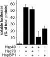
HspBP1 does not interfere with the stabilization of heat-denatured luciferase mediated by Hsc70. Luciferase was heat denatured in the presence of Hsp40, Hsc70 and HspBP1 as indicated and the amount of soluble luciferase was determined after removal of protein aggregates by centrifugation. Values were averaged from three independent experiments. Bars represent SEs.
Figure 4.
Figure 4.
HspBP1 increases the steady-state levels of coexpressed CFTR and CFTRΔF508 in HEK293 cells. (A) HEK293 cells were transiently transfected with CFTR-, HspBP1-, and CHIP-encoding plasmids in the indicated combinations. Amounts of pcDNA3.1-hspBP1 were one- and twofold of the amount of pcDNA3.1-CHIP when indicated. The ER-localized, core-glycosylated B-form (B) and the fully glycosylated, plasma membrane-localized C-form of CFTR (C) were detected by immunoblotting. Similarly, HspBP1 and CHIP levels were detected with specific antibodies. Immunodetection of Hsc70 and Hsp70 served as loading control. Each lane represents 60 μg of cellular extract. (B) Analysis of the cellular levels of CFTRΔF508 (ΔF508) in the presence of HspBP1 and CHIP as described under A.
Figure 5.
Figure 5.
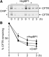
HspBP1 slows down the rate of CFTR degradation. (A) Kinetics of CFTR degradation was determined in pulse-chase experiments by using HEK293 cells transiently transfected with CFTR-, CHIP-, and HspBP1-expressing plasmids as indicated. At the given time points cell extracts were prepared and radiolabeled CFTR was immunoprecipitated and deglycosylated. (B) Data obtained in pulse chase experiments for control cells (squares) or upon coexpression of HspBP1 (circles) and CHIP (triangles) were quantified. Values for CFTR turnover in the presence of CHIP and HspBP1 were omitted because degradation kinetics was indistinguishable from the control reaction in the absence of cochaperones. Presented data were averaged from four independent experiments. Error bars represent SEs.
Figure 6.
Figure 6.
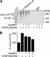
HspBP1 inhibits the CHIP-induced ubiquitylation of CFTR in vitro. (A) CFTR was in vitro translated in the presence of microsomes (IVT), followed by the addition of CHIP, UbcH5b, and HspBP1 at a one-, two-, and fivefold molar excess over CHIP as indicated. Samples were incubated for 1 h followed by immunprecipitation with an anti-ubiquitin antibody that specifically binds ubiquitin conjugates (α-ubiquitin IP). Control reactions received unrelated immunoglobulins (IgG). `IVT' represents 20% of the sample used for immunoprecipitation. (B) Densitometric quantification of ubiquitylated CFTR [ub(n)-CFTR] immunoprecipitated under A. Values were averaged from two independent experiments.
Figure 7.
Figure 7.
HspBP1 stimulates the sorting of CFTR to the plasma membrane. Glycosylated CFTR exposed on the surface of HEK293 cells was biotinylated and subsequently isolated by avidin affinity chromatography. Surface-biotinylated proteins and total cell extracts were analyzed by SDS-PAGE and immunoblotting with an anti-CFTR antibody. Cells were processed 24 h after transfection with CFTR-, HspBP1-, and CHIP-expressing plasmids in the indicated combinations. To monitor CFTR expression, 40 μg of cell extracts was analyzed by immunoblotting.
Figure 8.
Figure 8.
HspBP1 is essential for CFTR maturation. HEK293 cells were transiently transfected with CFTR-, HspBP1 shRNA-, and HspBP1-expressing plasmids as indicated. Expression levels were analyzed 72 h posttransfection after separation of 40 μg of cell extracts on an SDS-PAG and immunoblotting by using specific antibodies. Hsc/Hsp70 served as loading control. Cells that did not express HspBP1 shRNA received the pSUPER plasmid without an insert.
Figure 9.
Figure 9.
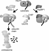
Model of the regulation of CHIP activity by the Hsc70 cochaperones BAG-1 and HspBP1. CHIP binds to the carboxy terminus of Hsc70 (C) and recruits the ubiquitin-conjugating enzyme UbcH5 to the chaperone complex. The two proteins cooperate to mediate the ubiquitylation of a chaperone client bound to the peptide-binding domain of Hsc70 (P). Simultaneous association of BAG-1 with the ATPase domain of Hsc70 (A) stimulates the CHIP-induced degradation of a subset of chaperone clients. On the other hand, HspBP1 inhibits the ubiquitin ligase activity of CHIP within the assembled chaperone complex.

Similar articles

Cited by

References

    1. Alberti, S., Demand, J., Esser, C., Emmerich, N., Schild, H., and Höhfeld, J. (2002). Ubiquitylation of BAG-1 suggests a novel regulatory mechanism during the sorting of chaperone substrates to the proteasome. J. Biol. Chem. 277, 45920-45927. - PubMed
    1. Ballinger, C.A., Connell, P., Wu, Y., Hu, Z., Thompson, L.J., Yin, L.Y., and Patterson, C. (1999). Identification of CHIP, a novel tetratricopeptide repeat-containing protein that interacts with heat shock proteins and negatively regulates chaperone functions. Mol. Cell. Biol. 19, 4535-4545. - PMC - PubMed
    1. Cardozo, C.P., Michaud, C., Ost, M.C., Fliss, A.E., Yang, E., Patterson, C., Hall, S.J., and Caplan, A.J. (2003). C-terminal Hsp-interacting protein slows androgen receptor synthesis and reduces its rate of degradation. Arch. Biochem. Biophys. 410, 134-140. - PubMed
    1. Connell, P., Ballinger, C.A., Jiang, J., Wu, Y., Thompson, L.J., Höhfeld, J., and Patterson, C. (2001). The co-chaperone CHIP regulates protein triage decisions mediated by heat-shock proteins. Nat. Cell Biol. 3, 93-96. - PubMed
    1. Dai, Q., et al. (2003). CHIP activates HSF1 and confers protection against apoptosis and cellular stress. EMBO J. 22, 5446-5458. - PMC - PubMed

Publication types

MeSH terms