

Commitment of C3H10T1/2 pluripotent stem cells to the adipocyte lineage
- PMID: 15210946
- PMCID: PMC470722
- DOI: 10.1073/pnas.0403100101
Commitment of C3H10T1/2 pluripotent stem cells to the adipocyte lineage
Abstract
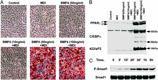
The increase of adipose tissue mass associated with obesity is due in part to an increase in the number of adipocytes. This hyperplasia results from recruitment of pluripotent stem cells present in the vascular stroma of adipose tissue. A model cell culture system has been developed that recapitulates this process both ex vivo and in vivo. After treatment of pluripotent C3H10T1/2 stem cells with bone morphogenic protein 4 (BMP4) during proliferation followed by differentiation inducers at growth arrest, the cells synchronously enter S phase and undergo mitotic clonal expansion, a hallmark of preadipocyte differentiation. Upon exiting the cell cycle, these cells express adipocyte markers and acquire adipocyte characteristics at high frequency. C3H10T1/2 cells treated with BMP4 in cell culture and implanted s.c. into athymic mice develop into tissue indistinguishable from adipose tissue in normal fat depots. We interpret the findings as evidence that BMP4 is capable of triggering commitment of pluripotent C3H10T1/2 stem cells to the adipocyte lineage.
Figures


Similar articles
-
BMP signaling pathway is required for commitment of C3H10T1/2 pluripotent stem cells to the adipocyte lineage.Proc Natl Acad Sci U S A. 2009 Aug 4;106(31):12670-5. doi: 10.1073/pnas.0906266106. Epub 2009 Jul 20. Proc Natl Acad Sci U S A. 2009. PMID: 19620713 Free PMC article.
-
Involvement of cytoskeleton-associated proteins in the commitment of C3H10T1/2 pluripotent stem cells to adipocyte lineage induced by BMP2/4.Mol Cell Proteomics. 2011 Jan;10(1):M110.002691. doi: 10.1074/mcp.M110.002691. Epub 2010 Aug 16. Mol Cell Proteomics. 2011. PMID: 20713452 Free PMC article.
-
Induction of EMT-like response by BMP4 via up-regulation of lysyl oxidase is required for adipocyte lineage commitment.Stem Cell Res. 2013 May;10(3):278-87. doi: 10.1016/j.scr.2012.12.005. Epub 2013 Jan 5. Stem Cell Res. 2013. PMID: 23395997
-
Cell line models for differentiation: preadipocytes and adipocytes.Exp Biol Med (Maywood). 2010 Oct;235(10):1185-93. doi: 10.1258/ebm.2010.010063. Epub 2010 Sep 23. Exp Biol Med (Maywood). 2010. PMID: 20864461 Review.
-
Regulators of adipocyte precursor cells.Poult Sci. 1997 Jan;76(1):118-23. doi: 10.1093/ps/76.1.118. Poult Sci. 1997. PMID: 9037698 Review.
Cited by
-
Shp2 suppresses the adipogenic differentiation of preadipocyte 3T3-L1 cells at an early stage.Cell Death Discov. 2016 Jul 4;2:16051. doi: 10.1038/cddiscovery.2016.51. eCollection 2016. Cell Death Discov. 2016. PMID: 27551539 Free PMC article.
-
The WNT inhibitor Dickkopf 1 and bone morphogenetic protein 4 rescue adipogenesis in hypertrophic obesity in humans.Diabetes. 2012 May;61(5):1217-24. doi: 10.2337/db11-1419. Epub 2012 Mar 23. Diabetes. 2012. PMID: 22447857 Free PMC article.
-
Indomethacin promotes adipogenesis of mesenchymal stem cells through a cyclooxygenase independent mechanism.J Cell Biochem. 2010 Nov 1;111(4):1042-50. doi: 10.1002/jcb.22793. J Cell Biochem. 2010. PMID: 20672310 Free PMC article.
-
CCN1/Cyr61 is regulated by the canonical Wnt signal and plays an important role in Wnt3A-induced osteoblast differentiation of mesenchymal stem cells.Mol Cell Biol. 2006 Apr;26(8):2955-64. doi: 10.1128/MCB.26.8.2955-2964.2006. Mol Cell Biol. 2006. PMID: 16581771 Free PMC article.
-
Mitochondrial dysfunction in obesity.Curr Opin Endocrinol Diabetes Obes. 2010 Oct;17(5):446-52. doi: 10.1097/MED.0b013e32833c3026. Curr Opin Endocrinol Diabetes Obes. 2010. PMID: 20585248 Free PMC article. Review.
References
-
- Hirsch, J. & Batchelor, B. (1976) Clin. Endocrinol. Metab. 5, 299-311. - PubMed
-
- Shepherd, P. R., Gnudi, L., Tozzo, E., Yang, H., Leach, F. & Kahn, B. B. (1993) J. Biol. Chem. 268, 22243-22246. - PubMed
-
- Yu, Z. K., Wright, J. T. & Hausman, G. J. (1997) Obes. Res. 5, 9-15. - PubMed
-
- Caplan, A. (1991) J. Orthop. Res. 9, 641-650. - PubMed
Publication types
MeSH terms
Substances
Grants and funding
LinkOut - more resources
Full Text Sources
Other Literature Sources
