. 2004 Jul;78(13):6723-34.
doi: 10.1128/JVI.78.13.6723-6734.2004.

A novel severe acute respiratory syndrome coronavirus protein, U274, is transported to the cell surface and undergoes endocytosis

Affiliations

A novel severe acute respiratory syndrome coronavirus protein, U274, is transported to the cell surface and undergoes endocytosis

Yee-Joo Tan et al. J Virol. 2004 Jul.

Abstract

The severe acute respiratory syndrome coronavirus (SARS-CoV) genome contains open reading frames (ORFs) that encode for several genes that are homologous to proteins found in all known coronaviruses. These are the replicase gene 1a/1b and the four structural proteins, nucleocapsid (N), spike (S), membrane (M), and envelope (E), and these proteins are expected to be essential for the replication of the virus. In addition, this genome also contains nine other potential ORFs varying in length from 39 to 274 amino acids. The largest among these is the first ORF of the second longest subgenomic RNA, and this protein (termed U274 in the present study) consists of 274 amino acids and contains three putative transmembrane domains. Using antibody specific for the C terminus of U274, we show U274 to be expressed in SARS-CoV-infected Vero E6 cells and, in addition to the full-length protein, two other processed forms were also detected. By indirect immunofluorescence, U274 was localized to the perinuclear region, as well as to the plasma membrane, in both transfected and infected cells. Using an N terminus myc-tagged U274, the topology of U274 and its expression on the cell surface were confirmed. Deletion of a cytoplasmic domain of U274, which contains Yxxphi and diacidic motifs, abolished its transport to the cell surface. In addition, U274 expressed on the cell surface can internalize antibodies from the culture medium into the cells. Coimmunoprecipitation experiments also showed that U274 could interact specifically with the M, E, and S structural proteins, as well as with U122, another protein that is unique to SARS-CoV.


Figures
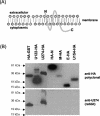
FIG. 1.
FIG. 1.
Rabbit polyclonal antibody specific for U274, the first ORF in the second largest subgenomic RNA of SARS-CoV. (A) Schematic diagram showing the predicted topology of U274; (B) Western blot analysis of various HA-tagged SARS-CoV viral proteins expressed in mammalian cells and probed with an anti-HA polyclonal antibody (upper panel) or the anti-U274 rabbit polyclonal antibody (lower panel).
FIG. 2.
FIG. 2.
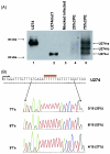
Expression and processing of U274 in SARS-CoV-infected Vero E6 cells. (A) Western blot analysis with anti-U274 rabbit polyclonal detected three protein bands in SARS-CoV-infected cells showing 25% (lane 4) and 75% (lane 5) CPE but not in mock-infected cells (lane 3). The highest (marked as U274-a) and lowest (marked as U274-c) molecular mass proteins in the infected cells migrated at the same rate as full-length U274 (lane 1) and U274mut1, which corresponds to the last 174 aa of U274 (lane 2), respectively. (B) Reverse transcription-PCR products obtained with RNA from infected cells were ligated into pCRII-TOPO vector and sequenced. Chromatographs showed that stretches of seven, eight, and nine T's were present in this region of sgRNA3, which corresponded to 16 to 21 bp downstream of the initiation codon (ATG) of U274. Analysis of the sequences of 18 different SARS-CoV isolates showed that all of them only have six T's in this region. The percentages of independent clones with the respectively number of T nucleotides are shown on the left.
FIG. 3.
FIG. 3.
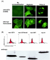
Cellular localization of U274 in infected and transfected Vero E6 cells as determined by indirect immunofluorescence. (A) SARS-CoV-infected cells stained with anti-U274 rabbit polyclonal antibody; (B) SARS-CoV-infected cells stained with anti-S mouse polyclonal antibody; (C) overlay of panels A and B showing cells that are expressing both S and U274 proteins (yellow); (D) SARS-CoV-infected cells (higher magnification); (E) Vero E6 cells transfected with untagged U274; (F) Vero E6 cells transfected with U274-HA; (G) Vero E6 cells transfected with myc-U274. Panels D to G are stained with anti-U274 rabbit polyclonal antibody.
FIG. 4.
FIG. 4.
Detection of myc-U274 on the cell surface by indirect immunofluorescence and FACS analysis. (A) Vero E6 cells transiently transfected with myc-U274 were stained with either the anti-U274 rabbit polyclonal antibody (upper panel) that recognizes the C terminus of the protein or anti-myc monoclonal antibody (lower panel) that recognizes N terminus of the protein. Cells were fixed with 4% paraformaldehyde and used directly (no permeabilization) or permeabilized with digitonin or permeabilized with Triton X-100 before incubation with antibodies. (B) FACS analysis of live Vero E6 cells transfected with myc-U274, myc-U274mut2, myc-GST, or myc-N. Cells were incubated with a myc-monoclonal antibody (unfilled histogram) or an irrelevant antibody of the same isotype (filled histograms), followed by the addition of FITC-conjugated goat anti-mouse F(ab′)2 antibody. Surface expression of myc-U274 caused an increase in surface fluorescence upon staining with myc-monoclonal antibody. The expression levels of the proteins were determined by Western blot analysis (with anti-myc antibody).
FIG. 5.
FIG. 5.
Internalization of antibodies by U274 and other cell surface receptors. Cells transfected with myc-GST (lanes 1 and 2 for Vero E6) or myc-U274 (lanes 3 and 4 for Vero E6 and lanes 11 and 12 for HeLa) were incubated with an anti-myc monoclonal antibody for 3 h at 37°C, followed by PBS washes or PBS-acid washes, and then the cells were harvested and lysed in Laemmli SDS buffer and subjected to Western blot analysis. The cell lysate for cells that were only washed with PBS would contain both surface-bound and internalized antibody (total). For cells that were subjected to an additional acid wash remove surface-bound antibody, the lysate would contain antibody that has been internalized (internalized). The heavy chains of the surface bound and internalized antibody (IgG heavy chains, upper panel) were detected by Western blotting. To compare the internalization efficiency of U274 with CD81 and TfR, the cells were overlaid with anti-human CD81 (lanes 5 and 6 for Vero E6 cells and lanes 13 and 14 for HeLa cells) or anti-human TfR (lanes 9 and 10 for Vero E6 cells transfected with a cDNA encoding for full-length human TfR and lanes 15 and 16 for HeLa cells) antibodies, and the amounts of total versus internalized antibodies were determined as described above. Expression levels of myc-GST or myc-U274 were determined by using anti-myc antibody (second panel). Expression levels of TfR were determined by using anti-TfR antibody (third panel). The level of endogenous actin protein was used to check for equal loading (lowest panel). This experiment was repeated three times, and similar results were obtained each time. A representative set of data is presented.
FIG. 6.
FIG. 6.
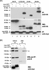
Interactions between U274 and other SARS-CoV proteins. (A) Cell lysates containing myc-GST or myc-U274 with either HA-N, U122-HA, E-HA, M-HA, or U274-HA proteins were immunoprecipitated with myc-polyclonal antibody and protein A-beads. The amounts of HA-tagged proteins that coimmunoprecipitated (IP) with myc-GST (lanes 1, 3, 5, and 7) or myc-U274 (lanes 2, 4, 6, and 8) were determined with an anti-HA monoclonal antibody (upper panel). Expression levels of the HA-tagged proteins were determined by subjecting aliquots of the lysates before immunoprecipitation to Western blot analysis (WB, middle panel). The first blot containing the immunocomplexes (i.e., upper panel) was stripped and reprobed with an anti-myc monoclonal antibody to determine the amount of myc-tagged proteins immunoprecipitated (lower panel). (B) Expression (before IP) and coimmunoprecipitation (after IP) of untagged S protein with myc-GST or myc-U274 were determined with an anti-S mouse polyclonal antibody (upper panel). Expression levels of myc-GST and myc-U274 in the lysates were determined by using anti-myc monoclonal antibody (before IP, lower panel).

Similar articles

Cited by

References

    1. Bannykh, S. I., N. Nishimura, and W. E. Balch. 1998. Getting into the Golgi. Trends Cell Biol. 8:21-25. - PubMed
    1. Baudoux, P., C. Carrat, L. Besnardeau, B. Charley, and H. Laude. 1998. Coronavirus pseudoparticles formed with recombinant M and E proteins induce alpha interferon synthesis by leukocytes. J. Virol. 72:8636-8643. - PMC - PubMed
    1. Bonifacino, J. S., and L. M. Traub. 2003. Signals for sorting of transmembrane proteins to endosomes and lysosomes. Annu. Rev. Biochem. 72:395-447. - PubMed
    1. Bos, E. C., W. Luytjes, H. V. van der Meulen, H. K. Koerten, and W. J. Spaan. 1996. The production of recombinant infectious DI-particles of a murine coronavirus in the absence of helper virus. Virology 218:52-60. - PMC - PubMed
    1. Bost, A. G., R. H. Carnahan, X. T. Lu, and M. R. Denison. 2000. Four proteins processed from the replicase gene polyprotein of mouse hepatitis virus colocalize in the cell periphery and adjacent to sites of virion assembly. J. Virol. 74:3379-3387. - PMC - PubMed

Publication types

LinkOut - more resources