. 2004 Jun 15;18(12):1423-38.
doi: 10.1101/gad.1200304. Epub 2004 Jun 2.

Phosphorylation of SMC1 is a critical downstream event in the ATM-NBS1-BRCA1 pathway

Affiliations

Phosphorylation of SMC1 is a critical downstream event in the ATM-NBS1-BRCA1 pathway

Risa Kitagawa et al. Genes Dev. .

Abstract

The ATM protein kinase is activated by intermolecular autophosphorylation in response to DNA damage and initiates cellular signaling pathways that facilitate cell survival and reduce chromosomal breakage. Here, we show that NBS1 and BRCA1 are required for the recruitment of previously activated ATM to the sites of DNA breaks after ionizing irradiation, and that this recruitment is required for the phosphorylation of SMC1 by ATM. To explore the functional importance of SMC1 phosphorylation, murine cells were generated, in which the two damage-induced phosphorylation sites in SMC1 are mutated. Although these cells demonstrate normal phosphorylation and focus formation of ATM, NBS1, and BRCA1 proteins after IR, they exhibit a defective S-phase checkpoint, decreased survival, and increased chromosomal aberrations after DNA damage. These observations suggest that many of the abnormal stress responses seen in cells lacking ATM, NBS1, or BRCA1 result from a failure of ATM migration to sites of DNA breaks and a resultant lack of SMC1 phosphorylation.


Figures
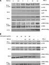
Figure 1.
Figure 1.
Dose dependencies of ATM activation and phoshorylation of ATM substrates. (A) Human primary fibroblast cells at early passage (<10) were exposed to indicated doses of ionizing irradiation and harvested 15 min later. Total and phosphorylated ATM and NBS1 were assessed by immunoblots after immunoprecipitation. Total and phosphorylated p53 and SMC1 were assessed by immunoblots of whole-cell lystates (WCE). Phophorylated histone H2AX was immunoblotted after acid cellular extraction (see Materials and Methods). (B) Human primary fibroblast cells at early passage (<10) were exposed to indicated doses of chloroquine for 4 h, then exposed as indicated to ionizing irradiation. Fifteen minutes after irradiation, cells were collected, and immunoblot analysis was performed as indicated.
Figure 2.
Figure 2.
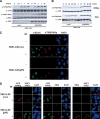
Influence of NBS1 on IR-induced phosphorylation and localization of ATM and ATM substrates. (A) NBS1-LB1 cells with [p95(NBS1)] or without (vector) stable retroviral complementation with the p95/NBS1 gene were exposed to indicated doses of ionizing irradiation and cellular extracts analyzed by immunoblotting. Total and phosphorylated ATM and SMC1 were assessed by immunoblotting after immunoprecipitation, and total cellular NBS1 was assessed by immunoblot. (B) Autophosphorylation of ATM in NBS1-deficient cells exposed to a low dose of ionizing radiation. Total and phosphorylated ATM was assessed by immunoblot after immunoprecipitation in cells collected at the indicated times after exposure to 0.5 or 2 Gy of IR. (C) Subcellular localization of H2AXγ and phophorylated ATM in NBS1-deficient or -proficient cells. Cells were fixed 15 min after 0 Gy (IR-) or 5 Gy (IR+) of γ irradiation, and then analyzed by immunofluorescence with the indicated antibodies. DNA was visualized by staining with DAPI. (D) Subcellular localization of phosphorylated ATM, phosphorylated SMC1, and NBS1 as a function of NBS1 status. Cells were fixed 15 min after 0 Gy (IR-) or 5 Gy (IR+), then costained with pairs of antibodies as indicated. DNA was visualized by staining with DAPI. For costaining with NBS1 antibodies, mouse monoclonal antibodies to ATMS1981p were used instead of the rabbit polyclonal antibodies that were used for other experiments in this study.
Figure 3.
Figure 3.
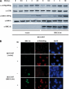
Influence of BRCA1 on IR-induced phosphorylation and localization of ATM. (A) HCC1937 (BRCA1-deficient) cells were transiently transfected with a human expression vector, carrying (BRCA1wt) or not carrying (vector) a wild-type BRCA1 gene. Following exposure to indicated doses of ionizing irradiation, total and phosphorylated ATM was assessed by immunoblot after immunoprecipitation, and total and phosphorylated SMC1 and total BRCA1 were assessed by immunoblot of whole-cell extracts. (B) Subcellular localization of BRCA1, phophorylated ATM, and H2AXγ in BRCA1-deficient or -proficient cells. Cells were fixed 15 min after 0 Gy (IR-) or 5 Gy (IR+) of γ irradiation, then labeled with antibodies or DAPI as indicated.
Figure 4.
Figure 4.
Interdependencies of IR-induced foci formation and pbosphorylation of NBS1 and BRCA1. (A,B) Subcellular localization of NBS1 and BRCA1 in HCC1937 (BRCA1-deficient) cells transfected with a transient expression vector carrying BRCA1 gene (BRCA1wt) or an empty vector (vector; A) and NBS1-LB1 (NBS1-deficient) cells stably transfected with a retrovirus vector carrying p95/Nbs1 gene (p95) or an empty vector (vec) were analyzed by immunofluorescence microscopy (B). Cells were fixed 15 min after 0 Gy (IR-) or 5 Gy (IR+) of γ irradiation, then labeled with anti-NBS1 (shown in red) or anti-BRCA1 (shown in green). DNA was visualized by staining with DAPI (shown in blue). (C) HCC1937 (BRCA1-deficient) cells were transiently transfected with a human expression vector, pcDNA3 carrying (BRCA1wt) or not carrying (vector) wild-type BRCA1 gene. Following exposure to indicated doses of ionizing irradiation, cellular extracts were subjected to immunoblotting. Cell extracts were blotted with antibody to phosphorylated Ser 343 of NBS1, then reblotted with anti-NBS1 antibody. (D) Cellular extracts of NBS1-LB1 cells stably transfected with a retrovirus vector carrying p95/NBS1 gene [p95(NBS1)] or an empty vector (vector) exposed to indicated doses of ionizing irradiation were separated on 5% Tris-acetate gel and blotted with anti-BRCA1 antibody.
Figure 5.
Figure 5.
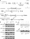
Targeted modification of the mouse Smc1 gene and effects on radiation responses. (A) Schematic of Smc1 knock-in procedure. (panel i) Genomic structure of the mouse Smc1 gene on chromosome X. The whole genomic Smc1 gene consists of 26 exons shown as boxes. Targeted region into which to insert the knock-in vector is shown as a thick bar. (panel ii) Targeting construct of the knock-in vector, pSmc1KI–Neotk, targeted locus, and targeted locus following cre-mediated recombination. Neomycin resistance and thymidine kinase genes (Neo-tk) flanked by loxP sites (shown as black triangles) are inserted into an intron between exon 18 and 19. In pSmc1KI–Neotk, two nucleotide exchanges to change serine to alanine at amino acid positions 957 and 966 are made in exon 19 (shown as a dark box labeled as 19*). Homologus recombination of the pSmc1KI–Neotk is depicted by the large “X”s. Positions of the flanking probe for genotyping and BstEII sites are indicated. (B) Wild-type (Smc1WT) or Smc1S957AS966A immortalized mouse fibroblast cells were exposed to 0 Gy (IR-) or 10 Gy (IR+) of ionizing irradiation and cellular extracts were prepared 30 min later. Whole-cell lysates were subjected to immunoblotting with antibodies as indicated. (C) Thirty minutes after exposure to 0 (-) or 10 (+) Gy of ionizing irradiation (IR), whole-cell lysates were prepared from wild-type or mutant knock-in fibroblasts were immunoblotted with anti-mouse NBS1 or BRCA1 antibodies. Duplicate samples of cell lysates from irradiated knock-in cells were treated with protein phosphatase (PPase+) prior to electrophoresis.
Figure 6.
Figure 6.
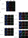
SMC1 phosphorylation is not required for IR-induced formation of foci containing phospho-ATM, H2AXγ, NBS1, 53BP1, phosphorylated CHK2, or BRCA1. Wild-type (Smc1WT) or Smc1 phosphorylation mutant knock-in (Smc1S957AS966A) fibroblast cells were fixed with 4% paraformaldehyde 30 min after 0 Gy (-) 10 Gy (+) of ionizing irradiation (IR), then subjected to immunofluorescence microscopy. (A) Staining with antibodies recognizing phosphorylated ATM or SMC1. (B) Staining with antibodies recognizing H2AXγ, NBS1, 53BP1, phosphorylated CHK2, BRCA1, and phosphorylated SMC1. For costaining of phosphorylated SMC1 (red) with BRCA1 (green), rabbit polyclonal anti-Ser957p antibody was used.
Figure 7.
Figure 7.
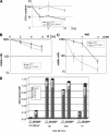
Radiation response abnormalities in Smc1 mutant knock-in cells. (A) Defect in the S-phase checkpoint. MEFs derived from wild-type (Smc1WT) or Smc1S957AS966A embryos were assessed for inhibition of DNA synthesis 30 min after exposure to indicated doses of IR. Error bars are average of triplicate samples. (B) Increased sensitivity to IR. Wild-type or knock-in immortalized mouse fibroblast cells were plated in 6-well culture dishes and irradiated with the indicated doses of ionizing radiation (IR). Cell viability was assessed 72 h after treatment, and is plotted as the percent viable cells relative to results for untreated control cultures. Each point represents the mean of three samples, with error-bars showing standard deviation. The radiosensitivity of Smc1 mutant knock-in fibroblasts was indistinguishable from that seen in ATM-null murine fibroblasts (data not shown). (C) Increased sensitivity to alkylating agents. Wild-type or Smc1 mutant knock-in immortalized mouse fibroblast cells were plated in 6-well culture dishes, and treated with methylmethane sulfonate (MMS) for 1 h. Cell viability was assessed 72 h after treatment, and is plotted as the percent viable cells relative to results for untreated control cultures. Each point represents the mean of three samples, with error-bars showing standard deviation. (D) Defect in chromosomal repair after IR. MEFs derived from wild-type or knock-in embryos were irradiated with 1 Gy of ionizing irradiation (IR), then treated with colcemid at indicated times after irradiation. Chromosome aberrations were scored in 100 metaphase spreads of each sample and numbers of chromosome gaps (csg), chromosome breaks (csb), chromatid gaps (ctg), and cromatid breaks (ctb) per cell were charted.
Figure 8.
Figure 8.
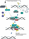
Proposed model for an IR-induced signaling pathway. Chromatin structure changes caused by DNA breakage or other mechanisms leads to intermolecular autophosphorylation of ATM dimers, resulting in release of phosphorylated and active ATM monomers. If DNA-strand breaks are present, several proteins, including NBS1 and BRCA1, are recruited to the sites of the breaks independent of the ATM activation process. After activation, monomeric ATM can phosphorylate nucleoplasmic substrates, like p53, and if NBS1 and BRCA1 have localized to DNA breaks, activated ATM is recruited to the break. At the DNA break, activated ATM can phosphorylate substrates, including SMC1. The phosphorylation of SMC1 reduces chromosomal breakage and enhances cell survival.

Similar articles

Cited by

References

    1. Alligood K.J., Milla, M., Rhodes, N., Ellis, B., Kilpatrick, K.E., Lee, A., Gilmer, T.M., and Lansing, T.J. 2000. Monoclonal antibodies generated against recombinant ATM support kinase activity. Hybridoma 19: 317-321. - PubMed
    1. Bakkenist C.J. and Kastan, M.B. 2003. DNA damage activates ATM through intermolecular autophosphorylation and dimer dissociation. Nature 421: 499-506. - PubMed
    1. Banin S., Moyal, L., Shieh, S., Taya, Y., Anderson, C.W., Chessa, L., Smorodinsky, N.I., Prives, C., Reiss, Y., Shiloh, Y., et al. 1998. Enhanced phosphorylation of p53 by ATM in response to DNA damage. Science 281: 1674-1677. - PubMed
    1. Canman C.E., Wolff, A.C., Chen, C.Y., Fornace Jr., A.J., and Kastan, M.B. 1994. The p53-dependent G1 cell cycle checkpoint pathway and ataxia-telangiectasia. Cancer Res. 54: 5054-5058. - PubMed
    1. Canman C.E., Lim, D.S., Cimprich, K.A., Taya, Y., Tamai, K., Sakaguchi, K., Appella, E., Kastan, M.B., and Siliciano, J.D. 1998. Activation of the ATM kinase by ionizing radiation and phosphorylation of p53. Science 281: 1677-1679. - PubMed

Publication types

MeSH terms

Substances